Korean J Med > Volume 89(3); 2015 > Article
당뇨망막병증 최신 지견

Abstract

Over the past decade, significant advances have been made in both the diagnosis and treatment of diabetic retinopathy. Ultrawide field fundus photography and spectral domain optical coherence tomography have allowed more accurate, convenient, and early diagnosis of diabetic retinopathy. Numerous randomized clinical trials have demonstrated the effectiveness of anti-vascular endothelial growth factor agents for the treatment of diabetic retinopathy, although more work is necessary in terms of long-term clinical outcomes and socioeconomic costs associated with these treatments.

서 론

당뇨망막병증은 당뇨병성 만성합병증 중 하나로 전 세계적으로 중요 실명 원인 중 하나이다[1,2]. 최근 당뇨망막병증의 진단 분야에서는 무산동 광각 안저 촬영(nonmydriatic ultra wide field fundus photography)과 스펙트럼 영역 빛간섭단층촬영(spectral domain optical coherence tomography)이 보편화되고, 치료에서 여러 가지 항혈관내피세포 성장인자들(vascular endothelial growth factor)의 유리체강 내 주사가 광범위하게 사용되고 있다. 또한 역학에서도 제5기 국민건강영양역학조사 결과가 발표되면서 당뇨망막병증의 역학에 대한 최신 결과들이 발표되었다[3]. 이에 당뇨망막병증의 역학, 진단, 그리고 치료에 대한 최신 자료들을 소개하고자 한다.

본 론

역학

의사와 환자들 모두에서 당뇨병성 만성합병증에 대한 인식이 호전되고 당뇨병의 치료제가 다양해지고 향상됨에 따라 제1형 당뇨병의 경우는 물론 제2형 당뇨병 환자들에서도 심한 형태의 당뇨망막병증의 빈도가 감소하였다는 보고들이 있다[4-9]. 하지만 그에 반해 당뇨황반부종의 빈도는 상대적으로 증가하여 이에 대한 진단과 치료에 대한 중요성이 부각되고 있다[4-9]. 한국인에서 최신 당뇨망막병증 역학연구 결과들에 따르면 당뇨병으로 진단받은 환자들에서 당뇨망막병증의 유병률은 약 11-19%까지 보고되고 있으며, 그중 당뇨황반부종이나 증식당뇨망막병증 등 심한 당뇨망막병증의 유병률은 약 5%로 보고되었다[3,10,11]. 그리고 이와 연관된 위험인자들로는 당뇨병 유병기간과 혈중 당화혈색소 수치가 있는 것으로 보고되었다[3,10,11]. 당화혈색소 수치가 8.1% 이상인 경우 정상군에 비해 약 3배 정도로 당뇨망막병증이 생길 확률이 높으며 당뇨병 유병기간이 11년 이상인 경우 약 15배 정도 당뇨망막병증이 생길 확률이 처음 진단받은 사람들에 비해 높은 것으로 발표되었다[3,10,11]. 그 외에도 이 두 가지 위험인자들은 당뇨망막병증의 발생과 더불어 이미 당뇨망막병증을 진단받은 환자들에서 심한 형태의 당뇨망막병증으로의 진행에도 관여하는 것으로 보고되어 당뇨망막병증의 발생과 진행에 혈당관리가 가장 중요함을 재차 확인할 수 있다. 또한 당뇨병 유병기간이 10년 이상이 된 경우 약 반수 이상에서 당뇨망막병증을 가지고 있어 고위험군들에 대한 당뇨망막병증 검진의 강조가 필요하다. 최근 외국의 여러 코호트 연구들에서는 당화혈색소 수치와 같은 기존의 위험인자 외에도 혈압, 지질 대사이상, 임신, 신증, 유전, 비만, 영양 상태 등도 위험인자들로 거론되고 있다. 한국인에서 이런 인자들이 어떤 영향을 주는지에 대한 추가 연구들이 필요하다[12-17].

진단

당뇨망막병증의 진단에 가장 중요한 검사는 안저검사이며 망막 주변부까지 관찰을 해야 하기에 환자들의 동공 산동은 필수적이다. 산동을 하게 되면 몇 시간 동안 독서나 운전 등 일상 생활에 지장이 있고 극히 드문 경우이나 폐쇄각 녹내장이 악화될 가능성이 있어 당뇨병 환자들에서 산동 검사가 힘든 경우가 종종 있어왔다. 최근 무산동 광각 안저 촬영기술이 개발되어 임상에서 사용되기 시작하면서 당뇨망막병증의 진단에 도움을 주고 있다[18-20]. 무산동 광각 안저 촬영은 환자들 산동이 필요 없으며, 기존의 안저 촬영에 비해 넓은 부위를 관찰할 수 있다는 장점들이 있어, 당뇨망막병증의 진단을 위한 환자들의 순응도와 검사의 민감도를 향상시킬 수 있다는 장점들이 있다[18-20]. 특히 당뇨망막병증 환자들에서 놓치기 쉬웠던 주변부 허혈 변화나 신생 혈관들의 진단에 있어 기존 검사에 비해 민감도가 많게는 4배 이상 좋은 것으로 보고되었다(Fig. 1) [18-20]. 하지만 아직은 상대적으로 낮은 황반부 해상도, 망막 외 병변들의 간섭이나 인공물(artifact) 등의 문제점들이 있어 무산동 안저 촬영과 검사만으로 당뇨망막병증을 진단하기에는 무리가 있다[18-20]. 앞으로 무산동 광각 안저 촬영이 당뇨망막병증의 진단에 보편적으로 사용되기 위해서는 이에 대한 보완이 필요하다.
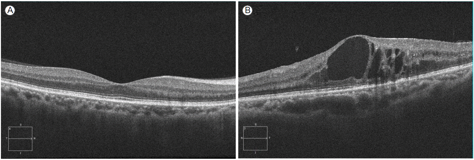
빛간섭단층촬영은 안과 진단 분야에서 21세기 최대의 발명이라 해도 과언이 아닐 정도로, 망막은 물론 모든 안과 영역에서 진단과 치료에 광범위하게 사용되고 있다. 빛간섭단층촬영은 몇 초라는 짧은 시간 내에 검사가 가능하며, 비침습적이며, 황반 상태를 실시간, 정량적으로 측정할 수 있다는 장점들이 있다. 특히 스펙트럼 영역 빛간섭단층촬영(spectral domain optical coherence tomography)이 개발되어 해상도가 월등히 좋아지면서 여러 망막질환에 있어서 진단이나 추적관찰에 필수검사로 사용되고 있다[21-23]. 최근 연구에 따르면 무증상의 당뇨황반부종을 찾아내는 데 있어 높은 민감도와 특이도를 보여주고 있으며, 레이저 광응고술이나 유리체강 내 주사 치료 전후에 있어서 진단에 필수적인 검사가 되었다(Fig. 2) [21-23]. 또한 당뇨황반부종 환자에서 중심 망막두께는 물론 망막 내외층의 특징적인 소견들을 분석하여 시력 예후를 예측하고자 하는 연구 결과들이 발표되고 있다[21-23].

치료

현재까지 당뇨망막병증의 치료에 있어서 가장 중요한 것은 혈당관리이다. 하지만 최근 당뇨망막병증을 치료하기 위해 많은 새로운 약이나 기구들이 개발되어왔다. 그중 임상연구들을 거쳐 현재 임상에서 보편적으로 사용되고 있는 새로운 약제들을 소개하고자 한다.

스테로이드 유리체강 내 주사

당뇨망막병증의 발생이나 진행에 있어 가장 중요한 역할을 하는 것은 혈관내피세포 성장인자이다[24]. 레이저 치료는 증식당뇨망막병증을 가진 눈에서 허혈 부위를 감소시키고 신생 혈관의 발생이나 출혈을 억제시키는 것으로 고위험 당뇨망막병증 환자에서 급격한 시력 상실을 막는 데 현재까지 보편적으로 사용되어온 치료이다[25]. 하지만 당뇨황반부종이 있는 경우 국소 레이저(focal laser) 치료 후 시력 호전이 어렵고 황반부종의 재발과 레이저 적응증이 제한되어 있어 이를 보완하기 위한 다른 치료 방법들이 집중적으로 연구되어 왔다. 스테로이드 유리체강 내 주사는 혈관내피세포 성장인자들을 억제하면서 당뇨망막병증 환자의 망막에서 만성적인 염증상태를 호전시키기 위해 널리 사용되고 있다[26,27]. 최근 안구 내 주사용으로 보존제 없는 덱사메타손 약이 개발되고 한 번 주사 후 장기간 효과를 나타내는 덱사메타손 유리체강 내 삽입물(dexamethasone intravitreal implant)이 임상에서 사용되기 시작하여 한 번의 시술로 장기간의 황반부종 감소효과를 기대할 수 있게 되었다[28,29]. 하지만 안압 상승이나 백내장 등 스테로이드 약 자체에 따른 부작용에 대한 치료나 이에 대한 보완이 필요하다[30].

항혈관내피세포 성장인자 유리체강 내 주사

당뇨황반부종 환자에서 유리체강 내 스테로이드 주사는 기존의 레이저 치료와 더불어 또는 단독으로 사용되어 시력 호전이나 시력 유지를 얻을 수 있다는 많은 결과들이 발표되어 당뇨황반부종 환자들에서 치료에 큰 도움이 되었다. 하지만 스테로이드 유리체강 내 주입 후 안압 상승과 백내장 진행 등 스테로이드의 부작용 때문에 다른 혈관내피 성장인자 억제제들의 도입이 필요해졌는데 현재까지 사용되는 대표적인 약들로는 bevacizumab, ranibizumab, aflibercept 등이 있다[31-39]. 이 중 ranibizumab (lucentis®, Genentech, Inc., South San Francisco, CA, USA)은 안과 영역에서 처음으로 개발된 혈관내피성장인자 억제제로 혈관내피세포 성장인자 A를 억제하는 단일클론 항체(monoclonal antibody)이다[31-35]. 많은 임상연구 결과들에서 ranibizumab은 혈관내피세포 성장인자를 억제하여 신생 혈관억제와 황반부종을 감소시켜 시력 향상이나 유지에 효과적인 것으로 발표되었다[31-35]. 그러나 주사 후 황반부종 재발이 흔해 반복 주사에 따른 비용이나 감염 등 여러 가지 제한점이 있어 앞으로 이에 대한 보완이 필요하다. Bevacizumab (avastin®, Genentech, Inc., San Francisco, CA, USA)은 비록 안과 영역에서 개발된 약은 아니나 다른 혈관내피세포 성장인자 억제제들과 비교하여 황반부종의 감소나 신생 혈관들의 억제에 있어 거의 동등한 효과를 나타내면서 비용이 낮아 현재 임상에서 많이 사용되고 있다[36]. 하지만 이 역시 문제점이 있는데 안구 내 투여 후 드물게 발생하는 전신 부작용 가능성에 대한 우려가 있으나 다른 약제와 비교해서 통계적으로 차이가 있다고 하기는 힘들다[37]. Aflibercept (Eylea®; Regeneron Pharmaceuticals, Inc., Tarrytown, NY, USA, and Bayer AG, Berlin, Germany)는 가장 최근에 개발되어 임상에서 사용되기 시작한 혈관내피세포 성장인자 억제제로 혈관내피세포 성장인자 수용체에 결합하는 재조합 융합 단백질(recombinant fusion protein)이다[38,39]. 황반부종의 감소에 있어 다른 혈관내피세포 성장인자 억제제들과 동등한 결과를 나타내면서 작용 기간이 상대적으로 길다는 장점이 있으나 이에 대해서는 장기간의 임상 결과들의 확인이 필요하겠다[38,39].

소절개 무봉합 유리체 절제술

최근 모든 수술 분야에서 소절개 수술이 각광을 받고 있는데 안과 영역에서도 예외는 아니어서 기존의 20게이지 절개창을 이용하는 유리체 절제술에 비해 수술 후 봉합이 필요 없는 23게이지나 25게이지 무봉합 유리체 절제술이 널리 시행되고 있다[40-42]. 기존의 유리체 절제술에 비해 수술 후 시력 회복이 빠르고 수술 시간도 단축될 수 있다는 장점들이 있다. 하지만 수술 후 무봉합 절개창의 누출로 인한 감염이나 저안압이 발생할 수 있어 이에 대한 주의가 필요하다[40-42].

결 론

최근 10년 동안 당뇨망막병증의 진단이나 치료에 있어서 획기적인 발전들이 있어 왔다. 하지만 아직도 완벽한 진단 방법이나 예방보다 더 만족스러운 치료 방법은 없다. 앞으로 당뇨망막병증의 치료에서는 단순히 당뇨망막병증을 치료하는 것보다는 당뇨망막병증 환자들의 시력 향상과 더불어 삶의 질까지 고려해야 할 것이다.

REFERENCES

1. Cheung N, Mitchell P, Wong TY. Diabetic retinopathy. Lancet 2010;376:124–136.
crossref

2. Yau JW, Rogers SL, Kawasaki R, et al; Meta-Analysis for Eye Disease (META-EYE) Study Group. Global prevalence and major risk factors of diabetic retinopathy. Diabetes Care 2012;35:556–564.
crossref pmid pmc

3. Yang JY, Kim NK, Lee YJ, et al. Prevalence and factors associated with diabetic retinopathy in a Korean adult population: the 2008-2009 Korea National Health and Nutrition Examination Survey. Diabetes Res Clin Pract 2013;102:218–224.
crossref pmid

4. Hovind P, Tarnow L, Rossing K, et al. Decreasing incidence of severe diabetic microangiopathy in type 1 diabetes. Diabetes Care 2003;26:1258–1264.
crossref pmid

5. Klein R, Klein BE. Is the prevalence of visual impairment rising or falling in the people with diabetes mellitus? It depends on who you study. JAMA Ophthalmol 2013;131:948–950.
crossref pmid

6. Klein R, Knudtson MD, Lee KE, Gangnon R, Klein BE. The Wisconsin Epidemiologic Study of Diabetic Retinopathy: XXII the twenty-five-year progression of retinopathy in persons with type 1 diabetes. Ophthalmology 2008;115:1859–1868.
crossref pmid pmc

7. Nordwall M, Bojestig M, Arnqvist HJ, Ludvigsson J; Linkoping Diabetes Complications Study. Declining incidence of severe retinopathy and persisting decrease of nephropathy in an unselected population of type 1 diabetes-the Linkoping Diabetes Complications Study. Diabetologia 2004;47:1266–1272.
crossref pmid

8. Klein R, Knudtson MD, Lee KE, Gangnon R, Klein BE. The Wisconsin Epidemiologic Study of Diabetic Retinopathy XXIII: the twenty-five-year incidence of macular edema in persons with type 1 diabetes. Ophthalmology 2009;116:497–503.
crossref pmid pmc

9. Klein R, Klein BE. Are individuals with diabetes seeing better?: a long-term epidemiological perspective. Diabetes 2010;59:1853–1860.
crossref pmid pmc

10. Park CY, Park SE, Bae JC, et al. Prevalence of and risk factors for diabetic retinopathy in Koreans with type II diabetes: baseline characteristics of Seoul Metropolitan City-Diabetes Prevention Program (SMC-DPP) participants. Br J Ophthalmol 2012;96:151–155.
crossref pmid

11. Kim YJ, Kim JG, Lee JY, et al. Development and progression of diabetic retinopathy and associated risk factors in Korean patients with type 2 diabetes: the experience of a tertiary center. J Korean Med Sci 2014;29:1699–1705.
crossref pmid pmc

12. Huang YC, Lin JM, Lin HJ, et al. Genome-wide association study of diabetic retinopathy in a Taiwanese population. Ophthalmology 2011;118:642–648.
crossref pmid

13. Sheu WH, Kuo JZ, Lee IT, et al. Genome-wide association study in a Chinese population with diabetic retinopathy. Hum Mol Genet 2013;22:3165–3173.
crossref pmid pmc

14. Dirani M, Xie J, Fenwick E, et al. Are obesity and anthropometry risk factors for diabetic retinopathy? The diabetes management project. Invest Ophthalmol Vis Sci 2011;52:4416–4421.
crossref pmid

15. Klein R, Klein BE, Moss SE. Is obesity related to microvascular and macrovascular complications in diabetes? The Wisconsin Epidemiologic Study of Diabetic Retinopathy. Arch Intern Med 1997;157:650–656.
crossref pmid

16. Tanaka S, Yoshimura Y, Kawasaki R, et al; Japan Diabetes Complications Study Group. Fruit intake and incident diabetic retinopathy with type 2 diabetes. Epidemiology 2013;24:204–211.
crossref pmid

17. Djuric Z, Ren J, Mekhovich O, Venkatranamoorthy R, Heilbrun LK. Effects of high fruit-vegetable and/or low-fat intervention on plasma micronutrient levels. J Am Coll Nutr 2006;25:178–187.
crossref pmid

18. Manivannan A, Plskova J, Farrow A, McKay S, Sharp PF, Forrester JV. Ultra-wide-field fluorescein angiography of the ocular fundus. Am J Ophthalmol 2005;140:525–527.
crossref pmid

19. Wessel MM, Aaker GD, Parlitsis G, Cho M, D’Amico DJ, Kiss S. Ultra-wide-field angiography improves the detection and classification of diabetic retinopathy. Retina 2012;32:785–791.
crossref pmid

20. Wessel MM, Nair N, Aaker GD, Ehrlich JR, D'Amico DJ, Kiss S. Peripheral retinal ischaemia, as evaluated by ultra-widefield fluorescein angiography, is associated with diabetic macular oedema. Br J Ophthalmol 2012;96:694–698.
crossref pmid pmc

21. Lim JI, Zelkha R, Niec M, Setlur V, Shahidi M. Inner and outer retinal thickness mapping of nonproliferative diabetic retinopathy by spectral-domain optical coherence tomography. Ophthalmic Surg Lasers Imaging Retina 2015;46:316–320.
crossref pmid

22. Boynton GE, Stem MS, Kwark L, Jackson GR, Farsiu S, Gardner TW. Multimodal characterization of proliferative diabetic retinopathy reveals alterations in outer retinal function and structure. Ophthalmology 2015;122:957–967.
crossref pmid

23. Bonnin S, Tadayoni R, Erginay A, Massin P, Dupas B. Correlation between ganglion cell layer thinning and poor visual function after resolution of diabetic macular edema. Invest Ophthalmol Vis Sci 2015;56:978–982.
crossref pmid

24. Penn JS, Madan A, Caldwell RB, Bartoli M, Caldwell RW, Hartnett ME. Vascular endothelial growth factor in eye disease. Prog Retin Eye Res 2008;27:331–371.
crossref pmid pmc

25. Photocoagulation for diabetic macular edema. Early Treatment Diabetic Retinopathy Study report number 1. Early Treatment Diabetic Retinopathy Study research group. Arch Ophthalmol 1985;103:1796–1806.
crossref pmid

26. Diabetic Retinopathy Clinical Research Network. A randomized trial comparing intravitreal triamcinolone acetonide and focal/grid photocoagulation for diabetic macular edema. Ophthalmology 2008;115:1447–1449.
crossref pmid pmc

27. Diabetic Retinopathy Clinical Research Network, Elman MJ, Aiello LP. Randomized trial evaluating ranibizumab plus prompt or deferred laser or triamcinolone plus prompt laser for diabetic macular edema. Ophthalmology 2010;117:1064–1077; e35.
crossref pmid pmc

28. Boyer DS, Yoon YH, Belfort R Jr, et al. Three-year, randomized, sham-controlled trial of dexamethasone intravitreal implant in patients with diabetic macular edema. Ophthalmology 2014;121:1904–1914.
crossref pmid

29. Callanan DG, Gupta S, Boyer DS, et al; Ozurdex PLACID Study Group. Dexamethasone intravitreal implant in combination with laser photocoagulation for the treatment of diffuse diabetic macular edema. Ophthalmology 2013;120:1843–1851.
crossref pmid

30. Lauer AK, Bressler NM, Edwards AR; Diabetic Retinopathy Clinical Research Network. Frequency of intraocular pressure increase within days after intravitreal triamcinolone injections in the diabetic retinopathy clinical research network. Arch Ophthalmol 2011;129:1097–1099.
crossref pmid pmc

31. Diabetic Retinopathy Clinical Research Network, Wells JA, Glassman AR, et al. Aflibercept, bevacizumab, or ranibizumab for diabetic macular edema. N Engl J Med 2015;372:1193–1203.
crossref

32. Nguyen QD, Brown DM, Marcus DM, et al; RISE and RIDE Research Group. Ranibizumab for diabetic macular edema: results from 2 phase III randomized trials: RISE and RIDE. Ophthalmology 2012;119:789–801.
crossref pmid

33. Brown DM, Nguyen QD, Marcus DM, et al; RIDE and RISE Research Group. Long-term outcomes of ranibizumab therapy for diabetic macular edema: the 36-month results from two phase III trials: RISE and RIDE. Ophthalmology 2013;120:2013–2022.
crossref pmid

34. Massin P, Bandello F, Garweg JG, et al. Safety and efficacy of ranibizumab in diabetic macular edema (RESOLVE Study): a 12-month, randomized, controlled, double-masked, multicenter phase II study. Diabetes Care 2010;33:2399–2405.
crossref pmid pmc

35. Mitchell P, Bandello F, Schmidt-Erfurth U, et al; RESTORE study group. The RESTORE study: ranibizumab monotherapy or combined with laser versus laser monotherapy for diabetic macular edema. Ophthalmology 2011;118:615–625.
crossref pmid

36. Michaelides M, Kaines A, Hamilton RD, et al. A prospective randomized trial of intravitreal bevacizumab or laser therapy in the management of diabetic macular edema (BOLT study) 12-month data: report 2. Ophthalmology 2010;117:1078–1086; e2.
crossref pmid

37. Moja L, Lucenteforte E, Kwag KH, et al. Systemic safety of bevacizumab versus ranibizumab for neovascular age-related macular degeneration. Cochrane Database Syst Rev 2014;9:CD011230.
crossref

38. Do DV, Schmidt-Erfurth U, Gonzalez VH, et al. The DA VINCI Study: phase 2 primary results of VEGF Trap-Eye in patients with diabetic macular edema. Ophthalmology 2011;118:1819–1826.
crossref pmid

39. Korobelnik JF, Do DV, Schmidt-Erfurth U, et al. Intravitreal aflibercept for diabetic macular edema. Ophthalmology 2014;121:2247–2254.
crossref pmid

40. Farouk MM, Naito T, Sayed KM, et al. Outcomes of 25-gauge vitrectomy for proliferative diabetic retinopathy. Graefes Arch Clin Exp Ophthalmol 2011;249:369–376.
crossref pmid

41. Park DH, Shin JP, Kim SY. Comparison of clinical outcomes between 23-gauge and 20-gauge vitrectomy in patients with proliferative diabetic retinopathy. Retina 2010;30:1662–1670.
crossref pmid

42. Ozone D, Hirano Y, Ueda J, Yasukawa T, Yoshida M, Ogura Y. Outcomes and complications of 25-gauge transconjunctival sutureless vitrectomy for proliferative diabetic retinopathy. Ophthalmologica 2011;226:76–80.
crossref pmid

(A) Standard fundus photography: normal. (B) Standard fundus photography: diabetic retinopathy with multiple retinal hemorrhage and exudates. (C) Ultrawide field fundus photography: normal. (D) Ultrawide field fundus photography: diabetic retinopathy with multiple retinal hemorrhage and exudates.
/upload/thumbnails/kjm-89-3-271f1.gif
Figure 1.
(A) Spectral domain optical coherence tomography: normal fovea. (B) Spectral domain optical coherence tomography: diabetic macular edema with cystic space and disruption of retinal layers.
/upload/thumbnails/kjm-89-3-271f2.gif
Figure 2.
TOOLS
METRICS Graph View
  • 1 Crossref
  •  0 Scopus
  • 10,104 View
  • 312 Download

Editorial Office
101-2501, Lotte Castle President, 109 Mapo-daero, Mapo-gu, Seoul 04146, Korea
Tel: +82-2-2271-6791    Fax: +82-2-790-0993    E-mail: kaim@kams.or.kr                

Copyright © 2024 by The Korean Association of Internal Medicine.

Developed in M2PI

Close layer
prev next