Korean J Med > Volume 81(1); 2011 > Article
약물 용출성 스텐트에서 발견된 신생내막 석회화 1예

요약

본 증례는 ST 분절 상승의 심근경색으로 약물 용출성 스텐트를 삽입한 환자에서 4년 후 발견된 스텐트 내강 안쪽을 둘러싼 원형의 두꺼운 석회침착소견에 대하여 신생내막과 관련된 새로운 죽상동맥경화증으로 추정하여 보고하는 바이다.

Abstract

Neointimal hyperplasia is the main mechanism of stent restenosis. Therefore, drug-eluting stents have replaced bare metal stents because there is less neointima and scar formation. Recently, some cases of stent restenosis after using a bare metal stent were found to involve calcification, not neointimal hyperplasia, and regarded as de novo atherosclerosis. We report unusual circular calcification inside a drug-eluting stent, which we called neointimal calcification. (Korean J Med 2011;81:98-101)

서 론

약물 용출성 스텐트(drug-eluting stent)는 기존 단순 금속 스텐트(bare metal stent)에 비하여 신생내막(neointima)과 반흔(scar) 형성이 적어 재협착을 감소시킨다고 알려져 있고[1-4], 2002년 약물 용출성 스텐트가 도입되면서부터 많은 양의 수요가 있어 왔다. 이에 따라 현재 관상동맥 내로 삽입된 약물 용출성 스텐트의 경과 및 신생내막 형성에 대한 연구가 혈관내초음파(intravascular ultrasonography, IVUS)와 광결합단층촬영(optical coherence tomography, OCT) 등을 통하여 이루어지고 있다. 저자들은 ST 분절상승 심근경색으로 경피적 관상동맥 조영술을 시행하여 약물 용출성 스텐트를 삽입한 환자에게서 4년 뒤 혈관내초음파를 시행하여 발견된 흔하지 않은 석회침착(calcification)을 경험하였기에 보고하는 바이다.

증 례

70세 남자 환자가 내원 3시간 전부터 시작된 흉통을 주소로 응급실 내원하였다. 고혈압, 당뇨로 경구약 복용 중이었으며, 4년 전 ST 분절 상승 심근경색으로 인근 대학병원에서 경피적 관상동맥 조영술을 시행하여 우관상동맥 근위부의 완전폐쇄 및 좌전하관상동맥 중간부위에 80%의 협착소견 발견되어 우관상동맥 근위부로 Paclitacel 용출성 스텐트(Taxus, Boston Scientific, Natick, MA, USA)를 삽입하고 5일 후 이차 관상동맥조영술 시행하여 좌전하관상동맥 중간부위에도 같은 용출성 스텐트를 삽입하였으며, 이후 연고지 관계로 본원으로 의뢰되어 외래에서 경과관찰 중이었던 분이다. 응급실에 내원하여 당시 시행한 심전도상 ST 분절에 이상소견 관찰되지 않고, 심근효소수치 정상으로 불안정성협심증 진단하에 내원한 다음날 관상동맥조영술을 시행하였다.
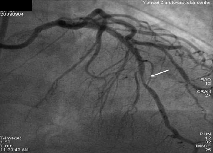
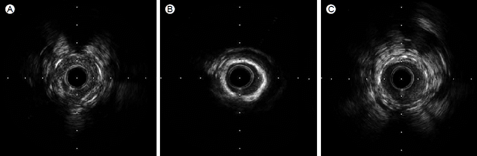
조영술 소견상 좌전하관상동맥의 중간부위에서 4년 전 삽입하였던 스텐트의 원위부에 80%의 재협착 소견(Fig. 1) 및 우관상동맥 원위부로 새로이 90%의 협착소견이 관찰되었다. 또한 4년 전 우관상동맥 원위부로 삽입하였던 스텐트에는 신생내막 과증식으로 인하여 20% 정도의 협착소견이 확인되었다. 좌전하관상동맥의 4년 전 스텐트 삽입한 부위에 혈관내초음파(IVUS)를 시행한 결과 스텐트 내강 안쪽을 둘러싼 원형의 두꺼운 석회침착소견이 관찰되었다(Fig. 2B). 좌전하관상동맥 스텐트 내강으로 협착된 소견 보인 부위와 우관상동맥 원위부에 새로 발견된 협착부위로 각각 Everolimus 용출성 스텐트(Xience, Abbott Vascular, Santa Clara, CA, USA)를 삽입하고 시술을 마쳤으며, 현재 환자 외래에서 흉통 없이 경과관찰 중으로 새로운 스텐트 삽입 후 9개월 뒤에 추적관찰을 위한 관상동맥조영술을 시행 예정이다.

고 찰

관상동맥에서 칼슘이 침착되는 현상은 20-30대 이후부터 일어나는 것으로 되어있으며 유전적 소인 및 임상적인 소인들이 작용하는 것으로 되어있다[5,6]. 관상동맥의 석회화는 관상동맥의 죽상동맥경화반의 총량과 비례하고 관상동맥의 협착을 예측하는 지표가 되며 허혈성 심장질환 발생의 독립적인 위험인자가 되는 것으로 알려져 있다[7-9].
1986년 관상동맥 질환에 처음으로 스텐트가 사용되면서 풍선확장술 후 심각한 합병증이었던 급성 또는 아급성 폐쇄를 현격히 감소시켰으며[10], 이에 따라 스텐트 삽입술 후 재협착을 감소시키는 데 초점이 모아졌다. 재협착의 주요 기전으로 1) 조기 탄력성 재수축(early elastic recoil) 2) 신생내막 과형성(neointimal hyperplasia) 및 3) 늦은 혈관 개형(late vessel remodeling)의 세 가지를 들 수 있는데 이 중 스텐트 삽입술 후에는 신생내막 과형성이 가장 중요한 재협착의 기전으로 알려져 있다. 신생내막 과형성은 시술 후 주로 6개월 내에 발생하며 proteoglycan matrix 내에 평활근 세포(smooth muscle cell)가 과도하게 증식하여 혈관을 재협착시키는 과정으로 이 과정의 촉발은 풍선확장술로 혈관 내막이 손상을 받은 직후부터 발생하는 염증단계에서 병변 내로 분비된 각종 염증세포, 혈소판 및 cytokine 들이 주 요인이라고 여겨진다[11-13]. 2007년 Doyle 등[14]이 4500여 명의 단순 금속 스텐트를 삽입한 환자를 대상으로 조사한 논문에 따르면 10년 내에 임상적으로 의미 있는 스텐트 재협착은 18.1%였으며, 그중 심근경색은 2.1%로 조사되었다.
하지만 Agostoni 등[15]은 11년 전 불안정성 협심증으로 단순 금속 스텐트를 삽입한 환자에게 운동부하검사 도중 발생한 ST 분절상승 심근경색으로 관상동맥조영술을 통해 이전 스텐트 삽입부위가 협착되어 흡착(aspiration)을 시행하였고, 흡착물에서 죽상동맥경화반(atherosclerotic plaque) 조각들을 발견하여 스텐트 주위로 새로운 죽상동맥경화증(de novo atherosclerosis)이 생겼음을 시사한다고 하였다. 또한 Fineschi 등[16]은 이전에 단순 금속 스텐트를 삽입한 안정성협심증 환자에게서 혈관내초음파를 시행하여 파열된 반(ruptured plaque)를 발견하여, 이를 스텐트내 협착증이 아닌 신생내막과 관련하여 생긴 죽상동맥증으로 추정하였다. 그리고 Appleby [17]는 안정성 협심증으로 단순 금속 스텐트를 삽입한 환자에게서 8년 뒤 협심증 증상으로 관상동맥조영술 재시행하면서 혈관내초음파로 스텐트 내강의 신생내막의 일부에서 석회화를 발견하였다고 보고하였으며 새로운 죽상동맥경화증이 스텐트와 관련하여 드물게 생긴 것으로 판단하였다.
본 증례의 경우 단순 금속 스텐트를 삽입한 위의 경우들과는 달리 약물 용출성 스텐트를 삽입한 환자의 혈관내초음파를 통해 스텐트 내강을 원형으로 둘러싼 특이한 형태의 석회화 소견을 찾아냈다. 이는 약물 용출성 스텐트를 삽입함으로써 발생빈도가 적을 것으로 받아들여졌던 신생내막 증식 및 새로운 죽상동맥경화증이 약물 용출성 스텐트와 관련하여 나타난 것으로 생각되고, 상기 병변에 대한 추적관찰이 필요할 것으로 생각한다.

REFERENCES

1. Colombo A, Drzewiecki J, Banning A, et al. Randomized study to assess the effectiveness of slow-and moderate-release polymerbased paclitaxel-eluting stents for coronary artery lesions. Circulation 2003;108:788–794.
crossref pmid

2. Stone GW, Ellis SG, Cox DA, et al. A polymer-based, paclitaxeleluting stent in patients with coronary artery disease. N Engl J Med 2004;350:221–231.
crossref pmid

3. Morice MC, Serruys PW, Sousa JE, et al. A randomized comparison of a sirolimus-eluting stent with a standard stent for coronary revascularization. N Engl J Med 2002;346:1773–1780.
crossref pmid

4. Moses JW, Leon MB, Popma JJ, et al. Sirolimus-eluting stents versus standard stents in patients with stenosis in a native coronary artery. N Engl J Med 2003;349:1315–1323.
crossref pmid

5. Wexler L, Brundage B, Crouse J, et al. Coronary artery calcification: pathophysiology, epidemiology, imaging methods, and clinical implications:a statement for health professionals from the American Heart Association: Wrighting Group. Circulation 1996;94:1175–1192.
crossref pmid

6. McGeachie M, Ramoni RL, Mychaleckyi JC, et al. Integrative predictive model of coronary artery calcification in atherosclerosis. Circulation 2009;120:2448–2454.
crossref pmid pmc

7. Mautner SL, Mautner GC, Froehlich J, et al. Coronary artery disease: prediction with in vitro electron beam CT. Radiology 1994;192:625–630.
crossref pmid

8. Rumberger JA, Simons DB, Fitzpatrick LA, Sheedy PF, Schwartz RS. Coronary artery calcium area by electron-beam computed tomography and coronary: atherosclerotic plaque area: a histopathologic correlative study. Circulation 1995;92:2157–2162.
crossref pmid

9. Sangiorgi G, Rumberger JA, Severson A, et al. Arterial calcification and not lumen stenosis is highly correlated with atherosclerotic plaque burden in humans: a histologic study of 723 coronary artery segments using nondecalcifying methodology. J Am Coll Cardiol 1998;31:126–133.
crossref pmid

10. Fischman DL, Leon MB, Baim DS, et al. A randomized comparison of coronary stent placement and balloon angioplasty in the treatment of coronary artery disease: Stent Restenosis Study Investigatiors. N Engl J Med 1994;331:496–501.
crossref pmid

11. Hoffman R, Mintz GS, Dussaillant GR, et al. Patterns and mechanisms of in-stent restenosis: a serial intravascular ultrasound study. Circulation 1996;94:1247–1254.
crossref pmid

12. Mehran R, Dangas G, Abizaid AS, et al. Angiographic patterns of in-stent restenosis: classification and implications for long-term outcome. Circulation 1999;100:1872–1878.
crossref pmid

13. Kastrati A, Hall S, Schömig A. Long-term outcome after coronary stenting. Curr Control Trials Cardiovasc Med 2000;1:48–54.
crossref pmid pmc

14. Doyle B, Rihal CS, O’Sullivan CJ, et al. Outcomes of stent thrombosis and restenosis during extended follow-up of patients treated with bare-metal coronary stents. Circulation 2007;116:2391–2398.
crossref pmid

15. Agostoni P, Vermeersch P, Knaapen M, Verheye S. Stent thrombosis is not always stent thrombosis: de novo atherosclerosis in a stented coronary segment. Int J Cardiol 2010;144:e19–21.
crossref pmid pmc

16. Fineschi M, Carrera A, Gori T. Atheromatous degeneration of the neointima in a bare metal stent: intravascular ultrasound evidence. J Cardiovasc Med (Hagerstown) 2009;10:572–573.
crossref pmid

17. Appleby CE. A calcified neointima: “Stent” within a stent. J Invasive Cardiol 2009;21:141–143.
pmid

Diagnostic angiography of the left anterior descending (LAD) artery shows approximately 80% restenosis of the distal portion of a drug (paclitaxel)-eluting stent placed in the middle of the LAD (arrow).
/upload/thumbnails/kjm-81-1-98-16f1.gif
Figure 1.
Intravascular ultrasonography of the left anterior descending (LAD), within the previously inserted (paclitaxel)-eluting stent: (A) end of the stent; (B) distal portion of the stent, with circular calcification inside the stent; and (C) proximal portion of the stent.
/upload/thumbnails/kjm-81-1-98-16f2.gif
Figure 2.
TOOLS
METRICS Graph View
  • 4,715 View
  • 89 Download

Editorial Office
101-2501, Lotte Castle President, 109 Mapo-daero, Mapo-gu, Seoul 04146, Korea
Tel: +82-2-2271-6791    Fax: +82-2-790-0993    E-mail: kaim@kams.or.kr                

Copyright © 2024 by The Korean Association of Internal Medicine.

Developed in M2PI

Close layer
prev next