서 론
Noonan syndrome (NS)은 전형적인 얼굴이형성증(dysmorphic face), 선천성 심장질환, 저신장 등으로 표현되는 상염색체우성 질환 중 하나로 알려져 있는데 이는 신생아 1,000-2,500명 당 1명 정도로 보고되고 있으며 이는 임상적 소견으로 진단한다. 이 질환의 진단은 매우 까다롭고 특히 성인에 있어서는 더욱 그러하다[1].
대략 80% 정도의 환자는 심혈관 질환을 동반한다[2]. 선천성 심장질환 중 폐동맥판막협착(pulmonary stenosis)이 가장 흔하고(25-35%) 이는 종종 심방중격결손증(atrial septal defect)을 동반한다. 그 외 비후성심근병증(hypertrophic cardiomyopathy, HCMP)과 부분 방실 결손(partial atrio-ventricular canal defect), 대동맥판막협착(aortic stenosis), 폐성고혈압(pulmonary hypertension), 동맥관개존증(patent ductus arteriosus), 가지 폐동맥 협착(branch pulmonary artery stenosis), 이엽성 대동맥판막(bicuspid aortic valve), 심실중격결손증(ventricular septal defect), 팔로사징후(Tetralogy of Fallot), 대동맥축착증(coarctation of aorta) 등도 동반되는 것으로 알려져 있다[2].
드물지만 다른 심혈관 질환으로는 대동맥박리(aortic dissection), 확장성심근병증(dilated cardiomyopathy), 승모판막이상(mitral valve anomalies), 폐동맥판폐쇄(pulmonary atresia), 대동맥뿌리확장(aortic root dilation), 제한성심근병증(restrictive cardiomyopathy), 삼첨판막의 엡스타인기형(Ebstein's anomaly of tricuspid valve), 관상동맥이상(coronary artery anomalies) 등이 보고되고 있다[2].
하지만 NS와 동반된 신동맥협착(renal artery stenosis)에 대한 보고는 아직 없으며 이에 본 저자들은 성인의 악성고혈압에 동반된 신동맥협착과 함께 폐동맥판막협착, 얼굴이형성증, 오목가슴(pectus excavatum)의 임상 증상으로 진단된 NS의 증례를 보고한다.
증 례
환 자: 27세 남자
주 소: 3년 전부터 시작되어 1달 전부터 악화된 호흡곤란
현병력: 환자는 내원 3년 전부터 New York Heart Association Functional Class II (NYHA Fc II)의 호흡곤란이 있었으며 최근 내원 1달 전부터 NYHA Fc III로 증상이 악화되어 외래 통해 내원하였다.
과거병력: 내원 3년 전 고혈압을 진단받았으나 약은 복용하지 않고 있었다.
가족력 및 사회력: 가족 중 특이 병력을 가진 사람은 없었으며 주 1회 회당 소주 1병, 10갑년 흡연력이 있었다.
약물복용력: 없음.
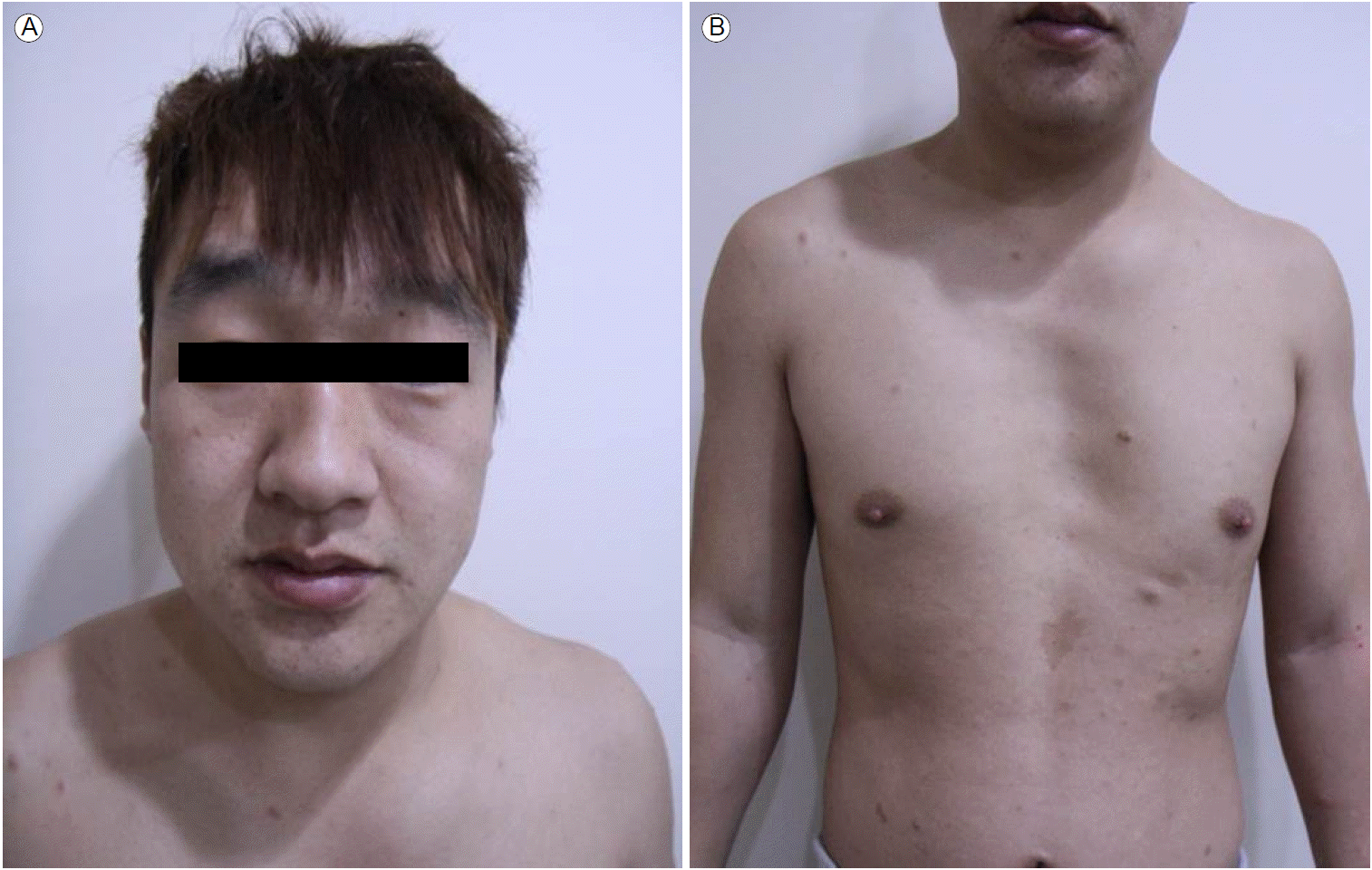
신체검진: 내원 당시 측정한 활력 징후는 혈압 187/113 mmHg, 맥박수 60회/분, 호흡 18회/분, 체온 36.7℃였으며 의식은 명료하였다. 흉부청진상 수축기 심잡음이 들렸다. 얼굴이형성증(hypertelorism, short neck, broad forehead 등)을 보였으며(Fig. 1A), 오목가슴(Fig. 1B), 저신장(161.6 cm, 5 percentile 미만)을 보였다[3].
검사실 소견: 말초 혈액 검사 소견은 정상범위 내의 potassium 4.1 mmol/L를 포함해 24시간 뇨 vanillyl mandelic acid 2.2 mg/day, metanephrine 105.7 μg/day, normetanephrine 236.6 μg/day, epinephrine 8.2 μg/day, norepinephrine 46.2 μg/day, 그리고 renin 2.78 ng/mL/hr, aldosterone 11.0 ng/dL 등으로 정상 범위였고, antinuclear antibody도 음성소견을 보였다. 갑상선 기능검사도 정상이었다.
흉부엑스선 소견: 양 폐에 폐혈관 음영의 증가 소견이 관찰되었으며, 왼쪽 심장경계의 직선화 소견을 보였다.
심전도 소견: 정상 동성 리듬이었으며 좌심실비대와 I, aVL, V3-6에서 T파 전위가 있었다.
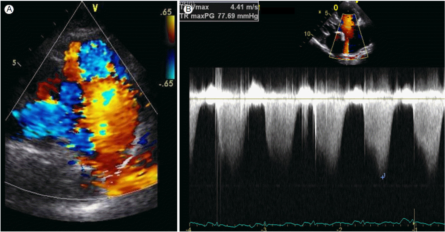
심초음파 소견: 정상 좌심실 수축기능을 보이며 심첨부 비후성심근병증(apical HCMP) 소견에 동반된 심첨부로 전위된 유두근(apically displaced papillary muscles) 소견을 보였다. 폐동맥판막의 비후와 함께 main pulmonary artery (MPA)와 left pulmonary artery가 늘어나 있었다. 삼첨판 역류의 최대속도가 4.4 m/s로 측정되어 우심실의 수축기 압력은 (4.42 × 4) + 우심방 압력(5 mmHg)으로 계산하면 82 mmHg로 계산되고 폐동맥 압력을 15/5 mmHg로 가정하면, 판막을 통한 압력차(transvalvular pressure gradient)는 67 mmHg로 중증의 폐동맥 판막 협착증(pulmonary valve stenosis, PS) 소견을 보였다(Fig. 2).
24시간 혈압 모니터링 소견: Non-dipper 형태의 고혈압소견을 보였으며 낮 평균 혈압은 167/92 mmHg, 밤 평균 혈압은 144/85 mmHg, 24시간 평균 혈압은 156/91 mmHg로 측정되었다.
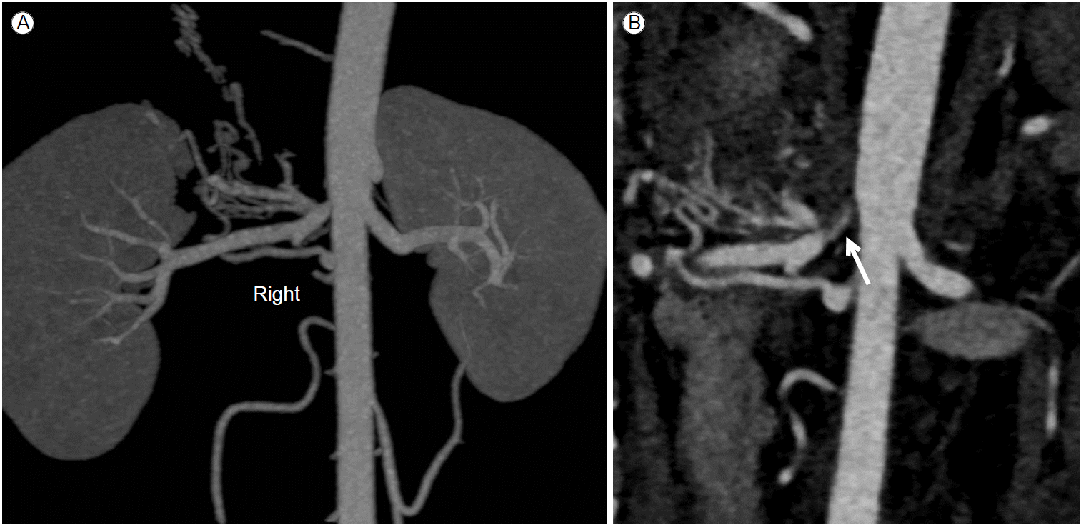
신동맥 CT 소견: 오른쪽 신동맥 입구에 심한 국소적 협착 소견을 보이고 있으며, 특징적인 염주알 형태(string of beads)의 섬유근육 이형성증(fibromuscular dysplasia) 소견을 보이고 있지는 않았다(Fig. 3).
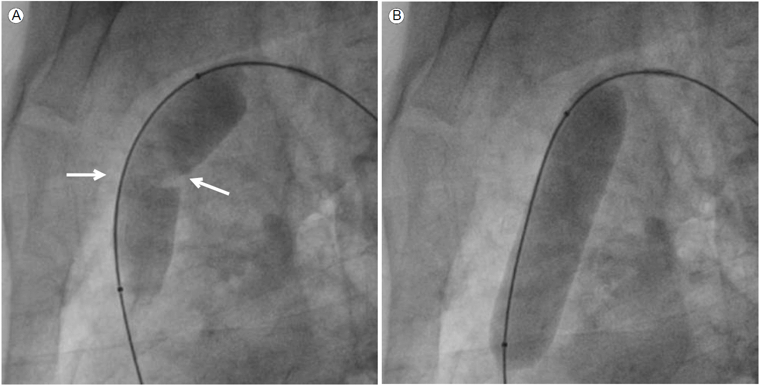
경피적 풍선 폐동맥판막성형술(percutaneous balloon pulmonary valvuloplasty): Maxi 풍선(20 × 60 mm, Cordis Corporation, Bridgewater, NJ, USA)을 사용하여 판막성형술을 시행했으며 시술 전 MPA의 압력은 15 mmHg, right ventricle 압력은 65 mmHg로 측정되었고 풍선이 완전히 부풀리기 전에 좁아진 판막에 의해 풍선 중간이 잘록해졌다가(“waist”) 완전히 부풀린 후 잘록한 부분이 없어진 것을 확인할 수 있었다(Fig. 4).
신동맥 혈관확장술: 신동맥협착소견에 대하여 시술을 시도했으나 카테터 진입 실패로 이에 대해서는 약물치료를 계획하였다.
심장 magnetic resonance imaging (MRI) 소견: HCMP 소견에 대한 평가를 위해 MRI를 시행하였으며 전반적인 심내막하의 조영 감소와 함께 심첨부의 비대소견으로 apical HCMP(apically displaced papillary muscle)에 합당한 소견을 보였다(Fig. 5).
치과 파노라마 엑스선 소견: 심한 치아상태 불량소견을 보였다.
유전자 검사 소견: NS 진단을 위해 시행한 검사에서 핵형(karyotype)은 46, XY였으며 NS와 연관된 유전자 중 PTPN11를 분석한 결과 정상 소견을 보였다.
고 찰
Noonan syndrome은 1962년에 처음 소개된 질환으로 독특한 얼굴생김새(dysmorphic face)와 선천성 심장질환을 포함한 다양한 비정상 소견을 그 특징으로 하였다. NS는 비교적 흔한 선천성 유전질환이며 남녀 모두에서 발생 가능하다. 소아청소년기에는 얼굴생김새 및 근골격계의 형태가 특징적으로 나타나 진단이 용이한 반면, 이 시기를 지난 성인에는 이러한 특징들이 애매하므로 진단을 하기 어려우며 그 진단은 일반적으로 van der Burgt 등[4]에 의해 제안된 임상적 진단 기준에 따른다(Table 1). NS로 확진하기 위해서는 전형적인 얼굴이형성증(typical face) 소견은 필수적이며 여기에 다른 1가지 major 소견 또는 2가지 minor 소견이 동반되거나, NS로 의심되는 얼굴생김새이상(suggestive face)과 함께 2가지 major 소견 또는 3가지 minor 소견이 필요하다[4]. 본 증례는 전형적인 얼굴이형성증, 오목가슴의 이학적 소견과 심초음파상 PS 및 HCMP의 소견을 동반하여 van der Burgt 등[4] 진단 기준에 따라 NS로 진단하였던 증례였다.
본 질환은 특징적으로 PS를 자주 동반하는 것으로 알려져 있으며(25-30%) [5], 본 증례에서도 PS가 발견되어 이를 경피적 풍선 폐동맥판막성형술을 시행하여 성공적으로 압력차를 감소시켰다.
일반적으로 7개의 유전자(PTPN11, SOS1, KRAS, RAF1, NRAS, BRAF 그리고 SHOC2)가 본 질환과 연관되어 있다고 알려져 있으며[5,6], 약 50% 이상의 환자에서 PTPN11 변이가 있다고 알려져 있으나 이는 절대적인 진단 기준은 아니다[7]. 본 증례에서도 대표적인 유전자인 PTPN11 변이를 조사하였으나 정상 소견을 보였다.
한편, NS는 자가면역질환과 드물게 연관되어 있다고 알려져 있어 전신 홍반성 낭창(systemic lupus erythematosus), 셀리악병(Celiac disease) 등의 증례 보고가 있으나[8], 명확한 연관 관계는 아직 알 수 없다. 본 증례에서도 자가면역질환이나 이외의 전신 질환의 증거를 찾을 수는 없었다.
본 질환은 신장이상을 10-11%에서 동반하는데[9], 주로 단신(solitary kidney), 신우확장(renal pelvis dilation), 중복된 집뇨계(duplicated collecting system) 등과 관련되어 있는 것으로 알려져 있다. 하지만 신동맥 협착과 연관되어 있다는 보고는 지금까지 없다. 본 증례는 신동맥 입구에 심한 협착 소견을 보이고 이로 이한 악성고혈압을 동반하였고 이와 함께 호흡 곤란으로 내원하게 되었으며 임상적으로 NS로 최종 진단되었다. NS와 신동맥 협착 사이의 병태생리학적 연관성에 대해서는 보고된 바가 없고 추가적인 연구가 필요할 것으로 사료된다.


 PDF Links
 PubReader
 ePub Link
 Full text via DOI
 Download Citation
 Print
Print


