과도한 음주력이 있는 당뇨병성 케토산증 환자에서 발생한 하만증후군 1예
Hamman Syndrome as a Rare Complication of Diabetic Ketoacidosis in a Patient with Chronic Heavy Alcohol Use: A Case Report
Article information
Trans Abstract
A 40-year-old man with altered mental status, abdominal pain, and vomiting was referred to our emergency department. Chest radiography revealed pneumomediastinum, raising concern for Boerhaave syndrome. Laboratory tests revealed high-anion-gap metabolic acidosis and hyperglycemic ketoacidosis, consistent with diabetic ketoacidosis (DKA). Endoscopic evaluation excluded esophageal perforation. Because the pneumomediastinum resolved with conservative management and DKA treatment, Hamman syndrome was diagnosed. The patient developed multiorgan dysfunction and required intensive care management, including mechanical ventilation and continuous renal replacement therapy. This case illustrates the diagnostic challenges of differentiating DKA from alcoholic ketoacidosis and Hamman syndrome from Boerhaave syndrome, highlighting the importance of a systematic approach and multidisciplinary critical care management for patients with severe metabolic derangements and multiorgan dysfunction.
INTRODUCTION
Diabetic ketoacidosis (DKA) is the most common acute hyperglycemic emergency in patients with diabetes mellitus. It results from absolute or relative insulin deficiency and elevated counterregulatory hormones, typically leading to hyperglycemia, ketosis, and metabolic acidosis. Although DKA most often occurs in individuals with uncontrolled type 1 diabetes mellitus (T1DM), it can also develop in adults with poorly controlled type 2 diabetes mellitus (T2DM) under stressful conditions, such as acute medical or surgical illnesses, or in adolescents with new-onset T2DM. The symptoms and signs of DKA include polyuria, polydipsia, weight loss, fatigue, Kussmaul breathing, vomiting, abdominal pain, and altered mental status [1].
Alcoholic ketoacidosis (AKA) is characterized by chron-ic heavy alcohol use and ketoacidosis. It has a different pathogenesis from DKA and develops in individuals with alcohol use disorder, often after nausea, vomiting, and acute starvation. A history of chronic alcohol consumption can complicate the differentiation between DKA and AKA. The blood glucose level is a key diagnostic feature that distinguishes DKA from AKA. Serum glucose levels above 250 mg/dL should prompt the consideration of DKA because most patients with AKA have low or normal glucose levels. Thus, ketoacidosis without hyperglycemia in a patient with chronic heavy alcohol use is virtually diagnostic of AKA [1,2]. The distinction between these two ketoacidotic states is essential for appropriate management.
Hamman syndrome, characterized by spontaneous pneumomediastinum without underlying pulmonary pathology, is a rare complication of DKA [3]. Boerhaave syndrome typically develops after forceful vomiting, which causes a sudden rise in intraluminal pressure and subsequent spontaneous esophageal rupture, and has occasionally been reported in patients with DKA [3]. It is often accompanied by pleural effusion, shock, and signs of sepsis [4]. Both Boerhaave syndrome and Hamman syndrome can present with pneumomediastinum, and their overlapping clinical manifestations, such as vomiting, chest pain, and abdominal pain, complicate the differential diagnosis [5]. Differentiating between these two conditions is crucial because misdiagnosis can lead to unnecessary invasive procedures or delayed treatment of a life-threatening condition.
Here, we report the case of a patient with chronic heavy alcohol consumption who developed DKA complicated by Hamman syndrome. This case illustrates the diagnostic challenges of differentiating DKA from AKA and Hamman syndrome from Boerhaave syndrome, highlighting the importance of a systematic approach and multidisciplinary critical care management for patients with severe metabolic derangements and multiorgan dysfunction.
CASE REPORT
A previously healthy 40-year-old man in a stuporous mental state was referred to the emergency department. He reported a 4-day history of epigastric pain, anorexia, nausea, and frequent vomiting. He also had a 20-year history of chronic heavy alcohol consumption, drinking approximately two bottles of soju, a Korean distilled liquor containing approximately 90 g of ethanol, almost daily, and a 40-pack-year smoking history. One month before presentation, the patient was diagnosed with fatty liver and gastritis at a primary clinic. He had no history of diabetes mellitus or asthma.
Upon arrival by emergency medical services, his vital signs were as follows: blood pressure, 60/40 mmHg; heart rate, 100 beats/min; body temperature, 36.7°C; respiratory rate, 24 breaths/min; and peripheral oxygen saturation, 95% on room air. Physical examination revealed a Kussmaul breathing pattern, epigastric tenderness, and abdominal distension. Arterial blood gas analysis showed a pH of 6.95, pCO2 of 18.3 mmHg, bicarbonate level of 4.0 mmol/L, and anion gap of 26 mmol/L, consistent with high-anion-gap metabolic acidosis. Laboratory results revealed an erythrocyte count of 4.72 × 10³/μL, hemoglobin level of 15.1 g/dL, hematocrit level of 41.4%, leukocyte count of 13.2 × 10³/μL, platelet count of of 4 × 10³/μL, sodium level of 105 mmol/L, potassium level of 6.1 mmol/L, chloride level of 75 mmol/L, phosphorus level of 7.7 mg/dL, uric acid level of 19.2 mg/dL, blood urea nitrogen level of 56 mg/dL, serum creatinine level of 2.44 mg/dL, C-reactive protein (CRP) level of 226.5 mg/L, alkaline phosphatase level of 269 IU/L, gamma-glutamyl transferase level of 299 IU/L, aspartate transaminase level of 205 IU/L, alanine transaminase level of 73 IU/L, total bilirubin level of 0.85 mg/dL, direct bilirubin level of 0.57 mg/dL, total protein level of 5.3 g/dL, albumin level of 3.3 g/dL, amylase level of 198 IU/L, lipase level of 51 IU/L, glucose level of 462 mg/dL, HbA1c level of 11.1%, serum osmolality of 286 mOsm/kg H2O, and positive serum total ketone bodies. A peripheral blood smear was negative for schistocytes. Urinalysis showed urinary ketones of 3+. The combination of hyperglycemia, ketosis, and highanion-gap metabolic acidosis was consistent with diabetic ketoacidosis.
Chest radiography revealed a linear opacity parallel to the left mediastinal border, consistent with pneumomediastinum (Fig. 1A), raising concern for the possibility of esophageal perforation. Therefore, chest computed tomography (CT) was performed for further evaluation and revealed aspiration pneumonia, mild emphysema, and pneumomediastinum, with air tracking along the esophagus (Fig. 2). Non-contrast abdominal CT revealed low hepatic attenu-ation, bilateral renal enlargement, and diffuse pancreatic swelling with peripancreatic fat stranding suggestive of severe fatty liver disease, acute kidney injury (AKI), and acute pancreatitis. CT showed no evidence of cirrhosis. Brain CT findings were unremarkable.
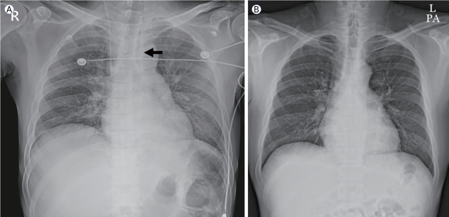
(A) Chest radiography reveals a linear opacity parallel to the left mediastinal border (black arrow), consistent with pneumomediastinum. (B) Follow-up chest radiography shows resolution of the linear opacity.
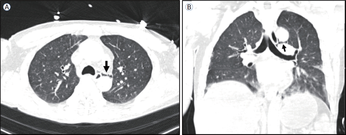
Chest computed tomography (CT) reveals aspiration pneumonia, mild emphysema, and pneumomediastinum with air tracking along the esophagus (black arrow) on axial (A) and coronal (B) views.
The patient developed hypercapnic respiratory failure and persistent altered mental status, requiring intubation with mechanical ventilation, and was transferred to the intensive care unit (ICU). Management of DKA included fluid resuscitation, continuous insulin infusion, and correction of serum electrolytes. Continuous kidney replacement therapy (CKRT) was initiated to treat the hemodynamic instability, AKI, and refractory metabolic acidosis. Broad-spectrum antibiotics were administered, and platelet transfusions were performed. On hospital day 2, upper gastrointestinal endoscopy revealed gastric ulceration but no evidence of esophageal perforation or Mallory-Weiss tear. Blood cultures later grew Klebsiella spp., and antibiotic therapy for intra-abdominal infection and pneumonia was adjusted. Subsequent blood cultures revealed no bacterial growth. The patient’s clinical course was favorable. His vital signs stabilized, blood glucose levels were controlled, and metabolic acidosis improved. CKRT was discontinued on hospital day 2, and extubation was performed on hospital day 3. Several days later, the patient experienced resolution of abdominal pain with no evidence of worsening pancreatitis, and oral feeding was initiated after transitioning to multiple daily subcutaneous insulin injections.
Both insulin and C-peptide levels were markedly low, indicating severe endogenous insulin deficiency. Autoimmune markers including antibodies against glutamic acid decarboxylase and anti-insulin antibodies were negative or within normal limits, consistent with idiopathic T1DM. Follow-up chest radiography showed improvement in the pneumomediastinum with conservative management after resolution of the underlying DKA. The patient was diagnosed with Hamman syndrome associated with DKA related to T1DM. Prior to discharge, laboratory testing demonstrated normalization of the white blood cell and platelet counts, reduced CRP levels, and improvements in both renal and hepatic function. Follow-up chest radiography revealed no evidence of pneumomediastinum (Fig. 1B).
Ethical approval for this study was obtained from the Jeonbuk National University Hospital Institutional Review Board (IRB No. CUH 2025-10-010). This case report was prepared in accordance with the ethical standards of the Declaration of Helsinki. Written informed consent for publication was obtained from the patient.
DISCUSSION
This case report describes the presentation and successful management of a rare case of DKA complicated by Hamman syndrome in a patient with a history of chronic heavy alcohol use. The combination of chronic alcohol use, the clinical presentation of abdominal pain and vomiting, and metabolic acidosis with ketosis initially led to the consideration of AKA. However, severe hyperglycemia with a serum glucose level of 462 mg/dL confirmed the diagnosis of DKA. DKA treatment focuses on insulin therapy, fluid resuscitation, and electrolyte replacement to correct acidemia, restore circulatory volume, and normalize blood glucose levels [1]. In contrast, AKA management emphasizes fluid resuscitation, glucose and electrolyte repletion, and symptomatic therapy, including antiemetics, with dextrose playing a crucial role [2]. This case underscores the importance of distinguishing between these two ketoacidotic states because their treatment approaches are fundamentally different.
Evaluation of liver cirrhosis can provide additional diagnostic insights in patients with chronic alcohol use presenting with ketoacidosis. Given the patient’s 20-year history of heavy alcohol consumption and the initial laboratory findings of severe electrolyte imbalance, thrombocytopenia, and renal dysfunction, underlying cirrhosis was considered. However, abdominal CT revealed severe fatty liver disease without definitive cirrhotic changes. Plasma volume depletion, a common complication in patients with decompensated liver cirrhosis or acute pancreatitis, can precipitate DKA or hyperglycemic hyperosmolar syndrome [6]. Although cirrhosis was not present in this case, plasma volume depletion resulting from acute pancreatitis, persistent vomiting, and poor oral intake likely contributed to the severity of DKA. Therefore, assessing the presence of cirrhosis and the patients’ volume status in patients with chronic alcohol use can help identify the precipitating factors of hyperglycemic crises.
Hamman syndrome is a rare complication of DKA, and its pathogenesis is complex and multifactorial. Respiratory compensation for metabolic acidosis, known as Kussmaul breathing, can increase alveolar pressure by 20-30 mmHg above normal inspiratory levels, predisposing patients to alveolar rupture. Additionally, DKA often induces severe vomiting, which is exacerbated by hyperglycemia-associated gastroparesis, thereby increasing intrathoracic pressure through the Valsalva maneuver [3]. Boerhaave syndrome is esophageal perforation caused by a sudden increase in esophageal pressure, typically resulting from severe vomiting or straining [7].
The patient’s alcohol history, vomiting, and pneumomediastinum identified on chest CT initially raised concern for Boerhaave syndrome, prompting further evaluation given its high mortality rate and potential requirement for emergent surgical or endoscopic intervention [8]. In contrast, Hamman syndrome associated with DKA generally has a favorable prognosis, is self-limiting, and resolves with the correction of ketoacidosis [3]. In this case, upper endoscopy revealed no evidence of esophageal perforation, thereby ruling out Boerhaave syndrome. With conservative management and resolution of the underlying DKA, follow-up chest radiography and CT confirmed resolution of the pneumomediastinum, leading to a diagnosis of Hamman syndrome. However, in patients with chronic alcohol use who present with DKA and severe vomiting, pneumomediastinum should not be presumed to be caused by Hamman syndrome without thorough investigation. Although this patient’s pneumomediastinum resolved spontaneously with DKA treatment and endoscopy excluded esophageal perforation, the overlapping clinical features of Hamman syndrome and Boerhaave syndrome, particularly in the setting of chronic heavy alcohol use and violent vomiting, necessitate comprehensive evaluation to avoid overlooking a potentially fatal esophageal perforation. Clinical suspicion for Boerhaave syndrome should remain high in such patients until the diagnosis has been definitively excluded by endoscopic examination or contrast esophagography. The distinction between these two conditions is essential for early intervention, accurate diagnosis and management, avoiding unnecessary invasive therapies, and preventing delayed treatment of life-threatening conditions.
Additionally, this case demonstrates the potential for DKA to progress to severe multiorgan dysfunction. Our patient presented with acute pancreatitis and developed concurrent thrombocytopenia and AKI, ultimately requiring ICU admission. This clinical course suggests that DKA can extend beyond metabolic derangements to cause life-threatening multiorgan complications that require advanced critical care.
In conclusion, this case highlights the importance of comprehensive evaluation with a broad differential diagnosis when evaluating pneumomediastinum in patients with ketoacidosis. Early recognition of Hamman syndrome can prevent unnecessary invasive procedures while ensuring appropriate conservative treatment. The potential for multiorgan dysfunction in severe DKA highlights the need for intensive monitoring and a multidisciplinary critical care approach to optimize patient outcomes.
Notes
CONFLICTS OF INTEREST
No potential conflict of interest relevant to this article was reported.
FUNDING
None.
AUTHOR CONTRIBUTIONS
Data curation, investigation, formal analysis, writing - original draft: Yujin Shin.
Data curation, formal analysis, validation: Sang Won Lee and Soo Jin Lee.
Conceptualization, data curation, supervision, writing - review & editing: Kyung Pyo Kang.
ACKNOWLEDGEMENTS
None.