


INTRODUCTION
Dehydration and rapid weight loss in pediatric patients are well-known precipitating factors for renal vein thrombosis (RVT). However, these factors rarely, if ever, cause RVT in adults. We report a case of a 31-year-old man with a history of hypertension who was taking antihypertensive medication without any complications, but who developed renal RVT and pulmonary thromboembolism when he reduced his weight by 8 kg in 2 weeks.
CASE REPORT
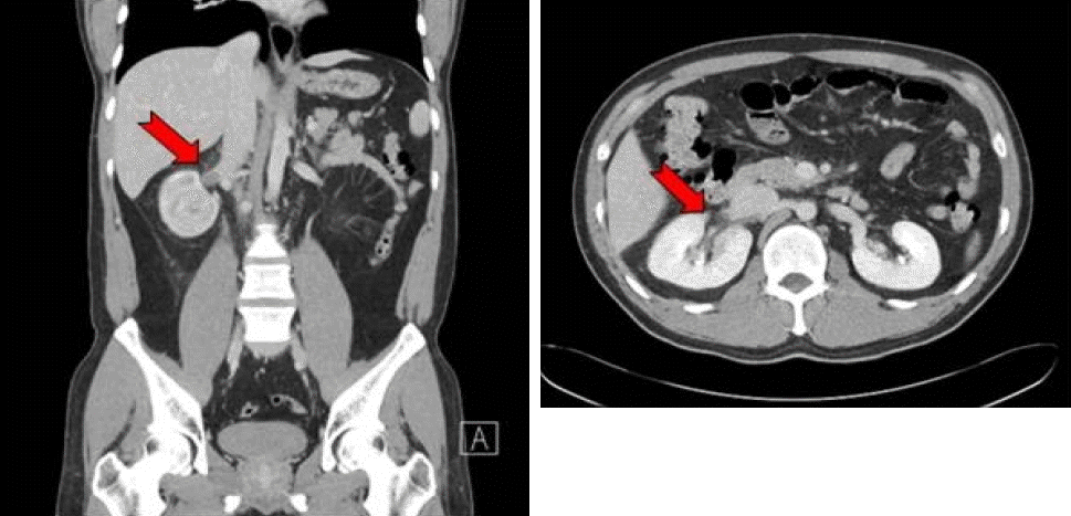
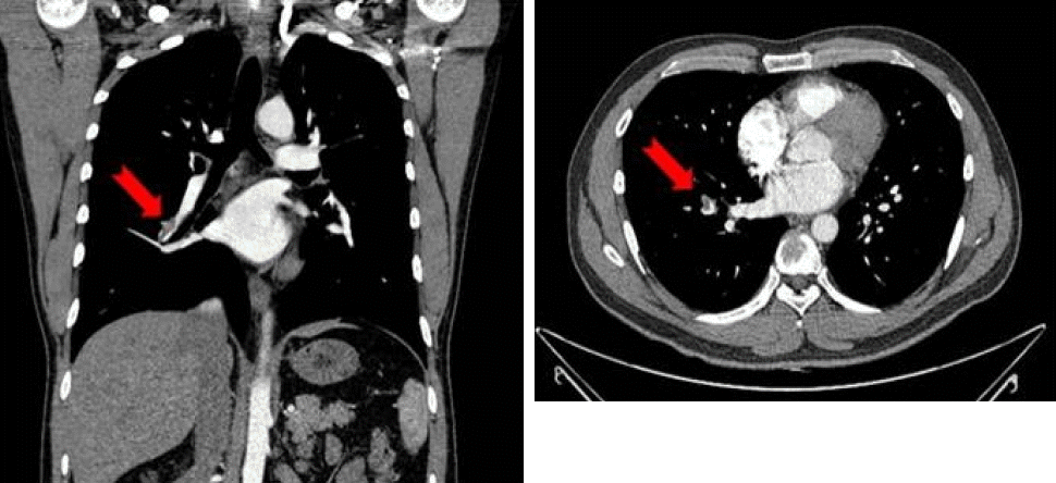
A 31-year-old Asian man, with a 3-year history of essential hypertension, was admitted to a local hospital because of severe right flank pain. The patient had been well until 6 days before admission, when a dry cough started. A dull, continuous, non-colicky right flank pain had developed 2 days prior to admission. He also felt severely thirsty and drank 2-3 L of water per day. His physician admitted him to the local hospital under the presumptive diagnosis of a right renal stone. The patient had been healthy all his life, except for a 20-kg weight gain in the previous 3 years, and his hypertension had been well-controlled for 3 years by an angiotensin receptor blocker and a calcium channel antagonist. He was a non-smoker and a social drinker. He thought that the reason for his hypertension was weight gain and decided to lose weight in a short period of time. He fasted, except for minimal water intake, and exercised hard for 2 weeks. As a result, he lost 8 kg in 2 weeks. A physical examination revealed a weight of 95 kg and height of 187 cm. His blood pressure was 100/75 mmHg in the supine position. His heart rate was 74 beats per minute and there was neither dyspnea nor chest discomfort. Pulmonary and cardiac auscultation were unremarkable, as was an abdominal examination. Bowel sounds were active, no tenderness, guarding, or organomegaly were noted, but the left costovertebral angle was tender to percussion. A contrast-enhanced computed tomography (CT) scan was performed at a local hospital. On the day of admission, his hemoglobin was 11.6 g/dL, hematocrit was 33.7%, serum sodium was 135 mM, blood urea nitrogen was 40.2 mg/dL, and serum creatinine was 3.4 mg/dL. Serum total protein and albumin were 7.2 and 4.4 mg/dL, respectively, and total cholesterol was 154 mg/dL. A urinalysis showed traces of albumin and one red cell and no white cells per high power field. Acute kidney injury was suspected, and the patient was transferred to our hospital. A CT scan showed two veins in the right kidney (Fig. 1). The lower vein had an intraluminal filling defect, and the middle and lower poles of the right kidney were congested, with an edematous perinephric space but without renal calculi. The upper renal vein was intact. The CT scan also showed multiple filling defects in the right pulmonary vasculature (Fig. 2). We diagnosed RVT and pulmonary thromboembolism. A Doppler ultrasound exam was performed in both lower extremities for further evaluation of the cause of the pulmonary thromboembolism. No evidence of deep vein thrombosis or significant arterial stenosis were found. Because we identified a collateral venous supply on the lesions, we started systemic anticoagulation instead of local thrombolytic therapy. Intravenous heparin (25,000 U/24 h) was continued until the patient was effectively anticoagulated with coumarin derivatives. We also started intravenous rehy dration to manage the contrast-induced nephropathy and dehydration. Serum creatinine decreased rapidly from 3.4 to 1.3 mg/dL over 10 days. Similarly, 10 days after admission, creatinine clearance was 110.5 mL/min and a follow-up CT scan showed that the previously noted intraluminal filling defect on the right lower pulmonary vasculature had almost disappeared, and that parenchymal congestion in the right renal vein had improved. Levels of anticardiolipin antibodies IgG (< 9.4 GPL) and IgM (< 9.4 MPL), protein C/S (91%/ 79%), antithrombin III (100%), factor V Leiden (normal) and homocysteine (12.9 μM) were measured in serum to evaluate hypercoagulability. All results were unremarkable.
DISCUSSION
RVT is rare and primarily observed in children with severe dehydration or in adults with nephrotic syndrome, renal tumors, hypercoagulable states, or after surgery or trauma to the renal vessels [1]. A diagnosis of RVT is rarely considered when it occurs in adults, as in this presented case; the initial presumptive diagnosis of our patient was a right renal stone. Acute RVT is usually symptomatic and associated with loin, testicular, or flank pain. Acute RVT is often accompanied by nausea and vomiting, and these symptoms might be confused with acute pyelonephritis or urolithiasis. Hematuria is nearly universal and most often microscopic. High venous pressure usually results in proteinuria. However, results can be negative for protein or hematuria, as in this case. In general, no specific laboratory tests are available to diagnose RVT [1]. When RVT causes a renal infarction, the distribution of the hypoperfused region tends to be medullary or subcortical, and renal impairment tends to be patchy and subtotal. A swollen kidney can occasionally rupture the capsule, resulting in a massive retroperitoneal hemorrhage [1]. Thrombosis of the longer left renal vein may also involve the ureteric, gonadal, adrenal, and phrenic branches that drain into the left vein, whereas the adrenal and gonadal veins drain directly into the inferior vena cava on the right side. This can be a risk factor for a pulmonary thromboembolism, as in the case presented here. The causes of RVT vary. In our patient, body weight increased over 3 years and was stable at 103 kg for several months; however, after 2 weeks of fasting, dehydration, and excessive exercise, his weight decreased to 93 kg. Regimens that promise rapid weight loss have great appeal for most overweight patients. Therefore, obese individuals may frequently undergo fasting and excessive exercise, as in this patient. However, several complications are related to rapid weight loss, such as dehydration, fatigue, and malnutrition, among others. Losing weight too fast may also increase the risk of gallstones and cardiac dysfunction as well as progress to sudden death [2,3]. The recommended safe target for weight loss is about 0.5-1 kg per week with proper hydration [4]. On admission to a local hospital, our patient had already consumed large volumes of water. Therefore, there was no clinical or laboratory evidence for dehydration and hemoconcentration. On admission to our hospital, he weighed 95 kg after hydration therapy, having lost 8-10 kg of bodyweight, likely due to fluid loss. It was concluded that this patient was at an increased risk for intravascular thrombi formation from severe dehydration caused by fasting and excessive exercise to lose weight. Dehydration is known to cause thrombotic complications by several mechanisms [5]. Due to reduced blood volume, regional blood flow may decrease and, thus, promote thrombi formation. In addition, hematocrit and protein concentrations increase as blood viscosity increases. Elevated blood viscosity is a known risk factor for vascular thrombotic occlusion. Ezzar [6] reported cases of acute RVT in seven adults. Three of the seven patients were dehydrated due to low fluid intake and hard physical effort in excessively hot weather (42-44℃) during a summer pilgrimage. Böhler et al. [7] reported a case of a young woman who tolerated an oral contraceptive agent without any complications, until she became severely dehydrated and consequently developed left RVT. However, compared with pediatric patients, dehydration and rapid weight loss are rarely recognized as risk factors for RVT in adults. Hypercoagulation states are also associated with RVT and have been documented in patients with nephrotic syndrome, causing loss of antithrombin III in the urine [8]. Insufficient blood concentrations of protein-C have also been associated with thromboembolic events [1]. Acquired or congenital antithrombin III or protein C deficiencies, antiphospholipid antibody syndrome, a factor V Leiden mutation, and hyperhomocysteinemia were excluded in this patient.
Even though the patient was an adult, the rapid weight loss with dehydration may have caused the RVT, and unusual thromboembolic events must be suspected to avoid diagnostic delay.
 PDF Links
 PubReader
 ePub Link
 Full text via DOI
 Download Citation
 Print
Print


