


INTRODUCTION
Sarcoidosis is a multisystemic granulomatous disease of unknown cause. The lungs, skin, eyes, liver, and lymph nodes are commonly affected, but muscular sarcoidosis is rare. Muscular involvement in sarcoidosis was first reported by Licharew in 1908 [1]. The symptomatic involvement of muscles occurs much less frequently than does asymptomatic involvement [1,2]. Three main clinical types of sarcoidosis have been reported for symptomatic muscular sarcoidosis: nodular, acute myositic, and chronic myopathic. The palpable nodular type reported by Licharew is the least common clinical type [3]. The nodular type can present with a soft-tissue mass that may be confused with a tumor, and magnetic resonance imaging (MRI) and muscle biopsy are useful methods for early diagnosis. The optimal management of the nodular type of muscular sarcoidosis is not well known, but surgical excision or corticosteroid treatment are initial options [4]. We report here the case of a woman with recurrent nodular muscular sarcoidosis who was treated with steroids.
CASE REPORT
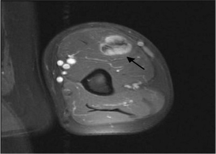
A 58-year-old Korean woman presented with a painless mass in her left arm. A large, firm, fixed mass was palpable in the biceps muscle. Radiographs of the arm revealed a soft-tissue shadow in the biceps. MRI demonstrated that the mass (18×2.3×1.6 cm) had irregular margins (Fig. 1) and enhanced heterogeneously with various signal intensities. The characteristics of this infiltrating mass were most consistent with a soft-tissue sarcoma or fibromatosis. An incision biopsy demonstrated chronic granulomatous inflammation with focal necrosis, suggesting sarcoidosis. Excision was thus performed.
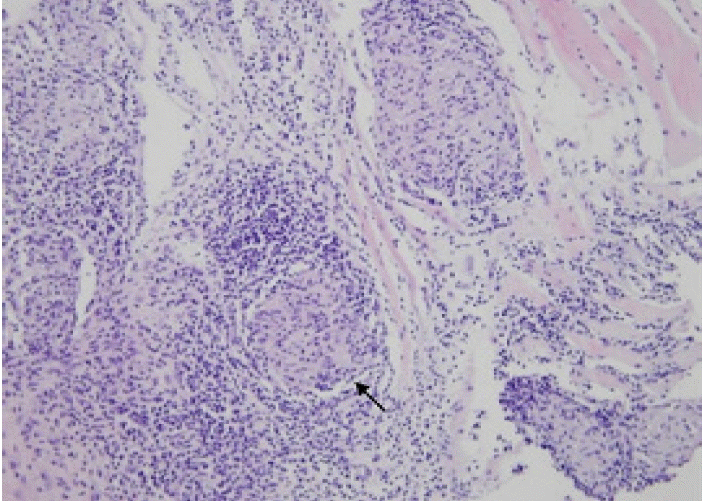
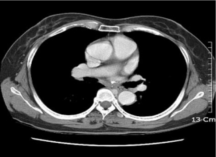
One year after excision, the patient returned to our hospital with multiple painless, mass-like lesions in her arms and legs. Ultrasonography demonstrated hypoechoic masses in the right upper arm (11×0.6×1.2 cm), left forearm (8.5×1×1 cm), right lower leg (6.3×1.2×2.4 cm), and right calf (13×1.5×2.6 cm). A gun biopsy was performed on the right leg mass. The biopsy revealed chronic granulomatous inflammation, compatible with sarcoidosis (Fig. 2). Acid-fast staining and Gomori’s methenamine-silver staining showed no microorganism. The chest radiograph was not remarkable, but the chest computed tomography image revealed bilateral hilar lymphadenopathy, consistent with stage I sarcoidosis (Fig. 3). Additional laboratory tests that measured blood cell count, serum angiotensin-converting enzyme level, serum electrolytes, serum calcium level, erythrocyte sedimentation rate, and C-reactive protein level were all within normal limits. The level of creatinine phospho-kinase and the creatine kinase-myocardial band were normal. Gallium uptake was detected in the upper and lower extremities by 67Ga-scintigraphy (Fig. 4). Corticosteroid therapy (prednisolone: 1 mg/kg/day) was initiated and the masses gradually decreased in size during the following week. The prednisolone dose was tapered until treatment was discontinued at 2 months. No sign of recurrence has been detected during 2 years of follow-up.
DISCUSSION
Asymptomatic muscle involvement occurs in 50~80% of sarcoidosis patients, but symptomatic involvement is rare [1,2]. Three forms of symptomatic muscular sarcoidosis have been described: nodular involvement, acute sarcoid myositis, and chronic sarcoid myopathy [5]. The nodular type is the least common type of symptomatic muscle involvement in sarcoidosis [3]. Although any skeletal muscle may be involved, the lower limbs are most frequently affected. The nodules are painless and are not accompanied by any limitation of movement or muscle weakness. However, contracture or myalgia may occur [5]. Serum creatine kinase levels and electromyographic results are typically normal. The excellent tissue contrast of MRI allows the detection of small nodules, and the MRI findings of the nodular type of sarcoidosis have been reported to be specific [6]. The cytological differential diagnosis of nodular muscular sarcoidosis includes tuberculosis, foreign-body granuloma, fungal or parasitic infection, and autoimmune diseases, such as rheumatoid arthritis [7].
Patients with nodular lesions and acute myositis can usually be effectively treated with corticosteroids at an initial dosage of 0.5~1 mg/kg/day [8,9]. Early initiation of treatment appears to increase its effectiveness. Although corticosteroid treatment was found to be rapidly effective for patients with nodular muscular sarcoidosis [9], relapse was observed in all patients several years later. The response to steroids is unpredictable in patients with the chronic myopathic type of muscular sarcoidosis.
Patient outcomes may be complicated by corticosteroid-induced myopathy [7]. Chloroquine, azathioprine, and methotrexate should be administered to steroid-dependent patients or those who fail to respond to corticosteroids [3,8,10]. New agents, including thalidomide and infliximab, may also be useful in selected cases [10]. Physicians should consider this unusual presentation of sarcoidosis and its responsiveness to steroid treatment when managing sarcoidosis patients.

 PDF Links
 PubReader
 ePub Link
 Full text via DOI
 Download Citation
 Print
Print


