


서 론
비침습적 검사에서 양성 소견을 보인 흉통 증상이 있는 환자에서 관상동맥 조영술을 시행하였을 때 혈관 내 직경의 50% 이상의 유의한 협착이 관찰되지 않는 경우는 레지스트리 연구에서 50% 이상, 많게는 70%까지 보고된다[1]. 이러한 관상동맥의 협착이 없는 허혈성 심질환(ischemic heart disease with non-obstructive coronary arteries, INOCA)은 환자의 삶의 질에 부정적인 영향을 미칠 뿐만 아니라[2] 경제적인 부담이 증가하고[3] 나아가서는 사망이나 심혈관계 사건의 발생을 증가시키는 것으로 알려져 있다[4]. 그럼에도 적절하게 진단되거나 치료를 받지 못하는 경우가 많다. 본 종설에서는 임상 연구 결과 및 전문가 합의 등의 문헌 검토를 통해 INOCA의 역학과 병태생리, 임상 양상, 진단 및 치료 등에 대해 살펴보고자 한다.
증 례
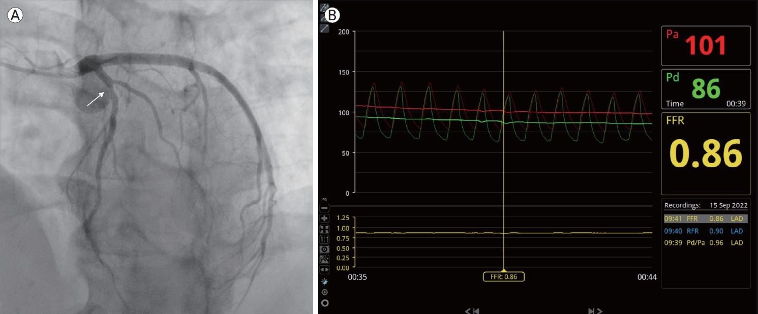
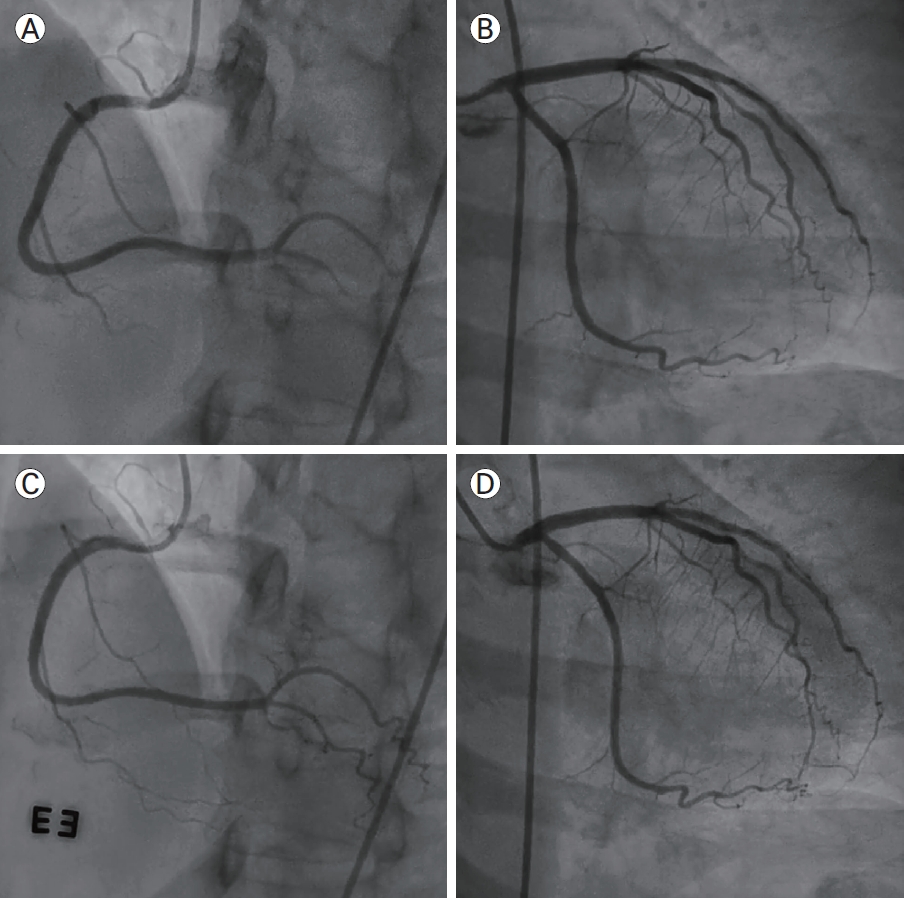
58세 여자 환자가 걷거나 계단을 오를 때 악화되는 흉통 및 호홉곤란을 주소로 외래에 내원하였다. 증상은 1년 전부터 발생하였고 수주 사이에 증상이 악화되었다고 하였다. 환자는 항고혈압제로 안지오텐신수용체길항제와 티아지드계 이뇨제를 복용 중이었다. 외래에서 시행한 경흉부 심초음파에서는 정상 심기능 소견이고 국소벽 운동 장애와 같은 소견은 확인되지 않았다. 환자는 입원하여 관상동맥 조영술을 시행하였다. 관상동맥 조영술에서 유의한 협착이 관찰되지 않았으며(Fig. 1) 양측 관상동맥 내에 에르고노빈을 투여하여 연축 유발 검사를 시행하였으나 유의한 관상동맥 내경의 연축이 관찰되지 않았다(Fig. 2). 미세혈관 저항지표(index of microcirculatory resistance, IMR)는 31이 확인되어 미세혈관기능 부전이 의심되었다. 이후 환자는 증상이 지속되어 정맥 주사 에르고노빈 유발 심초음파도 시행하였으나 음성 소견이 확인되었다. 환자는 현재 1년 이상 딜티아젬과 아토르바스타틴을 포함한 약물 치료를 유지하면서 흉통이나 호흡곤란의 증상 없이 안정적으로 지내는 중이다.
본 론
INOCA의 역학
국내외의 여러 레지스트리 분석에서 협심증이 의심되거나 부하 검사 양성 소견으로 관상동맥 조영술을 시행하는 환자에서 유의한 관상동맥의 협착이 발견되지 않는 경우는 40%에서 많게는 70% 내외까지 보고되고 있다[5-7]. 협착이 발견되지 않는 경우가 협착이 확인되는 경우에 비해 상대적으로 나이가 더 젊으며 여성의 경우에서 관상동맥 협착이 발견되지 않는 경우가 더 많은 것으로 알려져 있다[4,7].
한편 심근경색의 증거가 있으면서 관상동맥의 유의한 협착이 없는 경우에는 관상동맥의 협착이 없는 심근경색(myocardial infarction with non-obstructive coronary arteries, MINOCA)으로 정의할 수 있다. 2017년 유럽심장학회에서 MINOCA의 진단 기준에 대해 제시하였는데[8] 이에 따르면 1) 심근표지자 양성 소견이 동반된 임상 양상, 심전도상 ST 분절 변화, 좌각 차단 혹은 병적 Q파 및 심근생존능의 저하와 같은 허혈의 증거, 2) 관상동맥 조영술상 내경의 50% 이상의 유의한 협착이 없음, 3) 추가적 평가를 요하는 원인이 명백하게 존재하지 않는 경우로 정의하고 있다.
INOCA의 병태생리
동맥경화로 인한 관상동맥 내 협착은 심근허혈의 주된 요인이지만 INOCA의 경우 심근허혈이 관상동맥 미세혈관기능 부전(coronary microvascular dysfunction)이나 관상동맥의 연축과 같은 원인으로 관상동맥 내 혈류 공급과 심근의 산소 요구량의 불일치가 발생하여 발생할 수 있다[9,10]. 미세혈관기능 부전의 경우 관상동맥 미세순환의 구조적 혹은 기능적 이상으로 인해 발생하는 것으로 알려져 있다[11]. 구조적 이상의 경우 관상동맥의 소동맥의 내막층과 중막층의 증식과 비후로 인해 내강 대비 혈관벽이 두꺼워지는 리모델링이 발생하고 심근 내 모세혈관의 밀도가 감소하여 미세혈관의 산소 전달 능력이 감소하게 된다[12,13]. 여기에 급성 관동맥증후군을 겪거나 경피적 관상동맥 중재 시술을 받은 환자의 경우 미세혈전이나 동맥경화반의 파편이 미세혈관 색전을 일으켜 미세혈관 내강의 협착을 일으킬 수 있다[14]. 기능적 이상의 경우 아세틸콜린이나 기계적 자극에 대하여 혈관내피층은 산화질소와 같은 혈관 활성 물질이 매개하는 반응을 통해 혈관 확장 반응을 보이는데[15] 미세혈관기능 부전 상태에서는 이러한 혈관 활성 물질 매개 반응이 감소하거나 심지어는 혈관 수축 반응을 보이는 것으로 알려져 있다[16,17].
관상동맥의 연축은 흡연, 약물, 혈압 변화, 추위, 정서적 스트레스 등과 같은 자극에 대한 혈관의 과도한 수축으로 인해 발생하는 것으로 알려져 있다[18]. 또한 음주가 매우 강력한 위험인자로 알려져 있고 서양인과 동양인은 차이가 있다[19-21]. 이러한 관상동맥 연축은 관상동맥 내의 혈관평활근 세포의 과민 반응이 중요한 역할을 하는 것으로 보이며 일부 연구에서는 내피세포의 기능 부전이 연축에 일부 기여한다고 보고하기도 하였다[22].
위에서 기술한 미세혈관기능 부전이나 관상동맥 연축이 모두 존재할 수 있고 일부 환자에서는 동맥경화로 인한 관상동맥 협착과 미세혈관기능 부전 혹은 관상동맥 연축의 병태생리가 동반되어 증상이 나타나는 경우도 있다.
INOCA의 비침습적 진단
심근허혈의 평가를 위한 도구로 부하 심초음파, 심장 양전자방출 단층촬영, 심장 자기공명영상 등과 같은 검사가 있다[23]. 양전자 방출 단층촬영을 통해 미세혈관기능 부전에서 감소된 심근혈류를 정량적으로 평가할 수 있고 이러한 지표들과 예후와의 연관성이 다양한 연구를 통해 알려져 있다[24]. 또한 최근에는 심장 자기공명영상에서의 심근혈류의 정량적 분석 방법에 대해서도 연구가 진행되고 있다[25]. 이러한 검사는 비침습적이며 적은 방사선량 등으로 일부 검사가 선호될 수 있으나 해부학적인 정보를 제공하지 못한다. 따라서 INOCA를 진단하기에 불충분하다[26].
관상동맥 전산화 단층조영 검사(coronary computed tomography angiogram)는 관상동맥의 유의한 협착이나 동맥경화반을 진단하는 데 유용한 비침습적 진단 도구이다[27]. 그러나 관상동맥 전산화 단층조영 검사로는 심근허혈의 근거를 확인하기 어려우므로 INOCA의 진단에서는 제한적인 역할만을 수행한다(Fig. 2).
최근에는 관상동맥 전산화 단층조영 검사 시에 컴퓨터 모델링을 이용한 방법을 동원하면 심장혈관의 전체 용적 대비 심근량의 비율(ratio of luminal volume to myocardial mass)을 계산하여 미세혈관기능 부전을 정량화할 수 있다[28].
관상동맥 조영술을 기반으로 한 INOCA의 침습적 진단
관상동맥 조영술은 관상동맥의 협착을 진단하는 데 가장 정확한 방법으로 알려져 있다. INOCA의 진단을 위해서는 관상동맥 조영술로 유의한 협착이 있는지 확인하여야 하나 관상동맥 조영술 단독으로는 협착과 혈류의 제한 사이의 연관성을 알기 어려우므로[29] 압력철선을 이용한 분획 혈류 예비력(fractional flow reserve, FFR) 등의 혈관생리학적 평가가 필요하다[26,30]. FFR은 최대 충혈 시에 관상동맥 원위부의 압력과 대동맥압의 비를 계산한 것으로 FFR이 0.80 미만인 경우 관상동맥 내 혈류의 제한이 존재한다는 의미를 갖는다[31].
미세혈관기능 부전의 진단을 위해서는 관상동맥 혈류 예비력(coronary flow reserve, CFR)이나 IMR과 같은 지표가 사용된다. CFR은 도플러 철선이나 압력과 온도를 감지할 수 있는 열희석법(thermodilution method)이 가능한 철선을 사용하여 검사가 이루어진다[32]. CFR 값이 2.0 이하일 때 비정상 소견으로 진단할 수 있다. IMR의 경우 관상동맥 원위부의 압력과 최대 충혈 시 평균 통과 시간을 통해 계산할 수 있는데 25 이하가 정상 범위이다[33]. CFR은 관상동맥 협착과 미세혈관기능을 모두 반영하는 데 비해 IMR은 상대적으로 미세혈관기능 평가에 특이적인 지표이다[26]. 따라서 INOCA의 경우에는 FFR ≥ 0.8이고 CFR < 2.0인 경우에 해당한다. 특히 이 중에서도 IMR ≥ 25인 경우가 미세혈관기능 부전의 증거가 된다.
INOCA의 치료
약물 치료에 앞서 INOCA로 진단된 환자들에게는 금연, 체중 조절, 운동 등과 같은 생활 습관 조절이 필요하다. 아울러, 당뇨, 고혈압, 이상지질혈증과 같은 죽상경화성 심혈관계 질환의 위험인자의 적절한 조절 또한 필수적일 것이다.
INOCA로 진단된 환자들의 흉통의 치료는 주된 병태생리에 따라 개별화되어야 한다. 관상동맥의 연축이 있는 환자에서는 딜티아젬과 같은 칼슘통로길항제가 일차적으로 고려된다. 미세혈관기능 부전의 증거가 있는 환자에서는 베타차단제, 칼슘통로 길항제나 안지오텐신전환효소길항제가 고려된다. 니코란딜은 혈관확장제로 대체할 수 있는 선택지이나 종종 부작용이 보고되곤 한다[35]. 이외에도 라놀라진이나 이바브라딘, phosphodiesterase inhibitor와 같은 약제들이 미세혈관기능 부전의 개선에 연구되고 있으나 아직까지 많은 환자를 대상으로 긍정적인 결과를 보인 연구는 없는 실정이다. 라놀라진의 경우 일부 소규모 연구에서 증상의 개선을 보인 바 있으나[36] 국내에서는 처방에 제한이 있다.
 PDF Links
 PubReader
 ePub Link
 Full text via DOI
 Download Citation
 Print
Print


