항생제 약물알레르기의 진단과 관리

Diagnosis and Management of Antibiotic Allergies

Article information

Korean J Med. 2021;96(4):328-336
Publication date (electronic) : 2021 August 1
doi : https://doi.org/10.3904/kjm.2021.96.4.328
Division of Pulmonary, Allergy, and Critical Care Medicine, Department of Medicine, Hallym University Sacred Heart Hospital, Hallym University College of Medicine, Anyang, Korea
김주희
한림대학교 의과대학 한림대학교성심병원 호흡기-알레르기 내과
Correspondence to Joo-Hee Kim, M.D., Ph.D. Division of Pulmonary, Allergy & Critical Care Medicine, Department of Medicine, Hallym University Sacred Heart Hospital, Hallym University College of Medicine, 22 Gwanpyeong-ro 170beon-gil, Dongan-gu, Anyang 14068, Korea Tel: +82-31-380-3715, Fax: +82-31-380-3973, E-mail: luxjhee@hallym.or.kr
Received 2021 July 7; Accepted 2021 July 14.

Trans Abstract

Drug allergies encompass a spectrum of immune-mediated hypersensitivity reactions with various mechanisms and clinical presentations. β-lactam drugs are common causes of drug allergies. A detailed clinical history as well as skin and drug provocation tests, are essential to diagnose drug allergies. The key to successful treatment is avoidance or discontinuation of the offending drug, and replacing it with a safe alternative. Cross-reactivities among β-lactam antibiotics should be considered when choosing alternative medications. Proper management of β-lactam allergies is important at the individual and population levels, to reduce the likelihood of drug allergies and prevent antibiotic-related adverse outcomes.

서 론

약물이상반응(adverse drug reactions, ADRs)은 질병의 예방, 진단이나 치료를 위해 사용한 약물에 의해 발생한 유해하고 의도하지 않은 반응이며, 약물알레르기(drug allergy)는 ADRs 중 면역학적 기전, 즉 immunoglobulin E (IgE) 매개형 혹은 T 세포에 의해 발생하는 ADRs를 의미한다[1]. 본고에서는 임상에서 흔히 접할 수 있는 항생제 약물알레르기 증례를 바탕으로 진단과 치료 및 적절한 관리에 대해서 정리해 보고자 한다.

본 론

증례

52세 남자가 감기 증상으로 과거에 처방받았던 감기약 1봉지를 복용하고 30분 이내에 전신 두드러기와 호흡곤란, 어지럼증이 발생하여 응급실로 내원하였다. 응급실 내원 시 생체징후는 80/60 mmHg, 심박수 106회/분, 호흡 22회/분, 체온 36.0℃였으며, 산소포화도는 90%였다. 응급실에서 에피네프린(1:1,000) 0.3 mL 근주 후 메칠프레드니졸론 62.5 mg, 클로르페니라민 4 mg을 투여하고, 산소(nasal cannula 2 L/min)와 수액 공급을 시작하였다. 이후 환자의 증상은 점차 호전되어 응급실에서 24시간 관찰 후 귀가하였다. 3일 후 외래 방문 시 환자가 복용한 약은 erdostein (Sam Chun Dang Pharm. Co., Ltd. Seoul, Korea) 300 mg, cefaclor (Janssen Korea. Co., Ltd, Seoul, Korea) 250 mg, acetaminophen (Sam Chun Dang Pharm. Co., Ltd.) 500 mg, codaewon syrup® (Daewon Pharmaceutical Co., Ltd., Seoul, Korea)으로 확인되었다. 환자는 6개월 전 동일 약을 3일간 복용할 때에는 이상반응이 없었다고 하였다. Acetaminophen은 평소에도 편두통으로 자주 복용하던 약으로 원인 약일 가능성은 높지 않을 것으로 생각하였다. 혈청 cefaclor 특이 IgE가 51.7 KU/L (class 5)로 확인되어 cefaclor에 의한 아나필락시스로 진단하였다. 환자에게 의약품안전카드를 제공하고 세파클러를 포함하여 교차 가능성이 있어 회피해야 할 항생제와 사용 가능한 대체 항생제에 대해 교육하였다.

약물알레르기의 분류

약물알레르기는 발생 시점에 따라 약을 투여한 후 1시간 내에 발생하는 즉시형 과민반응과 1시간 후에 발생하는 지연형 과민반응으로 분류한다. 즉시형 과민반응의 증상으로는 두드러기, 혈관부종, 비염, 기관지 수축, 아나필락시스 등이 있으며, 1형 과민반응에 의해 발생한다[2]. 지연형 과민반응은 약물 투여 1시간 후 혹은 수일에서 수주후에 발생 가능하며, 반점구진성 홍반(maculopapular erythematous rash), 접촉피부염(contact dermatitis), 박탈성 피부염(exfoliative dermatitis), 급성 농포성발진증후군(acute generalized exanthematous pustulosis, AGEP), 드레스증후군(drug reaction [or rash] with eosinophilia and systemic symptoms, DRESS), 스티븐스-존슨증후군(Stevens-Johnson syndrome, SJS), 독성표피괴사융해증(toxic epidermal necrolysis, TEN) 등이 있다. 일반적으로 1시간을 기준으로 즉시형과 지연형 반응을 분류하지만, 드물게 즉시형 반응이 수시간(~6시간) 이내로 나타나기도 하며, 일부 환자들에서는 두 가지 반응이 중첩되기도 한다[3]. 지연형 반응은 4형 과민반응으로 분류되며 주로 관여하는 T세포와 사이토카인의 종류에 따라 표 1과 같이 4개의 아형으로 다시 분류한다.

Revised Gell and Coombs classification of drug reactions [9]

비알레르기성(nonallergic) ADRs는 과거 가성알레르기(pseudo allergic), 아나필락시스양(anaphylactoid) 반응으로 분류되었던 이상반응으로, 약물에 의해 비만세포나 호염기구가 직접 자극되어 다양한 매개물질이 분비됨으로써 1형 과민반응과 유사한 임상 증상이 나타난다. 아스피린과 비스테로이드성진통소염제(nonselective nonsteroidal anti-inflammatory drugs)에 의한 과민반응, 퀴놀론계항생제, 반코마이신, 마약성 진통제(opioid) 등에 의한 mas‐related G‐protein coupled receptor member X2 (MRGPRX2)의 경로를 통한 비만세포 자극 또는 탁센 계열 항암제 투여 시 발생하는 infusion reaction이 대표적이다[4].

약물알레르기의 진단

약물알레르기는 원인 약을 정확하게 진단해야 재발을 막을 수 있다. 의심 약물을 잘못 진단하면 향후 환자의 약 선택에 제한이 있고, 환자는 불충분하거나 부적절한 치료를 받게 될 가능성이 있다. 자세한 병력 청취가 필수적이며, 약물피부반응시험과 혈액 검사가 진단에 도움이 된다.

1) 병력 청취: 다음과 같은 항목들을 확인한다[2].

복용한 약 전체의 상품명과 성분명

첫 증상 발현 시기

증상이 나타난 신체 기관의 종류(피부, 간, 신장, 폐 등) 및 피부 발진의 양상

약 복용 후 증상 발현까지의 시간 및 중단 후 증상이 호전 될 때까지 소요 시간

해당 약물을 복용하게 된 이유

병용 약물

증상 호전을 위한 조치들(저절로 호전, 항히스타민제 복용, 응급실 방문 등)

동일 약물 혹은 교차반응성이 있는 약물의 복용력(감작 단계 확인)

증상 발현 이후 동일 약제 혹은 유사 계열 약제 노출 여부 및 증상 재현 여부

후천성면역결핍증후군 등 약물 과민반응 위험도가 높은 기저질환 동반 유무

2) 약물피부반응시험

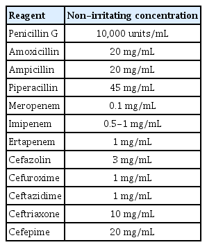
1형 과민반응: 피부단자시험(skin prick test)이나 피내반응시험(intradermal test)을 시행해 볼 수 있다. 베타락탐 항생제의 경우 피부반응시험의 검사법과 해석방법이 잘 정립되어 있어 널리 이용된다[5]. 피부반응시험은 검사하는 약제의 농도에 따라 위양성 또는 위음성 결과를 보일 수 있기 때문에 기존에 알려진 비자극농도(nonirritating concentration)를 이용하여 검사를 시행한다(Table 2). 일반적으로 피부단자시험을 먼저 시행하고 양성 반응이 나타나지 않으면 피내시험을 진행한다. 피내시험은 피부단자시험을 시작한 농도의 1/100로 시작하며 음성이면 10배씩 농도를 점차로 증가시킨다. 피내 시험은 드물게 전신알레르기 반응이 나타날 수 있어 주의가 필요하다. 피부시험 시행 10-15분 뒤에 3 mm 이상의 팽진과 발적이 발생하는 경우 양성으로 판정한다. 피부시험은 증상이 발생하고 3-6주 후에 시행하는 것을 권고한다[3].

Nonirritating penicillin and β-lactam concentrations [5,11]

4형 과민반응: 원인 약제를 진단하기 위해서는 증상이 회복된 후 첩포시험(patch test)이나 지연형 피내반응시험(delayed intradermal test)을 시행할 수 있다. 첩포 검사는 검사의 특이도는 높으나 민감도가 낮은 단점 (< 50%)이 있다[6]. SJS/TEN에서는 일반적으로 시행하지 않지만, maculopapular exanthem, AGEP, 또는 DRESS에서는 도움이 된다는 보고가 있다[7]. 첩포시험에 사용하는 약제는 생리식염수나 바셀린(petroleum)에 희석하여 10-30% 농도로 제조하여 사용하며 48시간과 72시간 후의 반응을 판독한다[3,8].

3) 검사실 검사

혈청 특이 IgE 측정법: 1형 과민반응의 경우 ImmunoCAP (ThermoFisher, Uppsala, Sweden)을 통해 penicilloyl G (C1), penicilloyl V (C2), ampicilloyl (C5), amoxicilloyl (C6), cefaclor (C7) 다섯 가지의 약물의 특이 IgE를 측정할 수 있다[5]. 이들 검사는 일반적으로 민감도(32-68%)는 낮으나 특이도(95-98%)가 높아 약물피부반응시험과 함께 사용하면 약물알레르기 진단의 민감도를 높일 수 있다[9].

Human leukocyte antigen (HLA) 대립유전자 검사: 특정 약물과 연관된 HLA 대립유전자들이 밝혀지면서 중증피부유해반응의 예방에 일부 활용되고 있다[9,10]. 국내에서도 알로푸리놀 투여 전 HLA-B*5801유전자 검사를 임상에서 시행할 수 있다.

호염기구활성화 검사(basophil activation test, BAT): 호염 기구 활성화 마커인 CD63, CD203c의 변화를 약물처리 전후 측정하여 즉시형 과민반응의 진단에 사용할 수 있으나 현재 연구목적으로 주로 시행된다[9].

림프구변환 검사(lymphocyte transformation test, LTT): T세포를 원인 약물과 함께 배양하여 약물 특이 T세포의 활성도와 증식도를 측정하여 T세포 매개 지연형과민반응 진단에 사용할 수 있다[9].

약물유발시험

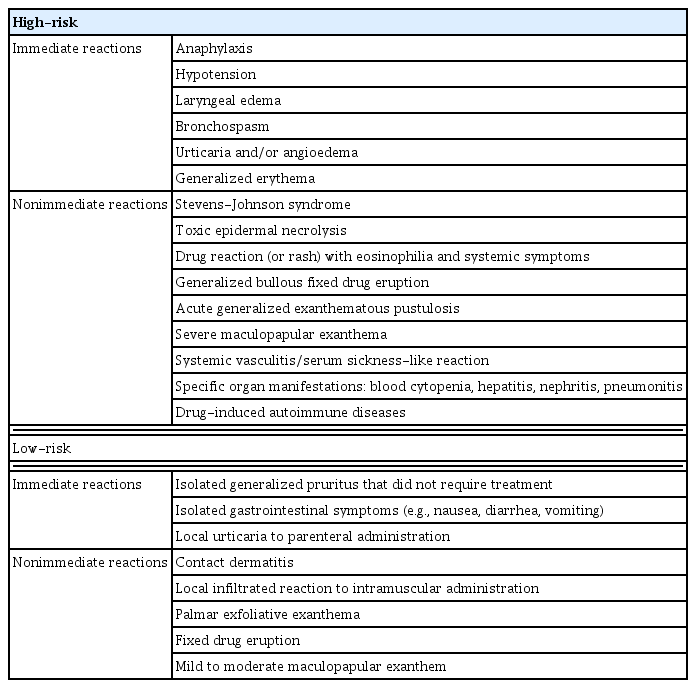
약물유발시험은 약제에 대한 이상반응을 확인하기 위해서는 가장 확실한 방법이지만, 잠재적 위험이 있어 표 3에서 제시하는 고위험군에서는 일반적으로 약물유발시험은 시행하지 않는다[3,5,11]. 그러나 임상 증상이나 검사 결과가 모호해서 약물알레르기를 배제하거나, 원인약 이외에 안전하게 사용할 수 있는 대체약을 탐색하는 경우 시행할 수 있다. 약물유발시험은 응급처치가 가능한 병원에서 알레르기 전문의의 감독 하에 시행하는 것을 권고한다.

Risk stratification of drug allergies according to the index reaction [3]

약물알레르기의 관리

1) 페니실린 알레르기 환자의 관리

고위험군: 표 3에서 제시한 고위험군에 해당하는 즉시형 과민반응 증상–아나필락시스, 호흡곤란, 전신 두드러기 등이 있을 경우 피부단자시험과 가능하면 혈청 IgE 검사를 시행한다. 둘 중 하나라도 양성일 경우 페니실린 알레르기로 진단하고 대체 항생제를 투여한다. 페니실린을 사용해햐 하는 경우에는 탈감작을 시행한다. 피부시험에서 음성을 보이면 약물유발시험을 시행하여 페니실린 알레르기 진단을 수정할 수 있다[5].

저위험군: 두드러기가 없는 가려움증이나 1주 미만의 반점구진상 발진 등의 병력이 있을 경우 피부반응 검사 없이 유발 검사를 시행해 볼 수 있다.

페니실린은 약물알레르기의 가장 흔한 원인 항생제로 알려져 있으나 병력만으로는 잘못 진단하여 광범위 항생제(broad spectrum antibiotics) 사용 빈도가 증가하고, 위막성 대장염 발생률과 항생제 내성균 출현의 위험도를 증가시키고, 의료비용의 상승을 초래하는 것으로 조사되었다[12,13]. 최근 미국감염학회와 보건의료역학회에서는 항생제 적정 사용을 목적으로 하는 통합적 전략 및 중재들을 항생제 스튜어드십(antimicrobial stewardship)이라 정의하고, 이를 실현하기 위한 다양한 전략, 약물감시, 교육을 포함하는 항생제 스튜어드십 프로그램(Antimicrobial Stewardship Program, ASP)을 개발하여 임상에 적용하고 있다. 이 활동의 일환으로 약물알레르기 환자를 체계적으로 평가하고 관리하며, 특히 페니실린 알레르기로 잘못 진단된 환자의 라벨을 지우는 활동(delabeling)이 강조되고 있다[12,14,15].

2) 페니실린과 세파로스포린의 교차반응성

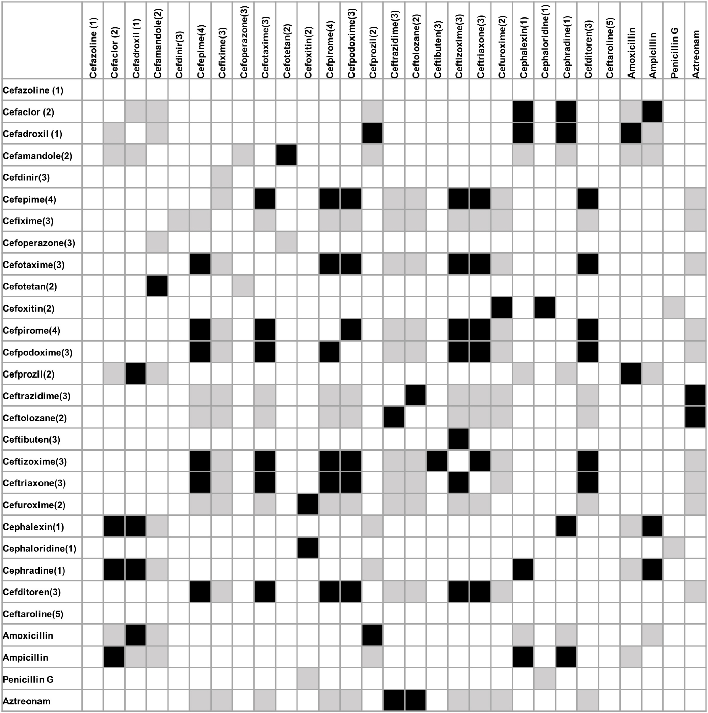
페니실린과 아미노페니실린, 세파로스포린 항생제는 베타락탐고리를 공통적으로 가지며 R기 곁사슬(R-side chain)이 유사할 경우 교차반응성을 보일 수 있다. 특히, 즉시형 과민반응에서는 베타락탐고리보다는 R1 곁사슬이 더 중요한 역할을 하는 것으로 알려져 있는데 페니실린, 아미노페니실린, 1세대와 2세대 세파로스포린의 상당수는 동일한 R1 곁사슬을 가지고 있다. 그러나 3세대 또는 4세대 세파로스포린이나 카바페넴(carbapenem), 모노박탐(monobactam)과의 교차반응성은 매우 드문 것으로 조사되었다(Fig. 1).

Figure 1.

Cross-reactivities between penicillins and cephalosporins (modified from reference [11]). This matrix shows similar (gray) and identical (black) side chains. The number in parenthesis is the cephalosporin generation. Empty boxes indicate a lack of side-chain similarity.

페니실린 알레르기 환자에서 아미노페니실린 투여

아미노페니실린을 이용한 피부시험이 음성인 경우 해당 항생제를 투여할 수 있으나 피부시험이 음성이라도 알레르기 반응이 나타날 수 있으므로 단계적인 약제투여(graded challenge)를 고려한다[16]. 단계적인 약제투여는 일반적으로 1/10 용량으로 시작하여 30-60분 간격으로 상용량까지 증량 후 1시간 정도 이상반응 여부를 관찰한다[3,11]. 저위험군에서는 1-2단계로 상용량에 도달해도 비교적 안전하다는 보고가 있다[17]. 피부시험이 양성인 경우는 대체 항생제를 사용하고, 적절한 대체 항생제가 없는 경우는 탈감작(desensitization)을 시행하여 투여한다.

페니실린 알레르기 환자에서 세파로스포린 투여

저위험군에서 피부반응검사 없이 유발 검사를 시행하거나 페니실린을 단계적으로 투여할 수 있다[11,18]. 그러나 즉시형 페니실린 알레르기 고위험군 환자에서는 1) 3, 4, 5세대 세팔로스포린을 단계적으로 투여하거나 2) 미생물균주에 적합한 대체 항생제를 투여하거나 3) 카파페넴이나 모노박탐계열의 항생제를 사용한다. 만일 페니실린이나 1, 2세대 세파로스포린 항생제를 사용해야 하는 경우에는 피부시험이 필요하며 알레르기 전문의와 상의할 것을 권고한다. 지연형 페니실린 알레르기 고위험군에서는 1) 아미노페니실린, 세파로스포린, 카바페넴 사용 모두 회피하거나 2) 대체 항생제를 사용한다. 베타락탐계항생제 사용이 필요한 경우 감염/알레르기 전문의와 상의할 것을 권고한다(Fig. 2).

Figure 2.

Management of patients with a history of hypersensitivity to penicillins (modified from references [11,18]). DRESS, drug reaction (or rash) with eosinophilia and systemic symptoms; PCN, penicillin; SJS, Stevens-Johnson syndrome; TEN, Toxic epidermal necrolysis.

세파로스포린 알레르기 환자에서 세파로스포린/페니실린 투여

저위험군에서는 R기 곁가지 구조의 유사성이 없는 다른 세대의 세파로스포린 항생제를 사용하거나, 페니실린을 단계적으로 투여해 볼 수 있다. 1세대 혹은 2세대 세파로스포린에 대한 즉시형 과민반응의 병력이 있으면 1) 구조가 다른 R기 곁가지를 가진 3, 4, 5 세대 세파로스포린 항생제를 단계적으로 투여하거나, 2) 카바페넴을 사용하거나, 3) 미생물균주에 적합한 대체 항생제를 투여한다. 3세대 혹은 4세대 세파로스포린 즉시형 과민반응의 병력이 있으면 1) 페니실린 혹은 구조가 다른 R1 곁가지를 가진 세파로스포린을 단계적으로 투여하거나, 2) 카바페넴을 사용하거나, 3) 미생물균주에 적합한 대체 항생제를 투여한다. 지연형 과민반응에서는 페니실린, 세파로스포린, 카바페넴 항생제 사용은 피하며, 베타락탐계항생제 사용이 필요한 경우 전문의와 상의한다(Fig. 3).

Figure 3.

Management of patients with a history of hypersensitivity to cephalosporins (modified from references [11,18]). DRESS, drug reaction (or rash) with eosinophilia and systemic symptoms; PCN, penicillin; SJS, Stevens-Johnson syndrome; TEN, Toxic epidermal necrolysis.

탈감작

탈감작은 약물에 대한 면역반응을 변형하여 일시적으로 관용을 획득하게 하는 방법이다. IgE 매개 즉시형 과민반응에서 탈감작은 이미 효과가 상당히 입증되었고, 페니실린 혹은 세파로스포린 탈감작 프로토콜은 잘 정리되어 있다(Table 4). Non-IgE 매개 즉시형 과민반응으로 나타나는 반코마이신에서도 탈감작으로 성공한 증례가 있다[19,20]. Trimethoprim-sulfamethoxazole, 항결핵제 사용 중 발생한 경증에서 중등증의 반점구진상 발진과 같은 지연형 과민반응에서도 탈감작을 성공한 증례과 프로토콜이 보고되었다[21-23]. 그러나 약열, 약인성 간염, 신장염 및 중증피부유해반응에서는 탈감작은 시행하지 않는다. 탈감작 시행 중 아나필락시스 등 중증 알레르기 반응이 나타날 수 있으므로 응급상황에 대비하여 시행해야 한다[11].

Example intravenous desensitization protocol for a β-lactam dose of 1 g [11,24]

결 론

항생제 알레르기는 정확하고 자세한 병력 청취를 바탕으로 피부반응시험, 혈청학적 검사를 종합하여 진단해야 한다. 또한 환자에게 원인 항생제와 교차반응성이 있는 유사계열의 항생제, 그리고 대체 가능한 항생제에 대한 정보를 함께 제공하고 교육하는 과정이 반드시 필요하다. 항생제 알레르기의 정확한 진단과 처치는 향후 재발을 막기 위한 가장 효과적인 방법이며, 사회적으로도 광범위항생제의 남용을 막고 의료비용을 절감하는 효과도 있을 것으로 기대된다. 그러나 항생제 알레르기를 진단할 수 있는 혈청학적 검사가 제한적이고 민감도가 낮아 이를 개선하기 위한 연구와 지원이 필요하다고 생각한다.

References

1. International drug monitoring: the role of national centres. Report of a WHO meeting. World Health Organ Tech Rep Ser 1972;498:1–25.
2. Solensky R, Phillips EJ. Drug allergy. In : Burks AW, Holgate S, O'Hehir R, et al, eds. Middleton's allergy: principles and practice 9th edth ed. Amsterdam: Elsevier; 2020. p. 1261–1282.e1261.
3. Romano A, Atanaskovic-Markovic M, Barbaud A, et al. Towards a more precise diagnosis of hypersensitivity to beta-lactams - an EAACI position paper. Allergy 2020;75:1300–1315.
4. Mayorga C, Fernandez TD, Montañez MI, Moreno E, Torres MJ. Recent developments and highlights in drug hypersensitivity. Allergy 2019;74:2368–2381.
5. Yang MS, Ban GY, Kim MH, et al. KAAACI Standardization Committee Report on the procedures and applications of the diagnostic tests for drug allergy. Allergy Asthma Respir Dis 2017;5:239–247.
6. Barbaud A, Collet E, Milpied B, et al. A multicentre study to determine the value and safety of drug patch tests for the three main classes of severe cutaneous adverse drug reactions. Br J Dermatol 2013;168:555–562.
7. Phillips EJ, Bigliardi P, Bircher AJ, et al. Controversies in drug allergy: testing for delayed reactions. J Allergy Clin Immunol 2019;143:66–73.
8. Blanca M, Romano A, Torres MJ, et al. Update on the evaluation of hypersensitivity reactions to betalactams. Allergy 2009;64:183–193.
9. Mayorga C, Celik G, Rouzaire P, et al. In vitro tests for drug hypersensitivity reactions: an ENDA/EAACI Drug Allergy Interest Group position paper. Allergy 2016;71:1103–1134.
10. Jung JW, Kim JY, Park IW, Choi BW, Kang HR. Genetic markers of severe cutaneous adverse reactions. Korean J Intern Med 2018;33:867–875.
11. Broyles AD, Banerji A, Barmettler S, et al. Practical guidance for the evaluation and management of drug hypersensitivity: specific drugs. J Allergy Clin Immunol Pract 2020;8:S16–S116.
12. Staicu ML, Vyles D, Shenoy ES, et al. Penicillin allergy delabeling: a multidisciplinary opportunity. J Allergy Clin Immunol Pract 2020;8:2858–2868.e16.
13. Shenoy ES, Macy E, Rowe T, Blumenthal KG. Evaluation and management of penicillin allergy: a review. JAMA 2019;321:188–199.
14. Vyles D, Antoon JW, Norton A, et al. Children with reported penicillin allergy: public health impact and safety of delabeling. Ann Allergy Asthma Immunol 2020;124:558–565.
15. Barlam TF, Cosgrove SE, Abbo LM, et al. Executive summary: implementing an antibiotic stewardship program: guidelines by the Infectious Diseases Society of America and the Society for Healthcare Epidemiology of America. Clin Infect Dis 2016;62:1197–1202.
16. Kim MH, Lee JM. Diagnosis and management of immediate hypersensitivity reactions to cephalosporins. Allergy Asthma Immunol Res 2014;6:485–495.
17. Iammatteo M, Blumenthal KG, Saff R, Long AA, Banerji A. Safety and outcomes of test doses for the evaluation of adverse drug reactions: a 5-year retrospective review. J Allergy Clin Immunol Pract 2014;2:768–774.
18. Blumenthal KG, Shenoy ES, Varughese CA, Hurwitz S, Hooper DC, Banerji A. Impact of a clinical guideline for prescribing antibiotics to inpatients reporting penicillin or cephalosporin allergy. Ann Allergy Asthma Immunol 2015;115:294–300.e2.
19. Chopra N, Oppenheimer J, Derimanov GS, Fine PL. Vancomycin anaphylaxis and successful desensitization in a patient with end stage renal disease on hemodialysis by maintaining steady antibiotic levels. Ann Allergy Asthma Immunol 2000;84:633–635.
20. Lee SH, Jo EJ, Mok JH, et al. Successful desensitization for vancomycin hypersensitivity. Allergy Asthma Respir Dis 2013;1:168–171.
21. Yoon D, Ahn H, Kim SY, Hwang SJ, Park HK, Kang HR. Successful rapid desensitization to trimethoprim-sulfamethoxazole-induced delayed hypersensitivity. Allergy Asthma Respir Dis 2015;3:155–158.
22. Ban GY, Jeong YJ, Lee SH, et al. Efficacy and tolerability of desensitization in the treatment of delayed drug hypersensitivities to anti-tuberculosis medications. Respir Med 2019;147:44–50.
23. Lee H, Kim MS, Yang HK, et al. Rapid desensitization to isoniazid and rifampin in an adolescent with active pulmonary tuberculosis. Allergy Asthma Respir Dis 2016;4:212–216.
24. Castells M. Rapid desensitization for hypersensitivity reactions to medications. Immunol Allergy Clin North Am 2009;29:585–606.

Article information Continued

Figure 1.

Cross-reactivities between penicillins and cephalosporins (modified from reference [11]). This matrix shows similar (gray) and identical (black) side chains. The number in parenthesis is the cephalosporin generation. Empty boxes indicate a lack of side-chain similarity.

Figure 2.

Management of patients with a history of hypersensitivity to penicillins (modified from references [11,18]). DRESS, drug reaction (or rash) with eosinophilia and systemic symptoms; PCN, penicillin; SJS, Stevens-Johnson syndrome; TEN, Toxic epidermal necrolysis.

Figure 3.

Management of patients with a history of hypersensitivity to cephalosporins (modified from references [11,18]). DRESS, drug reaction (or rash) with eosinophilia and systemic symptoms; PCN, penicillin; SJS, Stevens-Johnson syndrome; TEN, Toxic epidermal necrolysis.

Table 1.

Revised Gell and Coombs classification of drug reactions [9]

Type 1 Type II Type III Type IVa Type IVb Type IVc Type IVd
Immune reactant IgE IgG IgG INF-γ, TNF-α (Th1 cells) IL-5, IL-4/IL-13 (Th2 cells) Perforin/granzy me B (CTL) CXCL8 GM-CSF (T cells)
Antigen Soluble antigen Cell- or matrix-associa ted antigen Soluble antigen Antigen presented by cells or direct T-cell stimulation Antigen presented by cells or direct T-cell stimulation Cell-associated antigen or direct T-cell stimulations or direct T-cell stimulation Soluble antigen presented by cells or direct T-cell stimulation
Effector Mast cell activation FcR+ cells (phagocytes, NK cells) FcR+ cells Complement Macrophage activation Eosinophils T cells Neutrophils
Example of hyper-sensitivity reaction Allergic rhinitis, systemic anaphylaxis Hemolytic anemia, thrombocytop enia Serum sickness, arthus reaction Tuberculin reaction, contact dermatitis (with IVc) DRESS Maculopapular exanthema with eosinophilia SJS/TEN Bullous exanthema and fixed drug eruption Hepatitis AGEP

AGEP, acute generalized exanthematous pustulosis; CTL, cytotoxic T lymphocyte; CXCL8, chemokine; DRESS, drug reaction [or rash] with eosinophilia and systemic symptoms; GM-CSF, granulocyte macrophage colony-stimulating factor; IFN-γ, interferon-γ; IL, interleukin; NK, natural killer; PMN, polymorphonuclear neutrophil; SJS, Stevens-Johnson syndrome; TEN, toxic epidermal necrolysis; Th1, helper T cell type 1; Th2, helper T cell type 2; TNF-α, tumor necrosis factor-a.

Table 2.

Nonirritating penicillin and β-lactam concentrations [5,11]

Reagent Non-irritating concentration
Penicillin G 10,000 units/mL
Amoxicillin 20 mg/mL
Ampicillin 20 mg/mL
Piperacillin 45 mg/mL
Meropenem 0.1 mg/mL
Imipenem 0.5-1 mg/mL
Ertapenem 1 mg/mL
Cefazolin 3 mg/mL
Cefuroxime 1 mg/mL
Ceftazidime 1 mg/mL
Ceftriaxone 10 mg/mL
Cefepime 20 mg/mL

Table 3.

Risk stratification of drug allergies according to the index reaction [3]

High-risk
Immediate reactions Anaphylaxis
Hypotension
Laryngeal edema
Bronchospasm
Urticaria and/or angioedema
Generalized erythema
Nonimmediate reactions Stevens-Johnson syndrome
Toxic epidermal necrolysis
Drug reaction (or rash) with eosinophilia and systemic symptoms
Generalized bullous fixed drug eruption
Acute generalized exanthematous pustulosis
Severe maculopapular exanthema
Systemic vasculitis/serum sickness-like reaction
Specific organ manifestations: blood cytopenia, hepatitis, nephritis, pneumonitis
Drug-induced autoimmune diseases

Low-risk

Immediate reactions Isolated generalized pruritus that did not require treatment
Isolated gastrointestinal symptoms (e.g., nausea, diarrhea, vomiting)
Local urticaria to parenteral administration
Nonimmediate reactions Contact dermatitis
Local infiltrated reaction to intramuscular administration
Palmar exfoliative exanthema
Fixed drug eruption
Mild to moderate maculopapular exanthem

Table 4.

Example intravenous desensitization protocol for a β-lactam dose of 1 g [11,24]

Solution Volume of diluent (mL) Drug concentration (mg/mL) Total drug to be injected into each bottle (mg)
1 250 0.04 10
2 250 0.4 100
3 250 4 1,000
Step Solution Rate (mL/h) Time (min) Administered dose (mg) Cumulative dose (mg)
1 1 2 15 0.02 0.02
2 1 5 15 0.05 0.07
3 1 10 15 0.1 0.17
4 1 20 15 0.2 0.37
5 2 5 15 0.5 0.87
6 2 10 15 1 1.87
7 2 20 15 2 3.87
8 2 40 15 4 7.87
9 3 10 15 10 17.87
10 3 20 15 20 37.87
11 3 40 15 40 77.87
12 3 80 172.9 992.13 1,000
Total infusion time 337.9 min