피하낭종과 관절염의 형태로 나타난 Sweet 증후군 1예
A Case of Sweet’s Syndrome Presenting as Subdermal Cystic Lesions and Arthritis
Article information
Abstract
Sweet 증후군은 급성 열성 호중구성 피부질환으로 다양한 임상양상을 보일 수 있다. 피하 낭종을 포함하는 피부 병변, 관절염 및 구강내 궤양을 동반한 Sweet 증후군은 국내보고가 드물며 저자들은 초음파와 조직 생검을 통해 진단 후 스테로이드 전신 투여로 호전된 환자 1예를 경험하였기에 문헌고찰과 함께 보고하는 바이다.
Trans Abstract
Sweet’s syndrome is characterized by a combination of clinical and pathologic findings including fever, neutrophilia, tender erythematous skin lesions, and a diffuse infiltration of mature neutrophils in the upper dermis. Numerous diseases and clinical manifestations have been associated with the disease; however, Sweet’s syndrome associated with subdermal cystic skin lesions and arthritis is rare. A 71-year-old female patient presented with fever, erythematous plaques, multiple hypoglossal ulcers, and arthritis in both ankles. The skin lesions were variously sized areas of erythematous swelling on the forehead, back, and left shoulder. Musculoskeletal sonography revealed hypervascularity and a subdermal cyst in the erythematous plaque on her back. The results of a skin biopsy indicated the presence of mature neutrophilic infiltration in the dermis and thus led to the diagnosis of Sweet’s syndrome. We herein present an unusual case of Sweet’s syndrome presenting as erythematous subdermal cystic lesions, multiple hypoglossal ulcers, and bilateral ankle arthritis with a literature review. (Korean J Med 2012;83:150-155)
서 론
Sweet 증후군은 급성 열성 호중구성 피부질환(acute febrile neutrophilic dermatosis)으로 발열, 호중구성 백혈구증가증, 압통을 동반한 홍반성 부종성 판, 구진의 임상증상과 성숙한 호중구의 진피 침윤과 같은 조직학적 특징을 가지며 1964년 Sweet에 의해 처음 보고되었다[1]. 발병원인은 세균, 바이러스 및 종양 항원의 과민 반응, 내인성 사이토카인의 부적절한 분비, 호중구에 대한 항체의 발현, 그리고 종양과 관련된 과립구 콜로니 자극인자(Granulocyte-colony stimulating factor)의 형성 등이 라는 주장들이 있다[2,3]. Sweet 증후군은 피부 이외에도 심장, 간, 폐, 비장, 중추 신경계 및 근골격계에도 다양한 임상양상을 가지나 관절염과의 동반은 비교적 드물다. 저자들은 71세 여자에서 양측 발목의 관절염, 구강 궤양과 피하 낭종의 형태를 동반한 피부 병변의 Sweet 증후군 1예를 경험하고 성공적으로 치료하였기에 문헌고찰과 함께 증례 보고한다.
증 례
환 자: 여자, 71세
주 소: 발열, 양측 발목의 열감을 동반한 부종, 얼굴과 등의 피부 병변
현병력: 내원 4일 전부터 얼굴, 등, 왼쪽 어깨, 오른쪽 발목의 다양한 크기의 홍반성 판 및 구강내 궤양이 발생하였고, 내원 2일 전부터는 고열, 전신의 근육통 및 양측 발목의 심해지는 관절통이 발생하여 내원하였다.
과거력: 환자는 내원 10년 전 왼쪽 갑상선 절제술을 시행 받았으며, 동시에 당뇨병 진단되어 내원 당시 외래에서 Glimepiride (amaryl®) 6 mg, Metformin (Glupa®) 1,700 mg 경구 투여 중이다. 내원 3개월 전에는 왼쪽 가슴 흉통으로 우관상동맥(Right coronary artery)의 80% 협착으로 경피적 관상동맥 중재술을 시행 받은후 aspirin, clopidogrel 복용하면서 별다른 증상 없이 지냈다.
가족력과 사회력: 특이사항 없음.
진찰소견: 내원 당시 혈압 130/80 mmHg, 맥박수 분당 78회, 체온 38.8℃, 호흡수 분당 20회였으며, 경부 림프절이나 편도 비대 등의 특이 소견은 관찰되지 않았다. 흉부 및 복부에서는 특이 사항이 없었다. 피부소견은 얼굴, 등, 왼쪽 어깨, 오른쪽 발목에 경계가 명확하고 다양한 크기의 융기된 홍반성 판이 있었으며(Fig. 1A), 병변 부위의 통증을 호소하지는 않았지만, 압통이 있었다. 구강내 설하에는 다수의 다양한 크기의 통증성 궤양이 있었다(Fig. 1C).

(A) The patient had an erythematous plaque with a pseudovesicular appearance on her forehead (arrowheads). (B) The forehead plaque improved after corticosteroid therapy (arrowheads). (C) Multiple small aphthous hypoglossal ulcers were observed. (D) The hypoglossal ulcers improved after corticosteroid therapy.
검사실 소견: 혈액 검사에서 헤모글로빈 11.2 g/dL (12.0-16.0 g/dL), 백혈구 6,100/uL (4-8 × 10³/uL), 혈소판 163,000/uL (150-450 × 103/uL)이었으며, 적혈구 침강 속도(ESR)는 34 mm/hr (0-10 mm/hr), 단백면역화학 검사에서 C 반응성 단백질(C-reactive protein)은 14.30 mg/dL (0-5 mg/dL) 로 상승되었다. 혈청 생화학 검사에서는 혈당 202 mg/dL로 증가되었으며, 요침사 검사에서 당이 3+ 확인되었고 당화 혈색소(HbA1c)는 10.2% 였다. VDRL, 항HIV항체, 항핵항체, 류마티스인자, PT/aPTT, 항핵항체, 항호중구세포질항체, HLA-B51은 음성 혹은 정상 범위였다. 거대세포바이러스(Cytomegalovirus), 엡스타인바바이러스(Epstein-Barr virus) viral capsid antigen/early antigen, 리켓치아 쯔쯔가무시(Orientia tsutsugamushi), 렙토스피라 인터로간스(Leptospira interrogans), 한타 바이러스(Hantaan virus) 등에 대한 혈청 항체도 모두 음성이었다. Alpha-fetoprotein (a-FP)은 2.1 ng/mL (0.0-7.0 ng/mL), carcinoembryonic antigen (CEA)는 2.7 ng/mL (0.0-5.0 ng/mL), cancer antigen 125 (CA-125)는 14.21 U/mL (0.0-35 U/mL)였다. 말초 혈액도말검사상 독성 과립이 관찰되었으며, 호중구의 좌방이동(shift-to-left) 소견이 확인되었으나 비정형 골수모세포는 관찰되지 않았다. 갑상선 기능 검사 결과 T3 43.2 ng/dL (60-190 ng/dL), fT4 1.28 ng/dL (0.70-1.80 ng/dL), 그리고 TSH 2.62 uIU/mL (0.25-4.0 uIU/mL)이었다. 이상초과민 검사(pathergy test)는 음성이었다.
방사선학적 소견: 흉부 컴퓨터 촬영상 양측 폐내 다발성 소결절이 관찰되었으나 림프절 비대 및 흉수는 없었으며, 복부 컴퓨터 촬영에서는 특이 소견 없었다. 양측 발목은 경거(tibio-talar joint) 관절 초음파상 관절액의 저류가 관찰되었으며(Fig. 2A), 등의 구진성 판(Fig. 2C)에 대한 초음파 검사상 고혈류의 비전형적 침윤과 동반된 낭종성 병변이 확인되었다(Fig. 2D).

(A) Ultrasound showed synovial effusion of the tibiotarsal joint (arrowheads). (B) The effusion of the tibiotarsal joint improved after intra-articular corticosteroid therapy (arrowheads). (C) An erythematous plaque was present on the right lower part of the back (arrowhead). (D) Sonography revealed a subdermal cystic lesion (arrowhead).
피부 생검 조직의 병리학적 소견: 압통을 동반한 전신의 다양한 크기의 홍반성 판이 있어, 피하의 농양 가능성 고려하여 등의 병변에서 흡인 시행하였으나 농성 물질은 배출되지 않았다. 홍반성 판 하부 낭종 주위에서 시행한 생검 조직 검사 결과 진피 내 염증 소견과 함께 성숙한 호중구 침윤된 것이 확인되었다(Fig. 3).

(A) Diffuse neutrophilic infiltration in a skin biopsy of the back (HE stain, × 40). (B) Perivascular and dermal neutrophilic aggregation (HE stain, × 200).
치료 및 임상 경과: 고열으로 내원하였고, 흉부 컴퓨터 촬영상 양측 폐내 다발성 소결절이 관찰되어 폐렴에 의한 감염 고려하여 전신적 항생제 amoxicillin/clavulanate 3,600 mg/day을 투여 하였다. 항생제 투여 4일째에도 38.1℃의 발열이 지속되었으며 흉부 방사선상 변화가 없었다. 급성 열성 질환과 홍반성 피부 병변을 동반하였으며, 복부의 홍반성 판에서 시행한 조직 검사 결과 Sweet 증후군으로 진단하여 항생제는 중단하고 고용량 스테로이드(prednisol 62.5 mg/day)를 정주하였다. 또한, 양측 발목의 활액의 저류에 대하여 초음파 유도하 흡인을 하여 관절낭 내 활액의 흡인 검사 결과 WBC 7,000/mm3(Poly 90%, Monocytes 10%)으로 확인되었으며 그람 염색 결과 음성, 배양 검사 결과 동정되는 균은 없어서 화농성 관절염은 배제할수 있었다. 이에 스테로이드를 관절강 내 정주하였다. 일주일 후 이마 및 등의 피부 병변에도 병변내 스테로이드 주사 요법을 시행하였다. 투여 3일 후 발열 소실 되었으며, 7일 후 이마와 등의 피부 병변, 구강내 궤양이 뚜렷하게 호전되었다(Fig. 1B, 1D). 관절강내 주사후 추적관찰한 관절 초음파에서 경거 관절의 관절액 저류 또한 호전되었다(Fig. 2B). 이후 경구 스테로이드 용량을 점차적으로 감량하여 현재 외래에서 재발 없이 추적관찰 중이다.
고 찰
Sweet 증후군은 주로 얼굴, 목, 상체 및 사지에 통증이 동반된 홍반성 판 및 구진이 발생하는 임상적인 특징을 지니고 있다. 1986년 Su와 Liu에 의해 진단 기준이 제시된 이후, 1994년 von den Driesch에 의해 조정되었다[1]. Sweet 증후군은 1) 압통을 동반한 홍반성 판 또는 결절의 발생, 2) 혈관염의 증거 없이 성숙한 호중구의 진피 침윤의 두 가지 주 진단 기준을 만족하면서, 1) 38°C 이상의 발열, 2) 기저의 혈액학적 또는 고형 암종, 염증성 질환, 임신 또는 선행하는 상기도 감염, 위장관 감염, 또는 예방접종의 존재, 3) 스테로이드나 KI (potassium iodide)로 치료 후 빠른 호전, 4) 검사실 소견상 백혈구증가증(>8,000/mm3), 호중구증가증(>70%), ESR 증가(>20 mm/hr), CRP 양성의 네 가지 부 진단기준 중 두 가지 이상을 충족할 때에 진단된다. 피부 증상은 다양한 크기의 판의 형태이나 드물게 낭종이나 농포 또는 실제 액체가 채워지지 않은 형태의 낭포화 된 형태(pseudovesiculation)로도 보일 수 있다[1].
Sweet 증후군에서 관절통이 동반되는 경우는 32-62%로 흔하지만, 관절염이 동반되는 경우는 약 10-15%로 비교적 드물다[1,2]. 관절 증상은 피부 병변이 발생하기 전 또는 후에 발생할 수 있다. 대부분의 보고에서 관절염은 주로 비대칭적으로 무릎, 어깨, 엉덩이, 발목, 손목 관절과 같은 대관절을 주로 침범하였으나, 손과 발의 소관절을 침범함 경우도 있다[2]. 방사선학적 사진의 경우에 대부분 정상이며, 관절낭 활액의 경우 염증성 변화를 보일 수 있다. 본 증례의 환자에서는 초음파 결과 오른쪽 발목의 관절낭 내 활액의 저류, 동통을 동반한 열감, 부종으로 인하여 관절염이 동반되었다. 또한 환자의 이마, 등, 왼쪽 어깨, 오른쪽 발목에서 관찰된 홍반성 융기 판과 조직학적으로 확인된 호중구의 진피 침윤으로 인하여 Sweet 증후군의 피부증상에 합당하였다. 환자의 임상증상과 병력, 혈액 검사 결과를 바탕으로 주 진단 기준 두 가지와 부 진단 기준 세 가지를 만족하여 Sweet 증후군으로 진단하였으며 과거력상 Sweet 증후군과 관절염이 동반될 수 있는 기저 질환인 myelodysplastic syndrome, 간경화 및 약물 노출력은 없었다[3,4]. 관절염 및 구강 궤양은 고용량 스테로이드 및 병변 내 주입치료 이후 모두 호전을 보여 Sweet 증후군과 연관된 피부 외 소견으로 보인다.
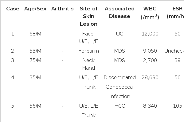
아프타성 궤양(aphthous-like ulceration), 궤양 구내염(ulcerative stomiatitis)은 Sweet 증후군에서 관찰될 수 있는 구강내 병변으로, 재발하는 구강내 궤양이 특징인 베체트 병과 감별이 필요하다. 베체트 병과 비교하여 Sweet 증후군은 피부 병변이 주이며, 구강 궤양은 약 3-30%의 경우에만 보고되어 있으며, 연관된 성기 궤양의 발생은 매우 드물게 보고되어 있다[5-6]. Sweet 증후군의 국내 보고는 이 등이 국내에서 발생한 Sweet 증후군 23예를 연구한 것이 있고, 여기에서 연관된 전신질환으로는 만성 염증성 장질환(6예)이 가장 흔하였으며 다음으로 급성 골수성 백혈병등의 혈액종양은 5예, 고형종양은 1예였다[7]. 질환별로 좀더 자세히 보면 궤양성 대장염, 골수 이형성 증후군, 임균 감염 및 간암 등과 같은 전신 질환 및 약제 와 연관된 Sweet 증후군이 보고되었으며[8-13], Jung 등[14]은 의식 혼탁으로 내원한 Neuro-sweet 환자 증례를 보고하였으나, 피하 낭종의 피부 병변 및 관절염이 확인된 국내 보고는 없었다. 이상의 국내에 보고된 증례의 임상적 특징을 표로 정리하여 보았다(Table 1).
Sweet 증후군은 치료를 하지 않을 경우 수 개월간 증상이 지속될 수 있다. 치료는 스테로이드의 전신적 투여이며, 치료 반응은 비교적 효과적이다. 스테로이드에 반응이 불충분할 경우 요오드화칼륨(potassiumiodide), 콜히친(Colchine), 인도메타신(indomethacin), 클로파지민(clofazimine), 사이클로스포린(cyclosporine), 답손(dapsone) 등을 병용 투여할 수 있다[15]. 또한 고령에서 재발을 반복하는 경우는 반드시 악성 종양의 동반여부를 확인하고 필요 시 면역억제제를 추가하여야 한다. 작은 크기의 단독 병변일 경우에는 외용 스테로이드의 사용이나 스테로이드(triamcinolone acetonide)의 병변내 주입으로 호전을 보인 사례들도 보고되었다[16]. 본 증례에서는 병변 내 스테로이드 주사 및 전신투여로 피하 낭종을 포함하는 피부 병변, 관절염 및 구강 내 궤양이 치료 후 호전되었고, 용량을 점차적으로 감량하여 재발없이 치료되었다.
