폐암 조직 검사 후 발생한 피하 결절
Subcutaneous nodule developed after needle biopsy for lung cancer
Article information
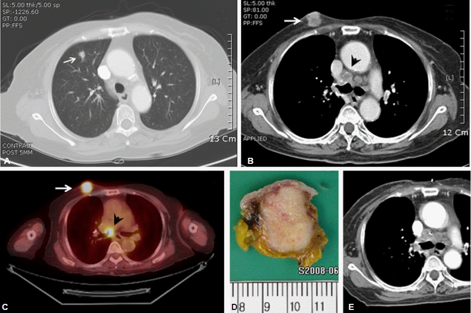
70세 여자 환자가 가슴 앞쪽 피부 밑에 생긴 결절을 주소로 내원하였다. 이 환자는 1년 전 1 cm 크기의 폐 종양이 생겨(그림 1A) 조직 검사를 시행하였고 비소세포폐암으로 진단되어 수술을 받았다. 수술 후 병기는 T1N0M0였다. 피부 밑에 만져지는 결절은 2 cm 크기로 흉벽에 고정되어 있지는 않았고 약간 단단한 느낌을 주었으나 결절을 덮고 있는 피부에는 이상 소견이 없었다. 통증 등 동반된 증상도 없었으나 1년 전 조직 검사 시 바늘이 들어간 자리와 일치하여 조직 검사 도중 일부 암세포가 피부 밑에 이식된 것으로 판단되었다. 흉부전산화 단층촬영에서 피부 밑 결절이 확인되었고 종격동에 여러 개의 커진 림프절이 발견되어 폐암도 재발되었음을 알 수 있었다(그림 1B). 18FDG-PET/CT에서 결절과 종격동 림프절의 SUV는 각각 9.7, 7.7이었다(그림 1C). 피부 밑 결절은 간단한 수술로 제거하였고(그림 1D, 1E) 종격동에는 방사선치료를 시행하기로 하고 퇴원하였다. 나이와 전신 상태 등을 고려하여 항암화학요법은 시행하지 않기로 결정하였다.

(A) A 1 cm-sized spiculated nodule suggestive of lung cancer was found in RUL. (B) The chest CT taken 1 year later revealed about 1.5 cm-sized subcutaneous nodule on anterior chest wall (arrow) and multiple mediastinal lymphadenopathy (arrowhead). (C) Hypermetabolic lesions indicating recurred cancer were noted on 18FDG-PET/CT. (D) The subcutaneous nodule was excised under local anesthesia. (E) The chest CT after surgery confirmed that the nodule had been completely removed.
세침 조직 검사는 비교적 간단하고 심각한 부작용도 적어 폐암을 진단하는데 매우 중요한 검사이다. 기흉, 출혈 등은 흔히 발생하지만 대부분 쉽게 해결되어 임상적으로 큰 문제가 되지 않는다. 이 환자처럼 바늘이 지나간 자리에 암세포가 이식되는 경우는 매우 드물어, Sinner와 Zajicek에 따르면 5,300예 중에서 0.02%의 빈도를 보였다1). 이식이 된 경우, 조직 검사 후 6~24개월 정도에서 발견이 되며 대부분 수술로 제거될 수 있다2). 하지만 흉막에 이식되어 치료가 어려울 수도 있다3). 액와부위에 근접하여 병변이 있는 경우에는 액와부 림프절도 모두 제거하는 것이 바람직한데 이는 액와부 림프절에 전이가 있었다는 보고들이 있기 때문이다4). 국소적 재발을 줄이기 위해 수술 후 방사선 치료를 시행하는 경우도 있으나 효과는 아직 검증되어 있지 않다.
바늘, 흉관, 내시경 등을 이용하여 암에 대해 어떤 시술을 행한 경우 드물지만 암세포가 이식될 수 있다는 점을 인지하여야 하며 환자가 그 부위에 이상 증상을 호소하거나 이상 소견이 발견되는 경우 이식형 전이의 가능성을 염두에 두고 검사를 시행하는 것이 바람직할 것으로 보인다.