Streptococcus Intermedius 패혈증이 동반된 광범위 문맥염과 간농양
Extensive Pylephlebitis and a Liver Abscess Combined with Streptococcus Intermedius Sepsis
Article information
Trans Abstract
Pylephlebitis (septic thrombophlebitis of the portal venous system) is a rare but serious complication of intra-abdominal infections that drain into the portal venous system. Its diagnosis is based on imaging; computed tomography may reveal a thrombus in the portal vein. Bacteremia may also be evident. As the symptoms are nonspecific, early clinical diagnosis is difficult, and delayed treatment can compromise outcomes. We report a case with extensive pylephlebitis and a liver abscess associated with Streptococcus intermedius sepsis; the case was treated successfully with antibiotics and anticoagulants. Such cases have not been widely reported.
INTRODUCTION
Pylephlebitis (septic thrombophlebitis of the portal venous system) is a rare but serious complication of uncontrolled infection in a region neighboring or drained by the portal venous system. In the West, the major causes of pylephlebitis are diverticulitis and appendicitis [1]. However, in one-third of cases, the cause remains ambiguous [2]. Early clinical diagnosis is sometimes challenging because the symptoms are nonspecific, and relies on a high level of clinical suspicion and adequate imaging. Delays in diagnosis and treatment can complicate the condition, raising the mortality rate to as high as 25% [3]. We herein report a rare case of an immunocompetent adult presenting with extensive pylephlebitis, a liver abscess, and sepsis caused by Streptococcus intermedius; the patient was treated successfully with antibiotics and anticoagulants.
CASE REPORT
A 43-year-old male was referred to Daegu Catholic University Medical Center (DCUMC) by a local clinic because of fever and worsening abdominal pain. Two weeks previously, he had developed pain over the entire abdomen and was admitted to a local clinic for examination. Contrast-enhanced abdominal computed tomography (CT) and laboratory tests revealed no specific finding. The patient received symptomatic therapy, but did not respond; he complained of worsening abdominal pain. He then developed new-onset fever and was referred to DCUMC emergency department. His medical history was uneventful, except for hyperlipidemia. He had smoked a pack of cigarettes daily for 20 years and had consumed 100 g alcohol weekly for 15 years.
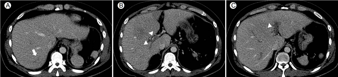
On admission, the patient’s vital signs were: blood pressure 100/60 mmHg, heart rate 89 beats/min, respiratory rate 25 breaths/min, body temperature 38.9℃, and room air oxygen saturation 93%. Physical examination revealed localized tenderness in the upper abdominal area and decreased bowel sounds. His initial laboratory tests revealed a white blood cell (WBC) count of 15,100/mm3 (normal: 3,600-9,600/mm3 ), an alanine aminotransferase level of 54 U/L (normal: < 40 U/L), an alkaline phosphatase level of 300 U/L (normal: 35-130 U/L), a gamma-glutamyl transpeptidase level of 150 U/L (normal: 8-61 U/L), a total bilirubin level of 2.9 mg/dL (normal: < 1.2 mg/dL), a C-reactive protein (CRP) level of 278.3 mg/L (normal: < 5 mg/L), a procalcitonin level of 12.17 ng/mL (normal: < 0.5 ng/mL), a prothrombin time (international normalized ratio) of 1.35 (normal: 0.8-1.2), a blood urea nitrogen level of 38.6 mg/dL (normal: 8.0-23.0 mg/dL), and a creatinine level of 2.1 mg/dL (normal: 0.8-1.2 mg/dL). Contrast-enhanced abdominal CT revealed an immature liver abscess in segments 7/8 (Fig. 1A) and extensive pylephlebitis; thrombi were visible in the lumina of the right, left, and main portal vein, accompanied by perivascular inflammatory changes (Fig. 1B, 1C).

Contrast-enhanced abdominal computed tomography images obtained at admission. (A) A poorly defined low-density lesion is evident in segments 7/8 (arrow). (B, C) Poorly attenuating thrombi are visible in the right and left portal vein (arrowheads).
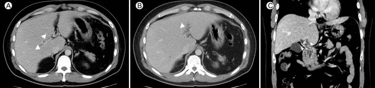
On hospital day 1, we commenced intravenous empirical antibiotics (cefotaxime 2 g every 6 hours and metronidazole 500 mg every 8 hours) and heparin. On day 3, the patient’s body temperature remained high, and Gram-positive cocci were detected in two blood cultures commenced soon after admission. On day 6, the cocci were identified as Streptococcus intermedius that was sensitive to cefotaxime. Therefore, intravenous cefotaxime and metronidazole were continued. The patient underwent echocardiography to evaluate bacterial endocarditis; no significant finding was evident. On hospital day 10, his fever subsided and he began to improve; he responded to the antibiotics. Oral warfarin commenced on day 10, and intravenous heparin was terminated on day 20. The WBC and the CRP level were within their normal ranges on day 15. Abdominal CT performed on day 25 showed interval improvement of the immature liver abscess in segment 7/8 (Fig. 2A), but the persistence of extensive portal vein thrombosis (Fig. 2B, 2C).
Contrast-enhanced abdominal computed tomography images obtained 25 days after admission. (A) The initial, poorly defined low-density lesion in segments 7/8 has essentially disappeared (arrow). (B, C) The combined volume of the poorly attenuating thrombi has decreased, but thrombi remain visible in the right and left portal vein (arrowheads).
As the laboratory data and physical examination showed that the patient had recovered completely, he was discharged on day 28. After discharge, he received 2 weeks of oral amoxicillin/ clavulanic acid and remained on warfarin. At the 3-month follow-up examination, repeat abdominal CT revealed complete resolution of the liver abscess and cavernous transformation of the portal vein attributable to the extensive pylephlebitis (Fig. 3). Warfarin was stopped after 3 months. The patient exhibited no sign of relapse during outpatient follow-up.
Contrast-enhanced abdominal computed tomography (CT) images taken 3 months after admission. (A, B) The right and left portal vein is almost obliterated (arrowheads). (C) Abdominal CT image showing numerous collaterals around the portal vein, consistent with cavernous transformation (arrowhead).
DISCUSSION
Septic thrombophlebitis of the portal vein is termed pylephlebitis, and is often associated with intra-abdominal infection. Common underlying diseases include appendicitis, diverticulitis, inflammatory bowel disease, pancreatitis, and liver abscess. In the pre-antibiotic era, the most common cause of pylephlebitis was appendicitis, but the etiologies shifted after the introduction of antibiotics. Recent reports from the USA show that the leading causes of pylephlebitis are pancreatitis (31%) and diverticulitis (19%) [4]. However, liver abscess (38%) and cholangitis (20%) remain common etiologies in Taiwan [5].
In the review of Kanellopoulou et al. [6] blood cultures were positive in 23-88% of pylephlebitis cases. The most frequently isolated pathogens were streptococci, Escherichia coli, Bacteroides fragilis, and Klebsiella pneumoniae [4,5]. Here, we detected Streptococcus intermedius in two blood cultures. Streptococcus intermedius is a member of the Streptococcus anginosus group (SAG), consisting primarily of mucosal commensals found commonly in the mouth and in the upper respiratory, gastrointestinal, and urogenital tracts. Despite the commensal nature of the SAG, the microorganisms sometimes act as pathogens of abscesses and blood [7]. Reported cases include intracranial infections caused by rhinosinusitis in pediatric patients, lung abscesses in adults, bacterial endocarditis, and sepsis associated with surgical infection. Here, we report a rare case of extensive pylephlebitis and a liver abscess associated with Streptococcus intermedius sepsis.
Pylephlebitis commonly presents with relatively nonspecific clinical features, such as fever, pain, nausea, vomiting, and anorexia. Thus, early clinical diagnosis is difficult, but delays in treatment can worsen outcomes. The most common symptoms are fever (86-100% of cases) and abdominal pain (74-82%) [6,8]. Diagnosis requires the confirmation of portal vein thrombosis, and evidence of bacteremia acquired by portal vein aspiration. However, such diagnostic methods are invasive, and thus rarely performed. Surrogate markers, including clinical features, imaging data, and the results of blood culture, are used instead. Portal vein thrombosis can be diagnosed via Doppler ultrasonography, CT, and magnetic resonance imaging of the abdomen. Ultrasonography reveals a hyperechoic lesion in the portal vein, and color Doppler imaging detects a decrease in blood flow. On abdominal CT, portal vein thrombosis presents as a filling defect in the contrast-enhanced lumen of the portal vein. In addition, abdominal CT may detect other intra-abdominal infective foci in patients with severe pylephlebitis; abdominal CT is thus the preferred first-line diagnostic modality [9].
When pylephlebitis is suspected or diagnosed, it is important that patients are treated immediately with broad-spectrum antibiotics for a recommended minimum of 4 weeks, given that a liver abscess may develop as a complication. Patients with confirmed macroscopic liver abscesses complicating pylephlebitis should be treated with antibiotics for least 6 weeks, with or without drainage [8]. The use of anticoagulants in patients with pylephlebitis remains controversial. Baril et al. [1] argued that anticoagulant therapy should be considered for patients with pylephlebitis of hypercoagulable status (because they had neoplasms or clotting factor deficiencies), but not for patients in whom clotting was normal and thrombi were confined to the portal vein. On the other hand, Plemmons et al [8]. reported 100% survival of patients treated with anticoagulants, in contrast to 60% survival of those who did not receive anticoagulants, although the difference did not attain statistical significance. Another study showed that early anticoagulant treatment for patients with pylephlebitis might reduce the incidence of serious complications and enhance recanalization [6]. Allaix et al [10]. recommended 3-6 months of anticoagulant therapy for patients with pylephlebitis but no underlying thrombotic disease. However, no consensus on the duration of anticoagulant therapy or follow-up has yet emerged. We treated a patient presenting with abdominal pain and fever caused by extensive pylephlebitis and a liver abscess; treatment with antibiotics and anticoagulants was successful.
Advances in imaging modalities and antibiotic therapies have reduced mortality from pylephlebitis. However, recent studies have documented a persistent 11% mortality rate and long hospital stays [4,5]. Thus, early diagnosis and appropriate treatment are essential. A high level of clinical suspicion is required for patients with fevers of unknown origin, intra-abdominal infections, or abdominal pain of unknown cause.