초음파를 이용한 어깨 통증의 진단
Ultrasonographic Assessment for Shoulder Pain
Article information
서 론
어깨 통증의 발생률은 일반 인구의 15-20%에 이를 정도로 비교적 흔하다[1]. 지속적인 어깨 통증의 원인으로는 회전근개 질환이 10%, 유착관절낭염(adhesive capsulitis)이 6%, 오목위팔관절(glenohumeral joint)의 골관절염이 2-5% 등이다[2]. 어깨 통증의 진단에 있어 초음파는 방사선 노출이 미미하고, 가격이 저렴하며, 진료실에 위치하여 진료 의사가 직접 수행하여 치료를 바로 결정할 수 있고, 움직임을 관찰할 수 있다는 장점이 있다. 대개 60 mm의 일자형 탐촉자를 이용하기 때문에 내과 진료시 갑상선 또는 경동맥을 관찰하는 초음파를 이용하여 쉽게 관찰이 가능하다. 다음은 초음파로 확인이 가능한 질환들에 대한 대표적 소견이다.
힘줄윤활막염(tenosynovitis)
힘줄집내 액체를 동반하거나 동반하지 않은 저에코성 또는 무에코성의 두꺼워진 조직이 탐촉자의 두 직각 면에서 관찰되고, 도플러 신호가 보일 수 있는 병변으로 정의된다[3]. 위팔두갈래근(biceps muscle)의 긴머리힘줄(long head)에서 주로 발생하며, 힘줄은 비교적 정상적인 두께와 에코를 보이지만 그 주변으로 무에코성의 액체 저류와 증가된 파워도플러 신호가 관찰된다(Fig. 1). 위팔두갈래근의 힘줄집은 오목위팔 관절과 통해있으므로 이 관절의 삼출이 이동한 것인지 힘줄윤활막염 단독 병변인지를 잘 구별해야 한다.
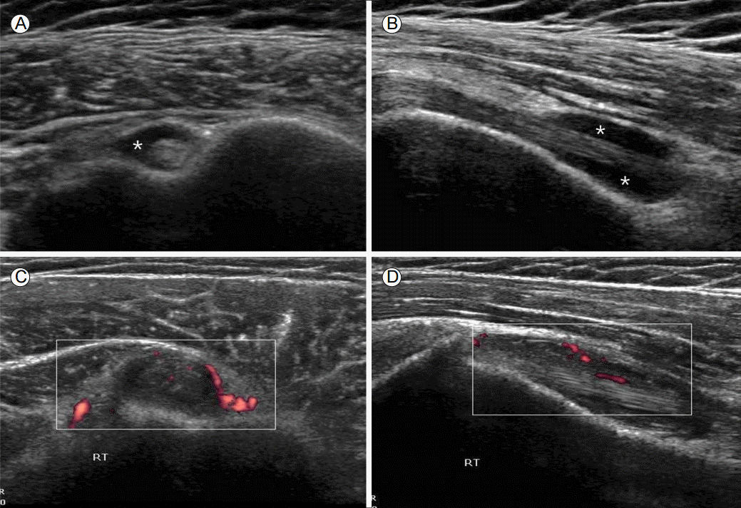
Tenosynovitis of the long head of biceps tendon. (A) Transverse and (B) longitudinal ultrasonographic (US) images demonstrate anechoic fluid (*) within the tendon sheath. The hyperechogenic thickened tendon sheath exhibits increased power Doppler signal in (C) transverse and (B) longitudinal US images.
힘줄염(tedinitis)와 힘줄병증(tendinosis)
힘줄염이 주로 염증(inflammation)에 의한 힘줄 병변이라면 힘줄병증은 퇴행(degeneration)에 의한 병변으로 초음파로 두 질환의 감별은 쉽지 않다. 가시위오목근은 균질한 실타래 모양의 에코를 보이며 어깨세모근과 비슷한 두께를 보이는 것이 정상이나, 힘줄염이나 힘줄병증이 있는 경우 불균질하게 증가 또는 감소하는 에코를 보이고 부종에 의해 힘줄의 두께가 증가하며(무증상의 반대측에 비해 2-2.5 mm 이상 증가될 때 고려) 미세석회화가 보이거나 도플러 신호가 증가하기도 한다(Fig. 2).
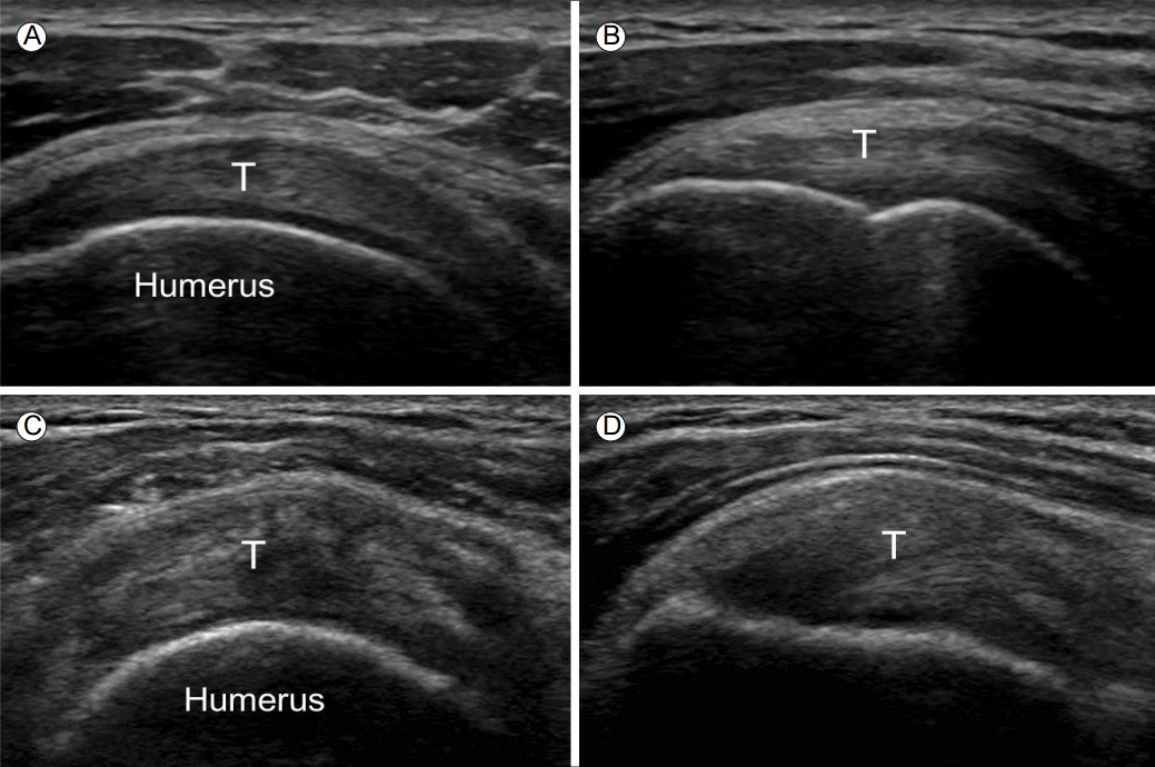
Tendinosis of supraspinatus tendon. In contrast of normal supraspinatus tendon (T) seen in (A) transverse and (B) longitudinal US images, there are heterogeneous echogenicity without definite defect and increased thickness of the tendon seen in (C) transverse and (B) longitudinal US images.
석회성건염(calcific tendinitis)
가시위오목근에서 흔하게 발생하며 골부착 1 cm 이내의 위험대에 잘 발생한다[4]. 석회화의 단계에 따라 다양한 초음파 소견을 보이는데, 약 79%에서 뚜렷한 후음향 음영(postacoustic shadow)을 동반한 고에코성 병변, 14%에서 후음향 음영이 희미한 고에코성 병변, 7%에서 후음향 음영이 없는 고에코성 병변으로 관찰된다(Fig. 3).

Calcific tendinitis of supraspinatus tendon. (A) Longitudinal and (B) transverse ultrasonographic images show hyperechogenic linear lesion (arrows) with faint posterior acoustic shadowing in the tendon.
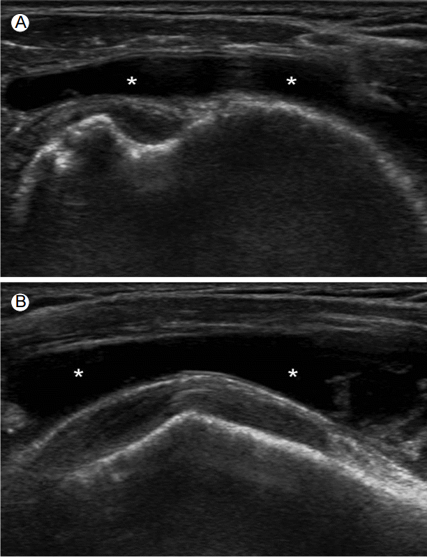
윤활막염(synovitis)
윤활막염의 대표적 초음파 소견으로는 삼출(effusion)과 윤활막 증식(synovial proliferation)이 있다. 삼출은 피하 지방과 비교하여 비정상적인 저에코성 또는 무에코성(때로는 동일 에코이거나 고에코일 수도 있는)의 관절내 물질로, 이동과 압박이 가능하나 도플러 신호를 보이지 않는다[3]. 어깨에서 삼출은 오목위팔 관절의 후면 또는 액와부에서 가장 잘 관찰된다(Fig. 4). 윤활막 증식은 피하 지방과 비교하여 비정상적인 저에코성(때로는 동일 에코이거나 고에코일 수도 있는)의 관절내 조직으로, 삼출과는 달리 이동하지 않고 도플러 신호를 보일 수 있는 병변이다[3].
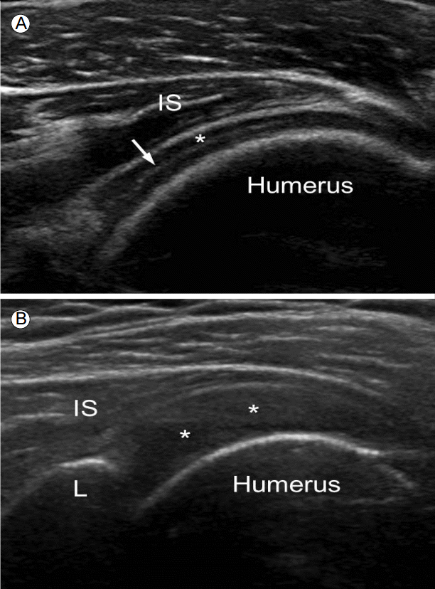
Synovitis of glenohumeral joint. Transverse ultrasonographic images at posterior glenohumeral joint shows (A) anechoic effusion (*) accentuates the appearance of the cartilage (double cortex sign; arrow) and (B) combined hyperechogenic synovial proliferation and relatively hypoechogenic effusion (**) enlarges the joint space between labrum (L), humerus and intraspinatus tendon (IS).
회전근개 파열
파열된 두께에 따라 부분층(partial-thickness) 파열과 전층(full-thichness) 파열로 나뉘고, 부분 파열은 파열된 부위에 따라 힘줄내(intrasubstance), 윤활낭면(bursal surface)과 연골면(articular surface) 파열로 나뉘며, 힘줄이 큰거친면(greater tuberosity)에 부착하는 부위에서 잘 발생한다. 초음파 소견으로는 고에코성의 균질한 실타래 에코가 갑자기 중단되어 국소적인 저에코성 또는 무에코성 병변이 존재하는 것이 탐촉자의 두 직각 면에서 모두 관찰되고 탐촉자에 의해 힘줄 사이가 압박이 될 수도 있다(Fig. 5) [5]. 그러나 출혈에 의해 파열 부위가 오히려 고에코성으로 관찰되거나, 뚜렷한 결함 없이 힘줄이 점진적으로 얇아지거나, 오래된 광범위 파열인 경우 힘줄이 끌어당겨져 관찰되지 않을 수도 있다.
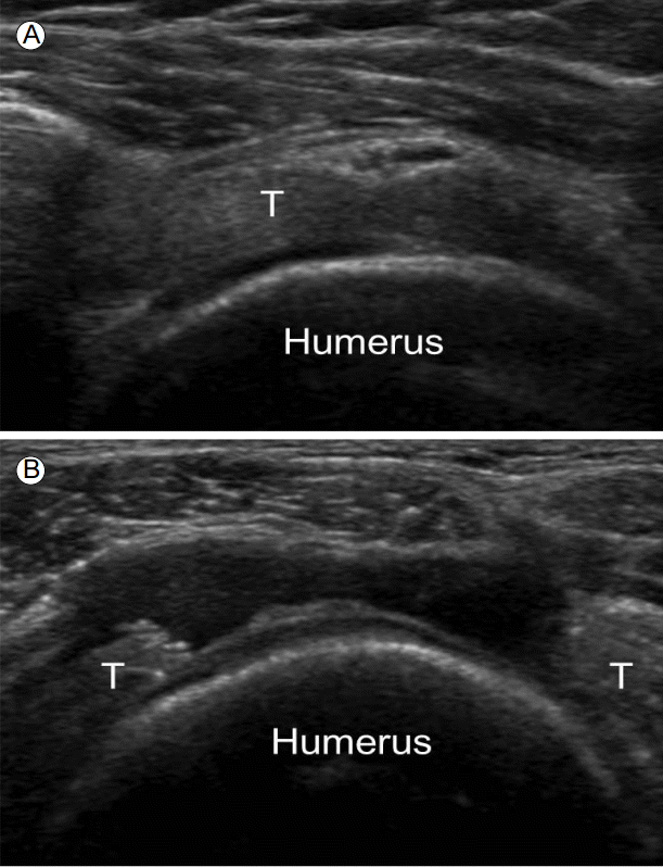
Rotator cuff tear. Transverse ultrasonographic images shows (A) bursal surface partial-thickness tear and (B) full-thickness tear of supraspinatus tendon making “cartilage interface sign”. T, supraspinatus tendon.
윤활낭염(subacromial subdeltoid bursitis)
세모근밑주머니(subdeltoid bursa)와 봉우리밑주머니(sabacromial bursa)는 어깨의 넓은 부위를 덮고 있는 인체에서 가장 큰 윤활낭들로 대부분 두 윤활낭이 통해져 있다. 정상적으로는 2 mm 이하의 두께의, 두 층의 고에코성 윤활낭 주변 지방층과 저에코성의 윤활낭액 선이 나란히 관찰되며 내부의 액체 저류는 보이지 않으나, 윤활낭염이 발생한 경우 이 부위의 액체 저류와 윤활낭벽의 두께 증가가 보이며 이는 어깨를 관찰하는 여러 각도의 스캔에서 쉽게 관찰이 가능하다(Fig. 6) [5].
Subacromial-subdeltoid bursitis. Transverse ultrasonographic image demonstrates well-defined, anechoic fluid (*) accumulates in the bursal space which is located between long head of biceps tendon and deltoid muscle.
결 론
이상에서 어깨 통증의 원인 질환 중 초음파로 쉽게 확인이 가능한 병변을 소개하였다. 각 질환의 특성을 이해하고 해부학적인 지식과 진찰이 선행된다면 어깨 통증에 대한 평가 및 치료가 가능할 것이다.