요역동학 검사로 확인된 자궁탈출에 따른 2차성 요로폐쇄성 패혈쇼크 1예
Septic Shock with Acute Kidney Injury due to Obstructive Uropathy by Uterine Prolapse, Documented in a Urodynamic Study
Article information
Trans Abstract
A 76-year-old woman with high fever and low blood pressure was admitted to the intensive care unit with a diagnosis of septic shock of unknown cause. A meticulous physical examination revealed a uterine prolapse with marked lower abdominal distention, suggesting urinary retention. After manual reduction of the uterine prolapse and insertion of a urinary catheter, the patient was managed with antibiotics for a presumed urinary tract infection. Escherichia coli was cultured on urine and blood culture media. Several days later the patient underwent a gynecological operation (anterior-posterior colporrhaphy) to correct the underlying cause of the obstructive uropathy. A preoperative and postoperative urodynamic study demonstrated marked urinary retention due to uterine prolapse. Pelvic organ prolapse including the uterus is not rare in older women. However, this common gynecological problem can cause lethal obstructive uropathy, such as uroseptic shock and acute kidney injury, if complications are present.
INTRODUCTION
Pelvic organ prolapse (POP) in older women is very common. It can cause symptoms in the lower genital, urinary, and gastrointestinal tracts that can affect daily activities and the quality of life, but does not often lead to morbidity or mortality [1]. However, occasionally there can be lethal complications such as septic shock and acute kidney injury due to obstructive uropathy [2]. Rapid and exact identification of the underlying causes of septic shock is important to reduce mortality rates [3]. A urodynamic study (UDS) could provide vital information about the physiological mechanisms of lower urinary tract dysfunction and facilitate treatment in patients with symptomatic or occult stress urinary incontinence. Women with symptomatic POP can be managed with conservative (pelvic floor muscle exercises or vaginal pessaries) or surgical (reconstructive or obliterative) strategies [4]. Although the role of a UDS before POP surgery is not universally recognized, selective use of a UDS would be helpful in the diagnosis and management of POP in critical situations [5]. In this article we report on the successful management of uroseptic shock combined with acute kidney injury due to obstructive uropathy by uterine prolapse, as documented in a UDS.
CASE REPORT
A 76-year-old woman visited the emergency room due to febrile sensations lasting several hours. The previous day she had developed gradually worsening shortness of breath that had been preceded by a 1-week history of dysuria, including urinary frequency and left flank pain. Her medical history included a 20-year history of hypertension and well-controlled diabetes mellitus and a 10-year history of unstable angina. She had five normal spontaneous deliveries and a 30-year history of prolapse after the vaginal deliveries. There was no history of urinary tract infection. Her vital signs showed a blood pressure of 84/58 mmHg, a pulse rate of 130 beats/min, a respiratory rate of 29 breaths/min, and a body temperature of 39.6°C. A physical examination revealed crackles in both lower lung fields, tenderness of the left costovertebral angle, lower abdominal distension, and a third-degree uterine prolapse. After manual reduction of the uterine prolapse, a urinary catheter was inserted and 450 mL of malodorous, turbid urine was drained. Due to the diagnosis of septic shock of unknown cause, an enhanced chest and abdominopelvic computed tomography (CT) scan was conducted. The CT scan revealed pulmonary edema with bilateral pleural effusion, hydronephrosis without ureteral stones (but with delayed contrast excretion), and thickening of the bladder wall (Fig. 1). The laboratory results were as follows: white blood cell (WBC) count of 14,400/mm3, C-reactive protein of 158.4 mg/L, blood urea nitrogen (BUN)/creatinine (Cr) ratio of 26.9/2.0 mg/dL (serum Cr level was 0.9 mg/dL 4 months previously), and hemoglobin A1c of 6.4%. Blood gas analysis revealed a pH of 7.52, PCO2 of 19 mmHg, PO2 of 53 mmHg, and bicarbonate of 15.5 mmol/L. Urinalysis demonstrated full-field bacteria and > 100 WBCs per high-power field. After performing blood and urine cultures, intravenous (IV) fluid replacement with empirical antibiotic therapy (ciprofloxacin at 400 mg IV q 12 h, piperacillin/tazobactam at 2.25 g IV q 6 h) were commenced. Fortunately, the patient’s blood pressure was recovered 5 days later without the use of vasopressors. Renal function also returned to normal levels (levels of BUN and Cr decreased to 15.8 and 0.8 mg/dL, respectively). Cultures of the blood and urine grew identical colonies with > 100,000 colonies/mL of Escherichia coli (ciprofloxacin-sensitive ≤ 0.25, piperacillin/tazobactam-sensitive ≤ 4). Piperacillin/tazobactam treatment was stopped on the fourth day, and ciprofloxacin was used for a period of 14 days. A urodynamic assessment was performed to evaluate the cause of the voiding difficulty and obstructive uropathy. The study revealed a functional bladder capacity of 95 mL. During the voiding test, her detrusor pressure was 20 to 30 cmH2O with increased rectal electromyography (EMG) activity, and her maximum urethral closing pressure (difference between the maximum urethral pressure and the intravesical pressure) was 19 cmH2O; these findings are compatible with a hypotonic bladder with a non-relaxing sphincter. Finally, marked urinary retention (a residual urine volume of 200 mL) before the reduction of the prolapsed uterus was noticed (Fig. 2A). On the basis of the UDS, a final diagnosis was made of lower urinary tract obstruction caused by bowstring effects induced by the uterine prolapse. To correct the cause of the obstructive uropathies described above, the patient underwent an anterior-posterior colporrhaphy in the gynecological department under spinal anesthesia due to her high medical risk. A postoperative follow-up UDS 1 month later showed a post-voiding residual urine volume of 58 mL. Four months after the operation, a follow-up UDS showed decreased EMG activity compared with previous examinations. The post-void residual urine volume was < 100 mL (Fig. 2B).
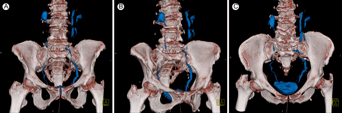
On nonenhanced abdominal CT scans after urinary catheter insertion, hydronephrosis without ureteral stones (but with delayed contrast excretion), bladder wall thickening, and opacified urinary tract were observed due to undrained contrast and urinary duplication. CT, computed tomography.
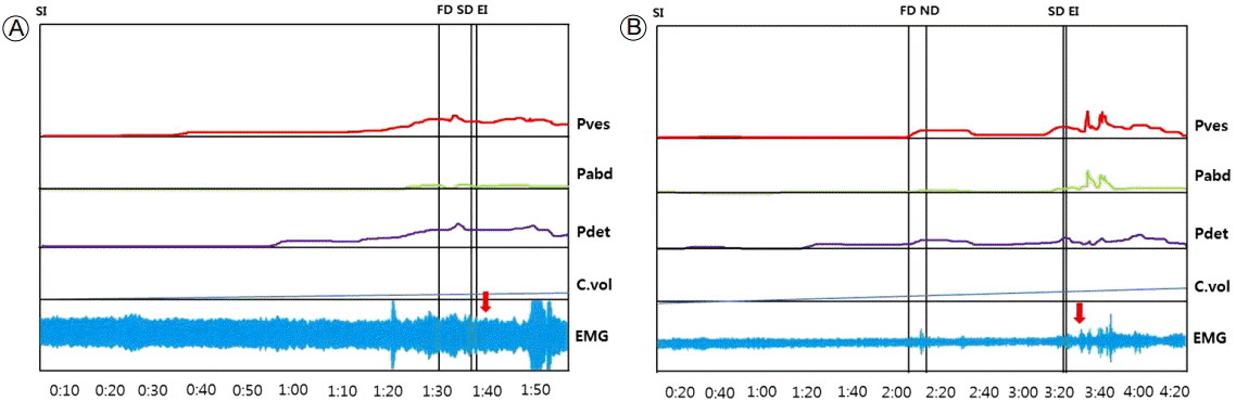
(A) A preoperative UDS showed a functional bladder capacity of 95 mL with increased rectal EMG activity. The post-voiding residual urine volume was >200 mL. (B) Four months postoperatively, the UDS showed a functional bladder capacity of 205 mL and decreased EMG activity (solid arrow) compared with the preoperative study. The change was due to improved bladder compliance. The post-voiding residual urine volume was <100 mL. SI, starting of saline infusion; FD, first desire; SD, strong desire; EI, ending of infusion; ND, normal desire; Pves, pressure of vesicle; Pabd, pressure of abdomon; Pdet, pressure of detrusor muscle; C.vol, capacity of bladder volume; UDS, urodynamic study; EMG, electromyography.
DISCUSSION
Urinary tract obstruction may cause urinary tract infections, including urosepsis and acute or chronic kidney injury, if not treated appropriately. There are many causes of urinary tract obstruction, such as ureteral stones, infection, blood clots, carcinoma of the ureter, prostate enlargement, and POP. Early detection of the exact cause of the urinary tract obstruction is important because most cases can be treated effectively [6].
POP is characterized by the downward descent of the female pelvic organ, resulting in protrusion of the vagina, uterus, or both. Patients with uterine prolapse generally present with symptoms related specifically to the prolapsed structures or with associated symptoms, including urinary, defecatory, or sexual dysfunction. As the prolapse advances, women may experience increased difficulty voiding. Advanced anterior or apical prolapse may “kink” the urethra, thereby resulting in obstructed voiding symptoms such as a slow urine stream, a sensation of incomplete emptying, and complete urinary retention in rare cases. A large posterior vaginal prolapse can also cause a mechanical obstruction by direct urethral compression. As many as 30% of women with third- or fourth-degree POPs have increased post-void residual volumes (> 100 mL) [7]. In this situation, urinary tract infections may develop. In addition, increased intratubular pressure may affect glomerular function and lead to renal failure [8]. Treatment is indicated for women with symptoms of a prolapse or any associated conditions including urinary, bowel, or sexual dysfunction. Obstructed urination or defecation or hydronephrosis from chronic ureteral kinking are all indications for treatment regardless of the degree of prolapse. Women with symptomatic POP who decline pessaries or in whom pessary treatment is unsuccessful are candidates for surgery [9].
Alongside an oral history and physical examination, a UDS helps to detect any concomitant urinary incontinence and aid our understanding of the physiological mechanism underlying lower urinary tract dysfunction. The UDS helps to improve the accuracy of diagnosis and facilitate targeted treatment, although several limitations remain, such as a lack of standardization of the technical details, inconsistent reproducibility of test results, and a wide range of normal physiological values [4].
To the best of our knowledge, this case of uroseptic shock with acute kidney injury due to obstructive uropathy by uterine prolapse is rare. Although the patient was an aged woman with a long history of diabetes, the possibility of diabetes-associated autonomic bladder dysfunction was low because her diabetes had been well controlled and her renal functions returned to normal after short-term treatment. In addition, the UDS showed the improvement of bladder compliance after surgical correction of the uterine prolapse.
In conclusion, we were able to successfully manage a patient with uroseptic shock and with acute kidney injury due to obstructive uropathy by uterine prolapse, which was documented in a UDS.