맹장암 간전이와 유사한 양상을 보인 개회충증 1예
A Case of Toxocariasis in a Patient with Cecal Cancer Mimicking Hepatic Metastasis
Article information
Abstract
호산구의 간침윤과 대장직장암의 간전이는 임상적으로 감별하기는 쉽지 않으며, 대장직장암에 있어 간전이의 유무의 확인은 치료방침을 결정함에 있어 반드시 필요한 과정이다. 혈청학적으로 호산구증가증과 컴퓨터 단층촬영상 대장암의 다발성 간전이를 보인 환자에서 자기공명영상 촬영 및 간생검을 통하여 개회충증에 의한 호산구 침윤으로 확인된 사례를 경험하였기에 문헌고찰과 함께 보고하는 바이다.
Trans Abstract
Toxocariasis is a common helminth infection and the most common cause of peripheral blood eosinophilia. The clinical presentation of liver toxocariasis is often confused with primary or metastatic hepatic tumors. Here, we report on a 52-year-old man admitted with cecal cancer and eosinophilia. Computerized tomography (CT) scans revealed multiple lesions with peripheral enhancement in the liver, and T1- and T2-weighted images revealed isointensity. The patient underwent right hemicolectomy and an exploratory laparotomy with a liver biopsy. Resection of the cecal specimen revealed an adenocarcinoma, while the liver biopsy revealed an eosinophilic abscess with no evidence of malignancy. A serologic test showed a marked increase in specific immunoglobulin G antibody concentrations against Toxocara canis and daily antiparasitic treatment with albendazole (800 mg per day for 7 days) was initiated. Follow-up CT scans indicated that all liver masses and eosinophilia were resolved.
서 론
개회충중(Toxocariasis)은 개회충(Toxocara canis)이나 고양이회충(Toxocara catis) 충란이 사람 장 내에서 부화하여 간, 폐 등 여러 장기에 침범하여 호산구성 육아종(eosinophilic granulomas)을 일으키는 질환이다[1]. 국내 성인 환자의 대부분이 간, 천엽, 육회나 동물의 내장을 생식한 경험이 있고[2], 호산구증가증(eosinophilia)의 가장 흔한 원인으로 알려져 있으며 과거 진단 기법의 부족으로 정확한 진단이 어려웠으나 IgG 항체 검사 및 toxocara canis enzyme linked immunosorbent assays (ELISA) 등 진단 기법의 발달로 진단율이 높아졌으며 국내에 상당수 존재하는 질환으로 보고되고 있다[1-5]. 대장직장암은 간전이가 가장 흔하며 대장직장암 환자의 약 50%에서 간전이를 보인다[6]. 또한 대장직장암의 간전이 병변은 임상적으로 호산구증가증에 의한 간침윤과 흡사하여 영상학적 구별이 쉽지 않다. 저자들은 복부 컴퓨터 단층촬영상 대장암의 간전이로 생각되었던 환자에서 혈청학적 검사, 자기공명영상 및 간생검을 통하여 개회충증에 의한 호산구 침윤으로 밝혀진 1예를 경험하였기에 문헌고찰과 함께 보고하는 바이다.
증 례
환 자: 모〇인, 55세 남자가 막 창자 및 간의 종양을 주소로 내원하였다.
현병력: 내원 5개월 전부터 우하복부 통증 발생하여 타 병원을 내원하여 시행한 검사상 맹장 및 간의 종괴 소견을 보여 수술적 절제 및 추가 검사를 위해 내원하였다.
과거력, 사회력 및 가족력: 고혈압, 70갑년 현재 흡연가(current smoker), 추간판 탈출증으로 척추체 고정술, 위암(부), 식도암(모)
신체검사 소견: 내원시 혈압은 120/70 mmHg, 맥박수 80회/분, 호흡수 18회/분, 체온 36.2℃였으며 의식은 명료하였다. 문진상 3개월 동안 8 kg의 체중이 감소(평상시 체중 60 kg)하였으나 혈변, 변비, 설사 등 배변습관의 변화는 없었고, 신체적 진찰상 우하복부에 압통 호소하였으나 반발통은 없었으며 두경부, 흉부 및 사지의 이상소견은 없었다.
검사실 소견: 내원시 말초혈액검사에서 백혈구 10,200/mm3 (호산구 18.7%, 1,907/mm3), 혈색소 12.2 g/dL, 혈소판 311,000/mm3이었다. 천식 및 알러지의 과거력은 없었으나 과거 육회, 간생식 및 회 섭취의 기왕력이 있었으며 IgE 1,500 IU/mL, 개회충에 대한 IgG는 25.91로 증가되어 있어 간병변이 개회충증의 침윤의 가능성이 있음을 시사하였다. 그 외의 간기능 검사, 신기능 검사, 급성기반응물질 검사, 및 소변검사는 정상범위 내의 값을 보였다. 암표지자 검사상 carcinoembryonic antigen (CEA) 7.59 ng/mL로 증가되어 있어 맹장의 종괴가 악성 종양임을 시사하였다.
방사선학적 소견: 내원시 흉부 및 복부방사선에서 과거 추체로 고정술을 받았음을 확인할 수 있었다. 병기설정을 위한 컴퓨터 단층촬영(computerized tomography, CT) 및 양전자 컴퓨터 단층촬영(positron emission tomography [PET]-CT)상 폐우하엽에 0.9 cm 크기의 결절과 함께 젖빛 유리음영을 보였고 이는 호산구 침윤으로 추정되었다. 또한 맹장과 상행결장 및 회맹판에 조영 증강되는 최대 직경 6.2 cm × 5.6 cm 크기의 종양과 그 주변으로 단경 0.5 cm 내외의 4-6개의 림프절 비대를 보이고 있었고 간우엽 비면(right hepatic dome)에 주변테의 조영증강(peripheral rim enhancement)을 보이는 다발성 병변이 관찰되었다(Fig. 1A). 간의 다발성 병변에 대하여 염증성 간병변과 전이 병변의 감별을 위해 자기공명영상(magnetic resonance imaging, MRI)을 시행하였고, 다발성 간병변은 조영제 주입 후 주변테의 조영증강을 보이고, hepatobiliary phase에서는 저음영의 불분명한(ill-defined) 비구형(non-spherical) 병변을 보이고 있었다(Fig. 1B). 동일 병변에 대한 T1- 및 T2-weighted 영상에서는 등강도(isointensity)를 보이는 최대 4.5 cm 크기의 다발성 결절들이 있어 다발성 전이병변보다는 염증성 간병변을 시사하였다(Fig. 1C and 1D).
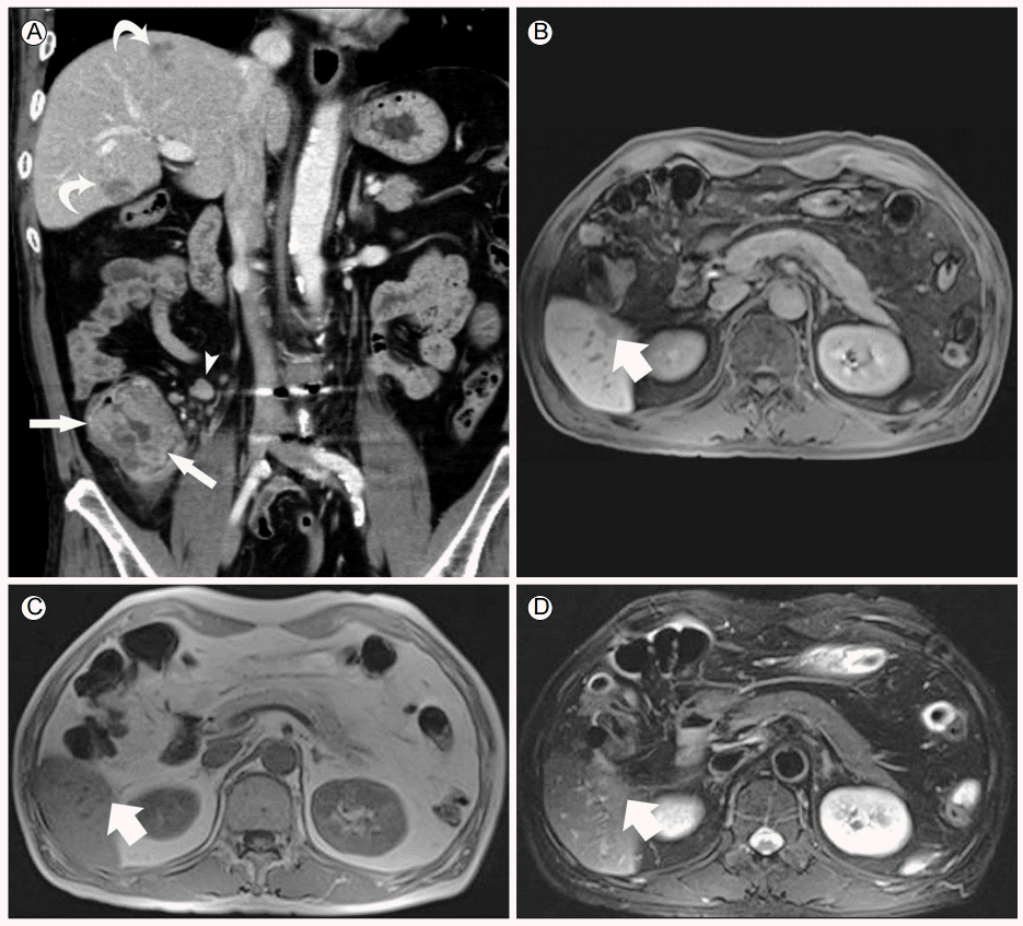
Hepatic abscess caused by toxocariasis mimicking metastasis in a 52-year-old man with cecal cancer. (A) Coronal contrast-enhanced CT image showing a heterogeneous enhancing mass (arrows) in the cecum with regional metastatic lymphadenopathies (arrowhead) suggestive of cecal cancer. The CT image also depicts several hypoattenuated nodules (curved arrows) in the liver, suggestive of metastases from cecal cancer. An axial gadoxetic acid-enhanced lesion (B) showed an ill-defined, non-spherical morphology. T1- (C) and T2 (D)-weighted images show an isointense nodule (arrow) in the right hepatic lobe. CT, computerized tomography.
임상경과 및 치료: 진단 및 치료 위해 우측 반대장절제술(right hemicolectomy) 및 시험적개복술(exploratory laparotomy)을 통한 간생검을 시행하였다. 맹장 및 상행결장 종괴에 대한 조직 검사상 중등도 분화도(moderately differentiated)를 보이는 선암(adenocarcinoma)이 확인되었으며 림프절 생검상 50개의 림프절 중 2개의 림프절에서 전이가 관찰되어 맹장암을 확진할 수 있었다(Fig. 2A). 간병변은 수술 당시 8번 분절, 5번 분절, 1번 분절에 각각 2 × 2 cm, 1.5 × 1 cm, 0.5 × 0.5 cm 크기의 노란색 병변이 관찰되어 신속동결조직검사를 시행하였고 검사상 염증병변으로 관찰되고, 수술 중 초음파 촬영술(intraoperative ultrasonography)에서도 종괴가 관찰되지 않아 절제하지 않고 조직검사만 시행하였다. 추후 간병변에 대한 조직검사 결과는 괴사를 동반한 급성 및 만성 염증소견(acute and chronic inflammation with necrosis)을 보여 다발성 간병변은 개회충증으로 인한 호산구증가증과 그로 인한 호산구의 간침윤으로 진단하였다(Fig. 2B). 수술 후 개회충증에 대하여 7일간 하루 2회 albendazole 400 mg/day을 복용하였으며 2개월 뒤 시행한 말초혈액검사상 백혈구 5,900/mm3 (호산구 0.2% 10/mm3)로 호산구 감소 및 복부 컴퓨터 단층촬영상 간우엽의 호산구 침윤의 감소를 확인할 수 있었다. 현재 환자는 맹장암에 대하여 5-Fluorouracil + Leucovorin + Oxaliplatin (FOLFOX) 보조항암화학 요법이 진행 중이다.
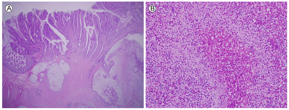
(A) Histopathology of the colon showing infiltrative atypical glands (adenocarcinoma) arising from the colonic mucosa. (H&E, ×20) (B) and geographic necrosis associated with numerous eosinophil infiltration (H&E, ×100).
고 찰
개회충증은 호산구증가증의 가장 흔한 원인이다. 호산구증가증을 보이는 환자에서 호산구의 간침윤은 약 30%에서 동반되며 농양이나 육아종의 형태로 나타날 수 있으며, 컴퓨터 단층촬영상 보통 2 cm 미만의 변연이 불분명한 다수의 저음영 결절을 보이는 것으로 알려져 있다. 그러나 호산구 침윤과 전이성 간병변이 혼재하는 경우도 상당수로 보고되고 있으며 컴퓨터 단층촬영만으로는 호산구의 간침윤과 대장직장암의 간전이와 임상적으로 구별하기는 쉽지 않다[3,7,8]. 이런 경우 자기공명영상을 통하여 두 병변을 비교적 명확히 구분할 수 있다고 보고되고 있다[7]. 호산구 간침윤으로 인한 염증성 병변의 경우 간전이와 비교하였을 때 1) 비구형, 2) 불분명한, 3) T1 weighted 영상에서 등강도의 특징을 보인다[7]. 이번 증례에서 보듯이 저자들은 대장암 환자가 호산구증가가 있고 컴퓨터 단층촬영상 다발성 간병변이 있다면 간병변에 대해 자기공명영상을 시행하여야 하고, 자기공명영상 소견상 간전이보다는 호산구의 간침윤을 시사할 경우 적절한 치료 시행을 위해 조직학적 확진을 위한 검사를 시행하여야 한다고 본다. 대장직장암에 있어 전이 병변의 유무는 치료 방침과 예후 판단에 있어 매우 중요한 인자이다. 대장직장암은 환자의 14-25%에서 진단 당시 간전이가 동반되어 있으며 질병 경과 중 60% 이상의 환자에게서 간전이로 발전하고 간전이를 동반한 대장직장암 환자의 5년 생존율은 평균 38% 정도로 보고되고 있으며, 10년 생존율은 평균 26% 정도로 보고되고 있다[9]. 대장직장암의 간전이의 경우 수술적 치료를 통하여 완치를 기대할 수 있는 질환이며 수술로 인한 사망률 또한 1-2%로 매우 낮으나 본 증례의 경우처럼 좌우엽을 포함한 다발성 간전이가 확인된 경우 수술적 절제는 불가한 것으로 알려져 있다[9].
본 증례는 개회충증으로 인한 호산구증가증을 동반하면서 컴퓨터 단층촬영상 맹장암의 간전이를 보인 환자로 자기공명영상 소견상 염증성 간병변의 소견이 보여 진단 및 치료 위해 우측반대장절제술 및 시험적개복술를 시행하였고, 최종적으로 맹장암 3기와 개회충증에 의한 호산구 간침윤으로 진단된 사례이다. 개회충증의 경우 공간점유병터(space occupying lesion)가 아니고 증상이 없다면 경과관찰이 가능하며, 증상이 있을 경우 abendazole 복용을 통하여 대부분의 사례에서 완치를 기대할 수 있는 질환이나, 대장직장암의 간전이의 경우 병변에 따라 수술적 치료 또는 항암화학요법을 시행하여야 하며 5년 생존율이 평균 38%에 불과한 질환이다[1,9,10]. 간병변의 정확한 진단은 치료와 예후에 있어 반드시 필요한 과정이며 불필요한 간절제술이나 항암 치료와 같은 침습적인 치료를 피함으로써 결과적으로 합병증 및 사망률을 감소시킬 수 있다.
호산구 증가증은 일반적으로 알려진 알러지 질환, 진균 및 기생충 감염뿐만 아니라 악성 종양에서도 동반될 수 있는 것으로 알려져 있다. 그러나 우리나라의 경우처럼 생식의 빈도가 높은 나라에서는 기생충 감염을 반드시 고려하여야 하며 특히나 본 사례의 경우처럼 생식의 기왕력, 호산구증가증을 보이는 대장직장암 환자에서 간의 결절을 동반하고 있을 경우 침습적인 치료에 앞서 자기공명영상을 통한 염증성 병변의 감별이 필요하겠으며 필요시 생검을 포함한 기생충에 대한 추가적인 검사가 반드시 시행되어야겠다.