심실빈맥의 발작으로 발현된 갈색세포종 1예
A Case of Pheochromocytoma Presenting as Ventricular Tachycardia Storm
Article information
Abstract
두통과 두근거림을 주소로 입원한 32세 여자 환자의 심전도 검사에서 단형 지속성 심실 빈맥이 확인되었다. 심실빈맥이 나타나기 전에 갈색세포종의 특징적인 증상 및 징후의 하나인 두통과 발한이 먼저 나타났으나 고혈압은 없었다. 심실빈맥의 발생을 억제하기 위해 베타 차단제를 투여하였으나 오히려 심실빈맥의 발생빈도가 증가하고 지속시간이 길어졌다. 24시간 보행 심전도 검사에서 다양한 형태의 QRS파를 가지는 심실조기박동, 가속 심실고유리듬, 심실빈맥이 순차적으로 출현하면서 맥박수가 빠르게 증가하는 것이 확인되어 증가한 자동능에 의해 심실빈맥이 유발되는 갈색세포종의 가능성을 고려할 수 있었다. 영상 및 내분비 검사를 통해 부신에서 발생한 갈색세포종을 진단하였으며 알파 차단제 투여를 시작한 후 심실빈맥의 발생이 억제되어 안전하게 갈색세포종 적출술을 시행할 수 있었다.
Trans Abstract
A 35-year-old woman was admitted for recurrent palpitations and headache with cold sweats. No structural abnormality was detected via cardiac imaging studies. A standard 12-lead electrocardiogram (ECG) revealed sustained monomorphic ventricular tachycardia (VT). Propranolol (120 mg/day) was administered; however, the frequency and duration of VT episodes increased rapidly. A 24-hr ambulatory ECG revealed frequent, successive, premature ventricular beats; accelerated idioventricular rhythms; and VTs with various cycle lengths and QRS complex morphologies. ECG findings suggested that the observed ventricular arrhythmias were driven by accelerated automaticity as their main electrophysiological mechanism. Based on clinical manifestations and ECG findings, pheochromocytoma was suspected. Solitary left adrenal pheochromocytoma was diagnosed by endocrine and imaging studies. Instead of propranolol, oral doxazosin (8 mg/day) was administered, and symptoms and VT attacks were successfully suppressed. After surgical resection of the pheochromocytoma, clinical VT was not observed in response to the high-dose isoproterenol provocation test.
서 론
갈색세포종 환자의 주요 증상 및 징후는 발한을 동반한 두근거림 및 두통이며 갑작스럽게 발현하는 것이 특징이다[1]. 드물지만 갈색세포종 환자의 초기 임상 양상이 심실성 빈맥이었던 사례도 일부 보고되었다[2-8]. 저자들은 베타 차단제에 반응하지 않는 심실빈맥 발작(ventricular tachycardia storm)으로 입원한 환자에서 갈색세포종(pheochromocytoma)을 진단하였기에 이를 문헌 고찰과 함께 보고한다.
증 례
환 자: 35세 여자
주 소: 두통과 발한을 동반한 두근거림
현병력: 2주 전부터 하루 2-3회 발작적으로 발생하는 두근거림 때문에 병원에 왔다. 두근거림은 5분 정도 지속되는데 두근거림이 있을 때 머리가 아프고 식은땀이 난다고 하였다.
과거병력: 특이 사항 없었다.
가족력 및 사회력: 특이 사항 없었다.
신체검진: 내원 당시 체온은 36.4℃, 호흡수 18회/분, 맥박수 70회/분, 혈압 100/60 mmHg였다. 입원 시 시행한 신체검사에서 특이소견은 없었다.
검사실 소견: 말초혈액검사에서 특이소견 없었고, 혈청 생화학 검사에서 심근 트로포닌-I 수치가 3.33 ng/mL로 증가한 것을 제외하고는 이상 소견 없었다.
단순 흉부 X-선 촬영: 심비대 및 폐 부종 소견은 없었다.
경 흉부 심초음파 검사: 좌심실 구혈률은 57%였고 구조적 이상 소견은 없었다.
최초 표준 12-유도 심전도: 입원 초기 무증상이었을 때의 심전도 소견은 분당 맥박수 80회의 정상 심율동이었으며 T파의 역위나 ST분절의 변화는 없었다.
발한을 동반한 두통이 있을 때의 심전도: 환자는 심실빈맥이 나타나기 몇 시간 전부터 간헐적으로 발한을 동반한 심한 두통을 호소하였으며 당시 시행한 심전도 검사에서 T파의 역위를 동반한 동성빈맥이 확인되었다(Fig. 1A).

Standard 12-lead ECG findings. An ECG was acquired after the patient had complained of palpitations, headaches, and cold sweats, but before the appearance of ventricular arrhythmias. (A) This analysis revealed marked sinus tachycardia of 120 bpm with T-wave inversions in the inferior leads. (B) An ECG acquired during VT episodes showed sustained monomorphic VT with inferior axis morphology, right bundle branch block configuration in the V1 lead, and increased Vi/Vt ratio, suggesting epicardial origin near the left ventricular outflow tract. ECG, electrocardiogram; VT, ventricular tachycardia.
심실빈맥이 나타났을 때의 표준 12-유도 심전도: 단형 지속성 심실빈맥은 V1 흉부유도에서 우각차단 형태의 QRS파를 보였고 전기적 축은 하향축이었다. 또한 V1흉부유도에서 R파/S파의 비율이 1보다 크고, R파의 전압과 지속시간이 길며, QRS파의 시작점부터 최고점에 도달하기까지의 시간이 긴 형태학적 특징을 가지고 있었다. 표준 12유도 심전도 소견을 근거로 심실빈맥이 좌심실 유출로 근처의 좌심실 바깥막에서 기원했을 것으로 추정하였다(Fig. 1B).
운동부하 심전도: 운동부하 심전도 검사 중 의미 있는 ST분절의 하강은 나타나지 않았고 심실성 부정맥도 유도되지 않았다.
24시간 보행 심전도: 24시간 보행 심전도 검사에서 지속성 심실빈맥의 발작이 하루 2-3차례 재발하여 5-10분간 지속되는 것이 확인되었다. 지속성 심실빈맥이 출현하기 전에 동성빈맥, 빈번한 심실조기박동, 가속 심실고유리듬(accelerated idioventricular rhythm)이 순차적으로 나타났으며, 이후 단일한 형태의 QRS파와 일정한 맥박수를 가지는 느린 심실빈맥이 출현하여 다양한 형태의 QRS파와 불규칙한 맥박수를 가지는 빠른 심실빈맥으로 짧은 시간 내에 진행하였다(Fig. 2). 24시간 보행 심전도에 기록된 심실빈맥은 맥박수 변화 및 QRS파의 형태변화가 심하여 심실빈맥의 발생기전이 증가한 자동능(automaticity)일 것으로 추정되었다.
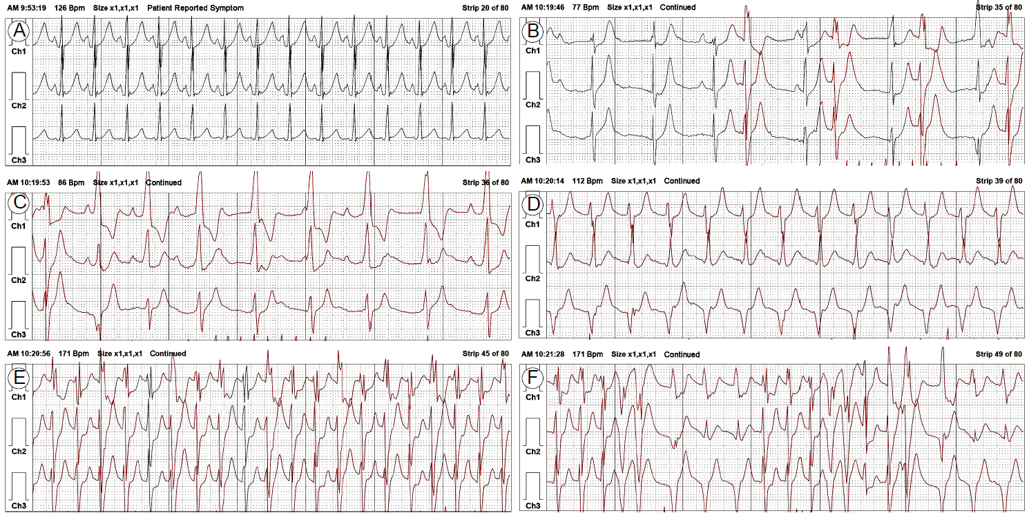
Twenty-four-hour ambulatory ECG monitoring findings. (A) The earliest abnormal ECG finding after the appearance of ventricular arrhythmia was marked sinus tachycardia with palpitations, headaches, and cold sweats. (B) Frequent premature ventricular beats were the first ventricular arrhythmia to appear. They were followed by (C) accelerated idioventricular rhythms of 60-100 bpm and (D) slow sustained monomorphic VT of 100-120 bpm. (E) Over several minutes, slow VT shifted to fast VT (> 50 bpm) with multiple QRS complex morphologies and rapidly changing VT cycle lengths. (F) Ventricular arrhythmias with various QRS complex morphologies and tachycardia cycle lengths suggested increased automaticity as their underlying electrophysiological mechanism. ECG, electrocardiogram; VT, ventricular tachycardia.
관상동맥 조영검사: 심근 트로포닌-I 수치가 증가해 있어 시행한 관상동맥 조영검사에서 관상동맥의 협착성 병변은 보이지 않았고 에르고노빈 투여 후에도 관상동맥의 연축은 유도되지 않았다.
심장 자기공명영상: 특이 소견 없었다.
경 과: 입원 3일째부터 심한 두통과 발한을 동반한 두근거림이 2-3일 간격으로 재발했다. 증상이 나타났을 때 측정한 혈압은 140/100 mmHg였다. 심실빈맥의 발생을 억제하기 위해 좌심실 유출로 근처에서 기원하는 특발성 심실빈맥에 준하여 경구 베타 차단제(propranolol 120 mg/일)를 투여하였으나 3일이 지나도 심실빈맥의 발생이 억제되지 않았으며, 오히려 심실빈맥의 발생빈도가 하루 7-8차례로 증가하고 지속시간이 20-30분으로 길어지는 등 임상양상이 빠르게 악화되는 경과를 보였다. 심실빈맥이 나타나기 몇 시간 전부터 발한을 동반한 심한 두통이 있다는 점과 24시간 보행 심전도 검사로 확인된 심실성 부정맥의 발생기전으로 증가된 자동능이 의심되는 점 등을 고려하여 갈색세포종에 대한 검사를 시행하기로 하였다.
진 단: 갈색세포종을 확인하기 위해 시행한 복부 전산화단층촬영 검사에서 왼쪽 부신의 위치에 3.8 × 3.7 cm 크기의 난원형 종괴가 발견되었다(Fig. 3A). 24시간 소변 검사상 메타네프린 12.5 mg/일(정상범위: 0.2-1.2 mg/일), 바닐만데릭산 17.5 mg/일(정상범위: 2-10 mg/일)였다. 123I-metaiodobenzylguanidine를 이용한 방사선 동위원소 촬영에서도 왼쪽 부신 종괴에 의한 동위원소 섭취증가가 확인되었고 부신 이외의 위치에서는 동위원소 섭취 증가가 없었다(Fig. 3B). 다발성 내분비샘 종양과 관련된 갑상샘이나 췌장의 종괴가 없음을 확인한 후 왼쪽 부신에서 발생한 고립성 갈색세포종으로 최종진단하였다.
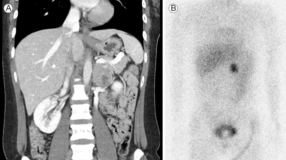
Abdominal CT and MIBG scan findings. (A) An abdomino-pelvic CT scan showed a 3.8- × 3.7-cm mass at the location of the left adrenal gland. (B) An 123I-MIBG scan also showed increased radioisotope uptake at the location of the left adrenal gland. CT, computed tomography; MIBG, metaiodobenzylguanidine.
치 료: 환자는 경구 알파 차단제(doxazosin 8 mg/day) 투여 2일째부터 발작성 심실빈맥의 발생이 극적으로 억제되기 시작했으며, 3일째부터는 심실빈맥이 재발하지 않아 알파 차단제 유지요법을 시행하면서 사고 없이 복강경하 부신 종양 적출술을 시행할 수 있었다. 조직 병리 결과는 양성 갈색세포종에 합당한 소견이었다. 수술 직후 알파 차단제 투약을 중단하였으며 연속 심전도 감시 중 심실빈맥의 재발은 확인되지 않았다.
수술 후 시행한 이소프로테레놀(isoproterenol) 유발검사 중 이소프로테레놀 정주량을 분당 10 μg까지 증량하였으나 심실빈맥이 재발하지 않았다. 환자는 종양 적출술 후 심실빈맥의 재발 없이 3년째 경과관찰 중이다.
고 찰
갈색세포종 환자의 주요 증상 및 징후는 발한을 동반한 두근거림 및 두통이며 갑작스럽게 발현하는 것이 특징이다[1]. 갈색세포종의 증상 및 징후는 과도하게 높아진 혈중 카테콜아민의 농도와 관련이 있으며 카테콜아민의 증가는 베타 수용체를 자극하여 부정맥을 유발할 수 있다. 갈색세포종 환자의 50-70%가 두근거림을 호소하는데 동성빈맥이 가장 흔한 리듬 이상이지만 심실빈맥이나 서맥 등 다양한 부정맥이 나타날 수 있다[9]. 저자들의 증례와 같이 갈색세포종의 초기 임상 양상이 단형 지속성 심실빈맥이었던 사례가 간헐적으로 보고되었고[3,4,6,7], 매우 드물지만, 치명적인 다형 심실빈맥이나 심실세동이 발생한 사례도 보고된 적이 있다[2,5,8]. 저자들의 사례의 경우 심실빈맥이 나타나기 전에 갈색세포종의 특징적인 증상 및 징후의 하나인 두통과 발한이 먼저 나타났다. 또한 심전도 검사상 다양한 형태의 QRS파를 가지는 심실성 부정맥들이 순차적으로 출현하면서 맥박수가 매우 빠르게 증가하였고, 심실빈맥 QRS파의 형태와 맥박수 변화가 심하여 증가한 자동능에 의해 심실빈맥이 유발되는 갈색세포종의 가능성을 고려할 수 있었다.
갈색세포종 환자에게 알파 차단제를 투여하지 않은 상태에서 베타 차단제를 투여할 경우 베타-1 수용체에 의한 골격근의 혈관 이완작용을 차단해서 혈압이 급격히 상승하는 고혈압성 위기가 올 수 있는 것으로 알려졌다[1]. 다행히 저자들의 사례에서는 베타 차단제 투여 후에도 고혈압성 위기가 오지는 않았지만 반대로 심실빈맥의 발생빈도가 증가하고 지속시간이 길어지는 등 임상경과가 빠르게 악화되었다. 비선택적인 β1, β2 수용체 차단제인 propranolol을 투여하였음에도 불구하고 심실빈맥의 임상경과가 악화된 원인은 불분명하다. 베타 차단제의 용량이 불충분하여 과도하게 분비된 카테콜아민이 베타 수용체에 미치는 영향을 충분히 차단하지 못해 자연경과적으로 악화되었을 가능성이 높다. 그러나 베타 수용체가 차단된 후 알파 수용체에 대한 카테콜아민의 영향이 상대적으로 과도해지면서 알파 수용체의 작용에 민감하게 반응하는 비정상적인 심근세포들이 과도하게 탈분극하여 심실빈맥이 유도되었을 가능성도 배제할 수 없다. 이전에 보고된 다른 증례들과 마찬가지로 베타 차단제 투여를 중단한 후 알파 차단제만 투여했음에도 불구하고 심실빈맥의 발생이 성공적으로 억제되었다는 사실[3-5]과 갈색세포종 적출술 후 시행한 이소프로테레놀 유발검사에서도 심실빈맥이 유도되지 않은 사실 등이 이러한 가설을 간접적으로 뒷받침한다.
갈색세포종은 정확한 진단과 적절한 내과 및 외과적 치료를 할 경우 완치할 수 있는 질환임에도 불구하고 저자들의 사례에서처럼 고혈압이 없거나 심실성 빈맥으로 발현되는 경우 진단이 지연될 수 있다. 갈색세포종의 특징적 증상 및 징후를 동반한 심실빈맥이 나타나는 환자를 대할 때는 갈색세포종이 숨어 있을 가능성을 고려해야 하겠다. 저자들은 심실빈맥의 발작으로 발현된 갈색세포종 1예를 문헌고찰과 함께 보고하는 바이다.