재발한 Churg-Strauss 증후군 치료 중 발생한 폐농양 1예
Lung Abscess During the Treatment of Relapsed Churg-Strauss Syndrome
Article information
Abstract
저자들은 오랜 기간 관해 상태를 유지하던 중 재발한 Chrug-Strauss 증후군 환자에서 시클로포스파마이드 및 경구용 스테로이드 치료 중 발생한 폐농양 1예를 경험하였다. 본 증례는 장기간의 관해 후에도 Churg-Strauss 증후군이 재발할 수 있고 치료 중 감염이 발생하였을 때 폐포자충폐렴이나 거대세포 바이러스 감염과 같은 기회감염 이외에 세균 감염도 발생할 수 있음을 보여주는 증례이기에 문헌고찰과 함께 보고하는 바이다.
Trans Abstract
Churg-Strauss syndrome (CSS) is a rare disease characterized by asthma, peripheral eosinophilia and vasculitis. A quarter of CSS patients with clinical remission have experienced a relapse. We report here a case of lung abscess in a patient with relapsed CSS. A 46-year-old man who achieved clinical remission of CSS was confirmed for relapse by the presence of peripheral eosinophillia, pulmonary lesions and perivascular infiltrate of eosinophils in colon pathology. After administration of systemic glucocorticoid and one cycle of cyclophosphamide pulse therapy, he complained of dyspnea, sputum and chest pain. There were necrotic masses containing internal air-fluid levels in the right upper and the left lower lobes of the lung on chest radiography. Percutaneous needle aspiration culture specimens revealed the presence of K. pneumoniae. The patient was successfully treated with a 5-week course of antibiotics. (Korean J Med 2014;86:106-109)
서 론
Churg-Strauss 증후군은 알레르기성 비염 및 천식, 호산구 증가증, 혈관 외 육아종 형성, 괴사성 혈관염 등을 특징으로 하는 드문 질환이다[1,2]. Churg-Strauss 증후군 환자들은 스테로이드 단독 또는 시클로포스파마이드 병합요법으로 적지 않은 수가 임상적 관해에 도달한다. 그러나 그 중 1/4은 재발하는 것으로 알려져 있다[2]. Churg-Strauss 증후군 환자들은 스테로이드 및 시클로포스파이드 치료로 인한 면역저하로 폐포자충 폐렴 및 거대세포바이스 감염 등의 기회 감염에 대한 노출될 가능성이 상대적으로 높다.
저자들은 장기간 관해 상태를 유지하던 중 재발한 Churg-Strauss 환자에서 전신 스테로이드 및 시클로포스파마이드 투여 후 발생한 세균성 폐농양 1예를 경험하였기에 문헌고찰과 함께 보고하는 바이다.
증 례
환 자: 이〇〇, 남자, 46세
주 소: 2주간 지속된 호흡곤란, 객담, 흉통
현병력: 환자는 9년 전 호흡곤란으로 내원하여 천식 병력, 말초혈액 호산구 증가증(16,786/mm3), 양측 폐의 미만성 경화 소견, 폐 조직의 혈관염을 동반한 호산구 침착 등을 근거로 Chrurg-Strauss 증후군을 진단받았다. 항호중구 세포질항체(antineutrophil cytoplasmic antibody, ANCA)는 음성이었다. 1년간 경구용 스테로이드 및 시클로포스파미드 치료(1,200 mg/회, 총 9회 강압요법)를 받았고 이후 임상적 관해 상태로 저용량의 스테로이드(prednisolone 5 mg/day)를 경구 복용하고 있었다. 천식은 seretide diskus 하루 2회 흡입하면서 잘 조절되고 있었고 류코트리엔 수용체 길항제를 사용한 적은 없었다.
환자는 2개월 전 혈변을 동반한 호흡곤란으로 내원하였고 말초혈액검사에서 호산구 7,632/mm3, 양측 폐의 흉막하경화 및 다초점성 중심소엽성 결절 소견(Fig. 1), 대장내시경 조직검사상 혈관염 등이 확인되어 Churg-Strauss 증후군 재발로 진단되었다. 환자는 2일간 메틸프레드니솔론 강압요법(1 g/day)를 시행받았고 이후 1회의 시클로포스파미드 강압요법(0.75 g/m2, 총 1,320 mg) 및 경구용 스테로이드(prednisolone 40 mg/day)를 투여받고 퇴원하였다. 퇴원 2주 후 기침, 가래, 우측 흉벽 통증 등이 동반되어 내원하였으며 발열, 오한, 호흡곤란 등은 없었다.
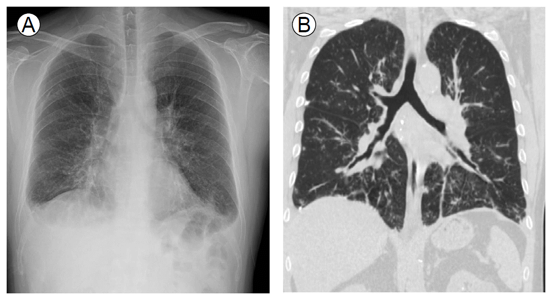
(A) Chest radiograph and (B) computed tomography after relapse of Churg-Strauss syndrome. Images show patchy centrilobular nodules, subpleural consolidation and diffuse bronchial wall thickening in both lungs.
과거력: 특이 소견이 없었다.
가족력과 사회력: 비흡연자로 특이사항 없었다.
진찰 소견: 내원 당시 활력 징후는 혈압이 120/86 mmHg, 맥박 수 분당 82회, 호흡 수 분당 20회, 체온은 36.8℃였다. 급성병색을 보이지는 않았으며 피부 결절과 자반 등의 피부 이상소견은 없었다. 흉부청진상 폐음과 심음 모두 정상이었다.
검사실 소견: 말초혈액검사상 백혈구 21,270/mm3 (호중구 88%, 호산구 0%), 혈색소 13.6 g/dL, 혈소판 337,000/mm3, ESR 57 mm/hr, CRP 7.17 mg/dL였다.
영상의학 소견: 단순흉부방사선사진상 우상엽에 공기 액체층을 포함하는 종괴, 좌하엽에 집락된 결절이 관찰되었고 전산화단층촬영에서도 같은 소견이 확인되었다(Fig. 2).
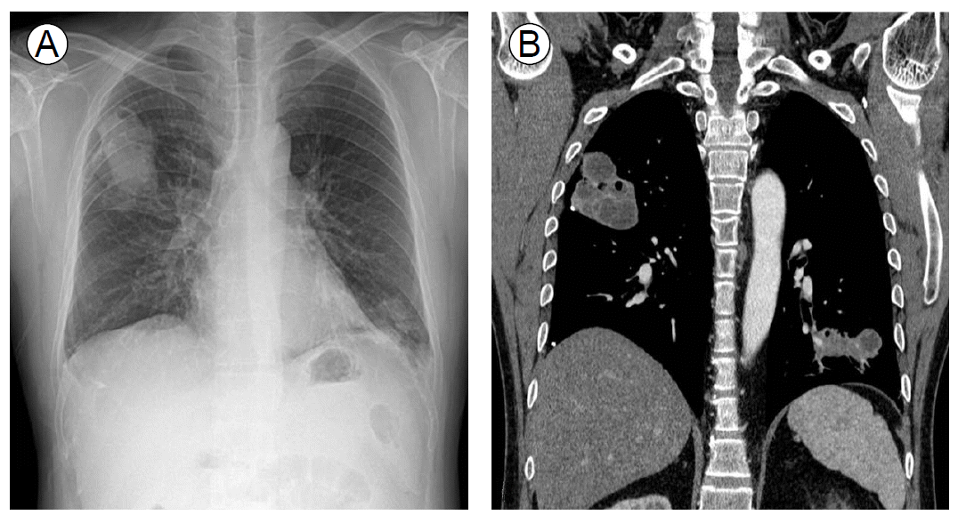
(A) The chest radiograph shows potato-like masses containing air-fluid levels in the right upper lung zone and nodular clustering in the left lower lung zone. (B) Computed tomography shows necrotic masses in the right upper and the left lower lung zones.
치료 및 경과: 폐 병변에 대해 경피적 바늘 흡인 검사로 얻은 검체에서 폐렴막대균이 확인되었다. 항생제 치료 3주 후 전산화단층촬영상 우상엽의 공동성 경화는 흔적만 남은 정도로 호전되었으며 좌하엽의 경화 병변은 거의 사라졌다(Fig. 3).

Chest radiograph after treatment with antibiotics. The extent of cavitary consolidation in the right upper lung zone decreased, and the lesion in the left lower lung zone nearly disappeared.
고 찰
Churg-Strauss 증후군은 천식, 호산구성 염증, 혈관 외 육아종 형성, 괴사성 혈관염 등을 특징으로 하는 질환이다. 원래 "알레르기성 육아종증 및 혈관염"으로 불렸으며 Wegener 육아종증, 현미경적 다발혈관염과 함께 소혈관을 침범하는 혈관염으로 분류되며 ANCA 연관 혈관염의 하나이다[1]. 1990년 미국류마티스협회에서 제시한 기준에 의하면 천식, 말초혈액의 백혈구 중 10% 이상의 호산구 증가, 단발 또는 다발 신경병증, 방사선학적 검사에서 발견된 이동성 또는 일시적인 폐침윤, 부비동 이상, 조직 생검상 호산구성 혈관염 소견 중 네 가지 이상을 보이는 경우 Churg-Strauss 증후군으로 진단할 수 있다[3].
본 환자는 9년 전 상기 기준의 네 가지를 만족하는 임상 양상으로 Churg-Strauss 증후군으로 진단되었고 1년간 경구 스테로이드 및 시클로포스파마이드 강압 요법 치료 시행 후 관해 상태에 도달하였다. 이후 8년간 저용량(Prednisolone 5 mg/day)의 경구용 스테로이드를 복용하며 별 문제없이 지내오던 중 Churg-Strauss 증후군이 재발하였다.
Churg-Strauss 증후군의 재발은 드문 것은 아니다. Guillevin 등[2]은 96명의 Churg-Strauss 증후군 환자를 32년간 추적 관찰한 결과 91.5%에서 임상적 관해에 도달했으며 이 중 25.6%에서 재발하였음을 보고한 바 있고 국내에서도 17명의 Churg-Strauss 증후군 환자를 9년간 추적관찰한 결과 94.1%에서 임상적 관해에 도달, 이 중 29.4% 환자가 재발하였음을 보고한 바 있다[4]. 그러나 이러한 재발은 대부분 관해 도달 후 2년 이내 발생하는 것으로 알려져 있다. 즉 본 증례처럼 임상적 관해 후 오랜 시간이 경과한 후에 재발하는 것은 드문 일이다. Guillevin 등[2]은 관해 상태에 있다가 재발한 Churg-Strauss 증후군 환자의 59%가 치료 2년 이내에 재발하였음을 보고하였고, Solans 등[5]의 연구에서도 재발 환자의 50%가 치료 2년 이내에 재발하였다.
본 환자의 경우는 관해 상태로 8년간 잘 지내오던 중 혈변을 동반한 호흡곤란, 말초혈액 호산구의 급격한 증가 등이 발생하였고 대장 조직 검사를 통해 재발이 확인되었다. 본 증례는 Churg-Strauss 증후군 환자가 관해 상태를 장기간 유지 중이라 하더라도 초진 당시와는 다른 임상 양상으로 언제든지 재발할 수 있음을 보여준다.
폐농양은 알코올 남용, 불량한 구강 위생, 발작 이상, 식도 운동성질환 및 연수 기능부전 등에 의한 흡인으로 인해 주로 발생한다[6,7]. 종양을 포함한 기저 폐질환 및 HIV 감염 등 면역 저하 상태에 의해 2차적으로도 발생한다[8].
본 증례의 환자는 상기 위험 인자들이 전혀 없었음에도 폐농양이 발생하였다. 일반적으로 Churg-Strauss 증후군 자체는 기회 감염의 위험인자로 간주되지 않는다. Churg-Strauss 증후군에서 흔히 동반되는 병변은 폐농양의 위험인자가 아니며 본 증례에서 발생한 폐농양의 경우에도 재발 당시에 발견된 폐 병변과 무관한 위치에 발생하였다. 따라서 본 증례의 폐농양은 일시적인 면역 체계의 기능 저하와 연관이 있을 가능성이 높다. 그러나 Churg-Strauss 증후군 치료에 의해 발생한 기회 감염 및 세균성 감염에 대한 보고는 지금까지 많지 않았다. Churg-Strauss 증후군 치료를 위해 고용량(prednisone 20 mg/day 이상)의 스테로이드를 1개월 이상 사용하는 경우 폐포자충 감염을 예방하기 위해 예방적 항생제(Trimethoprim-sulfamethoxazole) 사용만 추천되고 있다[9]. Guillevin 등[2]은 96명의 Churg-Strauss 증후군 환자를 추적관찰한 결과 스테로이드와 시클로포스파마이드에 의한 명확한 기회 감염의 발생은 확인되지 않았음을 보고하였고 국내에서도 Churg-Strauss 환자에서 스테로이드 치료에 따른 기회 감염 및 일반 세균에 대한 감염에 대한 증례보고는 없었다. 또한 장기간 저용량의 스테로이드 사용이 감염에 미치는 영향은 거의 없다고 여러 연구에서 밝혀져 있다[10]. 본 증례의 경우 재발한 Churg-Strauss 증후군에 대해 시클로포스파마이드 강압요법을 1회 시행하고 경구용 스테로이드(predenisolone 40 mg/day)를 2주간 복용 하던 중 세균감염에 의한 폐농양이 발생하였다. 단기간의 스테로이드 및 시클로포스파마이드 치료에도 폐농양이 발생될 수 있음을 보여주는 드문 증례라 하겠다.