당뇨환자에서 발생한 Candida glabrata 폐렴 1예
A Case of Candida glabrata Pneumonia in a Patient with Diabetes Mellitus
Article information
Abstract
입원 환자의 경기관지 분비물 배양 검사에서 효모형 진균 배양이 종종 보고되는데, 대부분의 경우 오염균이나 상재균으로 생각하는 경향이 있으나 본 예와 같이 기저질환으로 진균 감염에 취약한 상황에 있는 환자라면 Candida 감염 가능성을 생각하고 혈액 배양뿐 아니라 기관지내시경 등 보다 적극적인 검사 및 항진균제 사용으로 사망률 및 이환률을 낮춰야 하겠다[6].
Trans Abstract
Despite the increasing frequency of non-albicans candidemia, reported pneumonia cases due to Candida glabrata are rare. A 50-year-old diabetic woman developed cough and pleuritic chest pain, which were refractory to empirical antibiotics and anti-tuberculosis treatment for several months. The causative organism of the pneumonia was C. glabrata, which was cultured in bronchoalveolar lavage and blood culture specimens. Amphotericin therapy resulted in successful resolution of the pneumonia and candidemia. This is the first proven case of C. glabrata pneumonia in Korea. (Korean J Med 2013;84:577-580)
서 론
Candida 균혈증은 병원감염의 흔한 원인균으로 높은 사망률을 보이지만 호흡기계에서 상재균 또는 오염균으로 있는 경우가 많아 Candida 폐렴의 진단은 어려움이 있다. 그 중 Candida glabrata는 건강한 사람에 있어 비병원성균으로 알려져 있으나[1], 최근 항생제 또는 항진균제 등의 치료를 받는 면역 저하 환자들이 증가함에 따라 Candida glabrata에 의한 국소적 또는 전신적인 감염의 빈도가 증가하고 있다[2,3].
Candida glabrata에 의한 폐렴은 국외의 일부 보고[4-6]만 있을 뿐 폐렴으로 확진된 국내 보고는 없어 기관지 세척 배양검사 및 혈액배양 검사에서 Candida glabrata가 확인된 진균 폐렴 1예를 문헌고찰과 함께 보고하고자 한다.
증 례
환 자: 50세 여자
주 소: 우측 흉막 통증, 기침, 객담, 열감
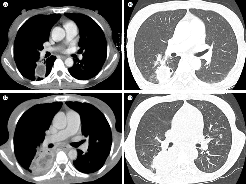
현병력: 3일 전부터 발생한 우측 흉막통을 주소로 2012년 2월 13일 본원 내원하였다. 내원 시 기침, 객담, 열감, 오한을 호소하고 있었다. 과거 병력상 내원 2년 전 외부병원에서 처음으로 우하엽 공동성 병변(Fig. 1A and 1B) 발견하고 항생제 및 경험적 항결핵치료 했으나 호전 없어 내원 5개월 전 본원으로 전원하여 기관지 내시경 검사 후 수술 준비 중이었으나 자의로 추적 중단했다가 흉막통의 악화로 재내원하였다.

(A, B) Initial chest computed tomography (CT) scans show 4.5 × 2.8 × 2.7 cm sized irregular cavitary consolidation with an irregular margin, internal cystic content, and some air foci in the superior segment of the right lower lobe. (C, D) Chest CT on admission shows increased extent of necrotic consolidation containing peripheral rim enhancing and clustered low attenuation areas.
과거력: 20년 전부터 당뇨병 있어 인슐린 사용 중이였다.
사회력 및 가족력: 15갑년의 흡연력이 있는 현재 흡연자였으며 가족력은 특이소견 없었다.
신체검사 소견: 입원 당시 혈압은 120/70 mmHg, 맥박 분당 84회, 호흡수 분당 20회, 체온 36.7℃였다. 의식은 명료하였고 결막의 창백이나 공막의 황달 소견은 보이지 않았으며 폐음 청진 시 수포음은 저명하지 않았다.
검사실 소견: 입원 당시 말초 혈액 검사는 백혈구 11,100/mm3 (중성구 85.6%), 헤모글로빈 10.6 g/dL, 혈소판 203,000/mm3였다. 생화학검사는 Aspartate transminase 18 U/L, Alanine transaminase 16 U/L, Alkaline phosphatase 126 U/L, albumin 1.9 g/dL, C-reactive protein 10.51 mg/dL, blood urea nitrogen 7.1 mg/dL, creatinine 0.7 mg/dL, 24hours urine protein 6.92 g, 혈당 523 mg/dL, hemoglobin A1c 14.4%였다.
방사선학적 소견: 입원 시 시행한 단순 흉부 방사선 검사상 우측 폐문부의 음영 악화 소견을 보이고 있었으며(Fig. 2A) 흉부컴퓨터 단층 촬영상 이전에 보였던 우하엽 병변은 크기와 괴사성 경화 모두 증가한 소견(Fig. 1C and 1D)이어서 영상학적으로 결핵을 완전히 배제할 수 없었다

Serial chest radiographs show gradual improvement in the right hilar area haziness (arrow). (A) The 1st hospital day, (B) The 7th hospital day at the intensive care unit, (C) The 80th hospital day, (D) 110th hospital day.
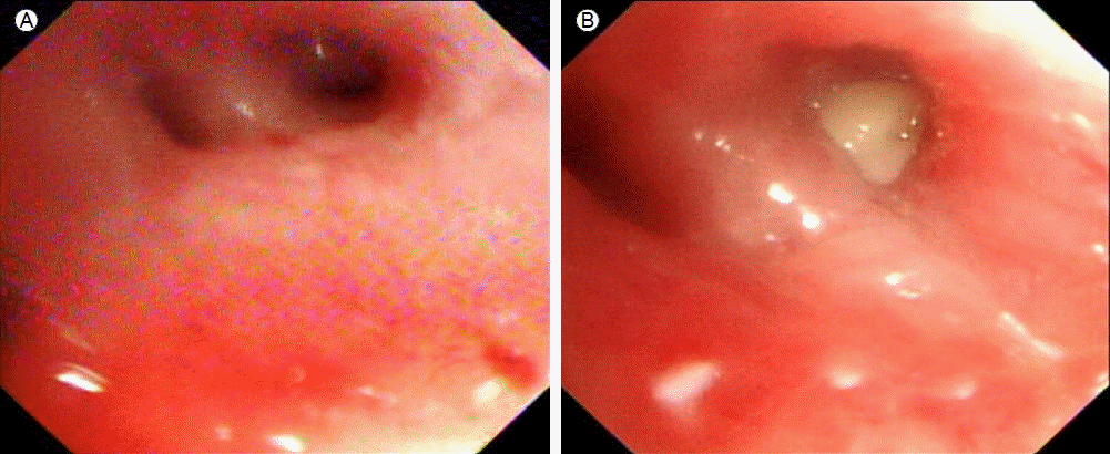
기관지 내시경 소견: 과거 입원 전 시행했던 기관지 세척술상 동정된 균은 없었으며, 경기관지 폐 생검도 특이소견 없었다. 2012년 2월 16일 기관지 내시경을 시행했으며 우하엽 입구에 농성 분비물(Fig. 3)이 다량 있었으나 기관지 세척액의 항산균 염색과 결핵 중합효소반응(polymerase chain reaction) 모두 음성이었고, 조직검사에서도 비특이적 만성 염증 소견이었다.

(A) Initial bronchoscopy shows mild erythematous mucosa in the superior segment of the right lower lobe. (B) Follow-up examination after admission shows white exudative material with more aggravated inflammatory change in the same area.
치료 및 임상경과: 세균성 폐렴에 준하여 ceftriaxone 정주하였으나 호전 없어 수술을 준비하던 중 입원 7일째 환자 상태 악화되어 기관 삽관 후 중환자실로 전실하였다. 이후 보고된 기관지 세척술 배양 검사 및 혈액 배양 검사에서 Candida glabrata가 동일하게 동정되었다. 또 중환자실 입실 후 객담 배양 검사에서는 다제내성 acinetobactor baumanii 균도 동정되어 colistin을 포함한 항생제(meropenem, teicoplanin 포함) 및 amphotericin B 사용 후 우하엽 병변은 점차 호전 추세를 보였고(Fig. 2B and 2D), 혈액배양 검사에서 candida 균 음전 상태로 총 21일 동안 amphotericin B를 유지하였다. 그 후 환자는 혈액 검사상 creatinine 상승 및 전신 부종으로 신장내과에서 당뇨성 신증에 대한 추가 치료 후 퇴원하였다.
고 찰
Candida glabrata에 의한 폐렴은 전세계적으로 극히 일부 보고[4-6]만이 있었으며, 국내에서는 폐렴 이외에 Candida glabrata에 의한 식도염, 이하선 농양, 요로 감염, 슬관절 감염, 엉덩허리근 농양등의 보고 및 단일기관에서 Candida glabrata 균혈증에 대한 연구[7]는 있었으나 폐렴에 대한 보고는 아직까지 없었다.
Candida glabrata는 Candida 속(genus)의 홑배수체 효모균(haploid yeast)으로 비이형성 진균(non-dimorphic fungus)이다. 이전에 보고된 바에 따르면 동물실험 모델에서 Candida albicans에 비해서 치사율이 낮은 것으로 알려져 있으나 최근 면역 저하 환자가 증가하면서 그 빈도가 증가하고 있다[8]. Candida glabrata의 또 다른 특징은 에너지 의존성 약물 방출 기전(energy dependent drug efflux mechanism)에 의한 azole 계열에 대한 저항성이 있는 것이다. 감염과 관련된 위험인자로는 혈액 암, 간경화, 당뇨병, 스테로이드 치료, 면역억제치료, 지속적인 항생제 사용 등이 있다[9]. 본 증례의 환자는 당뇨를 기저질환으로 가지고 있었으며 혈액배양 검사상 Candida glabrata가 동정되었으며 기관지세척배양검사에서 역시 동일한 Candida glabrata가 동정되어 오염균이나 상재균보다는 원인균으로 보는 것이 타당할 것으로 보인다.
Candida glabrata 감염 시에 가장 효과적인 항진균제에 대한 보고는 드물다. 여러 보고에 따르면 amphotericin B와 rifampin을 이용하여 치료한 예도 있고 항진균제 치료를 하지 않았으나 영상학적으로 호전된 경우도 보고된 바 있다[10]. Candida glabrata에 의한 진균혈증은 Candida albicans에 의한 진균혈증보다 사망률이 높아서 Komshian 등의 보고에 따르면 Candida glabrata 진균혈증 환자 12명에서 100% 사망률을 보였다[3]. 따라서 Candida 폐렴 환자에서는 최근 새롭게 개발된 voriconazole이나 정맥주사용 itraconazole 등과 같은 새로운 azole 계열의 항진균제를 일찍 사용하는 것도 고려해보아야 할 것이다[5].