구강내 전이로 진행한 전이성 간세포암 1예
A Case of Hepatocellular Carcinoma with Progressive Metastasis to the Oral Cavity
Article information
Abstract
본 증례는 간세포암으로 진단받고 경동맥 색전술을 5차례 받았으나 추적관찰 중 5개월 후에 폐, 림프절, 뼈 등 여러 장기로 전이되었고, 이후 2개월이 지나 경구개와 양쪽 편도에서 모두 종양이 생겨 구강 출혈을 주소로 내원하였다. 실시한 조직 검사에서 경구개와 편도에서 전이소견을 보였으며, 방사선 치료로 더 이상 출혈은 없었으나 60일 후 다발성 장기부전으로 사망한 증례를 경험하여 문헌고찰과 함께 보고한다.
Trans Abstract
Hepatocellular carcinoma (HCC) usually metastasizes to the lung, breast, lymph nodes, gastrointestinal tract, bone, kidney, and/or adrenal gland. Metastasis of HCC to the hard palate and tonsils is a rare phenomenon. Thus, we report a case of HCC with metastasis to the hard palate and both tonsils in a 45-year-old man. The patient was admitted to our hospital with a complaint of oral bleeding. Physical examination revealed a bleeding tumor located in the hard palate and histological examination showed metastasis from HCC. The patient died 2 months later due to multiple organ failure, although bleeding was controlled by radiotherapy. (Korean J Med 2012;83:268-271)
서 론
간세포암은 전 세계적 뿐만 아니라 국내에서도 흔한 악성종양으로 우리나라에서 전체 암 사망 원인의 2위를 차지한다[1]. 간세포암에 대한 진단 및 치료법의 향상으로 생존율이 향상되어 근치적 절제술 후 국내 누적 생존율이 27-56%까지 보고되고 있다[2]. 그러나 치료 후에도 간에 재발 및 다른 장기로 전이에 의해 예후가 여전히 불량하다. 2005년 Park 등[3]이 발표한 국내 연구결과에 의하면 간세포암은 약 15%에서 진단 당시 이미 원격 전이를 보였고, 전이가 흔한 장기로는 폐, 뼈, 부신, 복강, 흉벽 순이었다. 최근까지 전 세계적으로 보고된 간세포암의 구강내 전이는 주로 하악골과 잇몸이며[4], 경구개와 편도 전이는 드물다고 알려져 있다[5]. 저자들은 폐와 뼈, 림프절에 전이성 간세포암으로 진단받은 45세 남자 환자가 구강 출혈을 주소로 내원하여 경구개와 편도 전이로 진단된 증례를 경험하여 이를 보고하고자 한다.
증 례
환 자: 한〇〇, 남자 45세
주 소: 구강 출혈
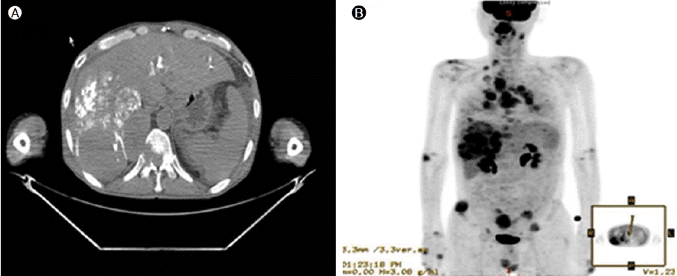
현병력: 내원 8개월 전 복부 팽만을 주소로 시행한 복부 전산화 단층촬영에서 간세포암이 발견되어 5차례 경동맥 색전술을 받았으나 관해가 되지 않았으며, 내원 3개월 전 추적 양전자 방출 단층촬영에서 폐와 뼈, 림프절 등 다발성으로 전이되어(Fig. 1) 넥사바를 복용 중, 내원 1개월 전 경구개에 종양이 생기고 크기에서 증가 소견을 보여 넥사바 복용을 중단하였고, 이후 외래에서 경과관찰 중 내원 당일 발생한 구강 출혈을 주소로 본원 응급실에 내원하였다.

Positron emission tomographic-computed tomographic images showing hypermetabolic mass lesions (maximum standardized uptake value = 5.3) in the right lobe with lipiodol uptake (A) and hepatocellular carcinoma with multiple distant metastases (B).
과거력: 내원 24년 전 만성 B형 간염 바이러스 보유자로 진단되었다.
가족력: 어머니-만성 B형 간염 바이러스 보유자
진찰 소견: 내원 당시 환자는 만성 병색이었으며, 의식은 명료하였고 활력징후는 정상이었다. 결막은 창백하지 않았으며, 공막은 황달 소견을 보이지 않았다. 신체 검사에서 호흡음은 깨끗하였고 심음은 규칙적이었으며 심잡음은 들리지 않았다. 복부는 팽만이 되어 있었고 간은 심와부에서 2 횡지로 촉지가 되었으며 비장종대는 관찰되지 않았다.
후두 내시경 소견: 후두 내시경에서 양쪽 편도에 0.5 cm 사이즈의 종양, 경구개에 3 cm 사이즈의 종양이 보였으며, 경구개에서 출혈소견이 보였다(Fig. 2).
검사실 소견: 내원 당시 응급실에서 검사한 말초혈액검사에서 백혈구 5,000/mm3, 혈색소 10.1 g/dL, 헤마토크리트 29.2%, 혈소판 130,000/mm3였고, 생화학적 검사에서 총 단백 5.5 g/dL, 알부민 2.6 g/dL, BUN 7.6 mg/dL, creatinine 0.7 mg/dL, AST 191 IU/L, ALT115 IU/L, alkaline phosphatase 150 IU/L, 총 빌리루빈 0.7 mg/dL, 프로트롬빈 시간 11.7초(121%, INR 0.90)였다. 간염 바이러스 표지자는 HBsAg 양성, anti-HBs 음성, HBeAg 음성, anti-HBe 양성, anti-HCV 음성이었고, 혈청 AFP는 115.3 ng/mL였으며, 소변 검사는 정상이었다.
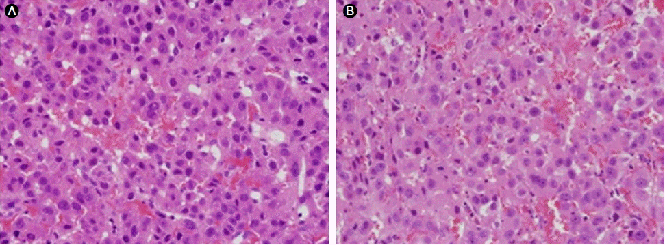
조직 소견: 경구개와 양쪽 편도에서 펀치생검을 실시하였고 간세포암에서 보이는 조직학적 소견을 보여 경구개와 편도 전이로 진단되었다(Fig. 3).

Microscopic findings of the hard palate (A) and tonsil (B). Large polygonal cells show atypical trabecular arrangement with abundant eosinophilic granular cytoplasm and centrally located round-to-oval nuclei with prominent nucleoli, features of hepatocellular carcinoma (hematoxylin and eosin staining, × 400 magnification).
치료 및 경과: 지혈제 등의 치료로 출혈이 멈추지 않아 고식적인 방사선 치료(2000 cGY)를 시행하여 종양의 크기는 50% 감소하였고, 출혈은 더 이상 없었다. 하지만 이후 병변은 다시 커지기 시작하였고 전이가 진단된 이후 약 60일 뒤 다발성 장기 부전으로 사망하였다.
고 찰
전이성 간세포암의 구강내 전이는 주로 하악골과 잇몸이며[4], 경구개와 편도의 전이는 드물다고 알려져 있다[5]. 구강내 전이는 Dick 등[6]은 하악에 전이한 간세포암을 처음 보고한 후 50여 증례가 보고되었으며, 이들 대다수가 골전이였고, 1/3만 치은전이였다[7]. 간세포암은 구강내 전이에 있어 인종에 따른 차이가 있는데 50%가 아시아인이나 원인은 알려져 있지 않다[4]. 대부분이 고령의 남자로 이러한 특징은 간세포암의 호발연령 및 성별을 반영하지만, 간세포암의 남녀성비 3:1과는 차이가 있으며 그 이유는 알려져 있지 않다[8]. 간세포암의 구강내 전이는 두 가지 혈행성 경로를 통하는데, 첫째는 간문맥 혈행성 경로로 간동맥, 간문맥을 침범하고 폐로 전이가 동반된 뒤 구강내 전이되는 경로이며, 다른 하나는 “Batson's plexus”라는 척추 정맥총 경로로 대정맥을 우회하여 구강내 전이되는 경로이다[8]. 본 증례의 경우는 폐로 전이가 동반된 뒤 경구개와 편도에 전이가 된 경우로 간문맥 혈행성 경로가 구강내 전이에 기여한 것으로 판단된다. 간세포암의 구강내 전이는 대개 무증상이고, 증상이 있을 경우 통증과 부종, 감각이상 등이 나타나며, 종괴는 구강에서 빠르게 자라고 궤양을 동반하며 쉽게 출혈되는 특징이 있다[4]. 본 증례는 통증은 없었으나 경구개에 위치한 종양 자체와 출혈로 인해 기좌호흡과 정상적인 식사가 불가능하여 삶의 질이 매우 떨어져 보였다. 구강내 전이 시 치료는 수술 및 방사선 치료, 항암제 치료를 할 수 있으며, 가능하다면 증상의 호전을 위해 수술적 치료가 삶의 질 측면을 향상시킬 수 있다[9]. 본 증례에서는 환자가 수술적 치료가 적합하지 않아 지혈을 위해 방사선 치료를 실시하였으며, 종양의 크기가 50% 정도로 감소되었다. 이후 재출혈 소견은 보이지 않았으나 다시 종양의 크기가 커지기 시작하였다. Kanazawa 등[4]은 구강으로의 전이 후 생존기간은 2주에서 2년이고, 평균 생존기간은 21주로 보고하였고, 본 증례의 환자는 60일만에 다발성 장기 부전으로 사망하였다.
