관절염의 영상 검사
Imaging of Arthritis
Article information
Trans Abstract
Imaging study of joint is important to differentiate various kinds of arthritis, and to evaluate the treatment response of the arthritis. Radiograph is the basic and first line imaging study of the joint, but there are overlapped imaging findings between arthritis. The objective of this review is to present a simplified approach to radiographic evaluation of arthritis and to help in making adequate decision to choose a further imaging study among ultrasonography, CT and MRI. (Korean J Med 2012;83:178-189)
서 론
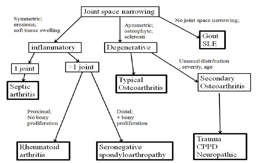
관절염의 종류에 따라 관절을 이루는 관절연골과 연골하골, 활액막, 힘줄과 인대의 변화가 각각 다른 양상으로 나타나므로 임상소견과 더불어 영상소견은 관절염의 감별진단에 도움이 되며, 영상 검사가 관절염의 진행 정도를 평가하고 치료경과를 관찰하는 데 중요한 역할을 한다. 영상검사에서 관절 연골손상에 따른 관절강 협착(joint space narrowing)과 대칭성(symmetricity), 관절주위 골다공증, 관절주위 골 경화(sclerosis), 골 미란(bone erosion), 연골하 낭종(subchondral cyst), 관절주위 연부조직 변화, 석회질 침착, 관절 모양의 변화, 침범관절의 개수와 분포 등을 평가한다.
본 론
관절염을 감별하는 데 있어서 여러 가지 방법들이 있으나 영상소견을 중심으로 정리한 Jacobson 등[1]이 보고한 것을 기본으로 몇 가지 수정하여 소개하고자 한다(Fig. 1) [1,2]. 그림 1의 흐름도는 전형적이고 특징적인 소견을 기준으로 한것이므로 비 전형적인 경우와 각 질환의 초기소견은 포함하지 않고 있기 때문에 임상소견과 더불어 판단하는 것이 필요하다. 여기서 가장 중요한 것은 세균성 관절염(septic arthritis)을 다른 관절염과 감별하여 조기에 적절한 치료를 하는 것인데, 급성 단일 관절 염증인 경우에는 임상소견으로 세균성 관절염을 다른 질환과 감별하는 것이 중요하다[3]. 관절염이 단일 관절인지 아닌지 평가하는 데 있어서 손목과 같이 한 부위에 여러 구획이 존재하는 경우에는 한 구획을 단일 관절로 평가해야 한다(Table 1) (Fig. 2) [4].

Major compartments of the wrist
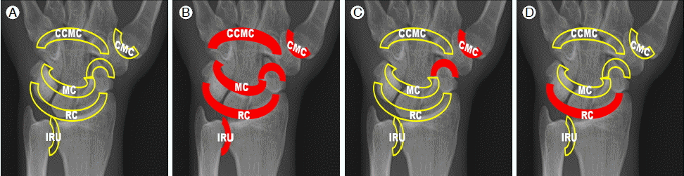
(A) Compartments of the wrist. The pisiform-triquetral compartment is not shown. The trapezioscaphoid region of the midcarpal joint is separated from the remainderof this joint. CCMC, common carometacarpal compartment; CMC, first carpometacarpal compartment; IRU, inferior radioulnar compartment; MC, midcarpal compartment; RC, radiocarpal compartment. (B) Rheumatoid arthritis. (C) Osteoarthritis. (D) CPPD (modified from Resnick D, Kransdorf MJ. Target area approach to articular disorders. In: Bone and Joint Imaging. 3rd ed. Philadelphia: Elsevier Saunders, 2005:519-539 [4]).
세균성 관절염의 경우 단일 구획에서 시작하여 치료가 적절하게 되지 않은 경우 주변 구획으로 진행되고, 류마티스 관절염에서는 초기에 양쪽 손목에 대칭적으로 여러 구획에 관절염이 생기고 결국 모든 구획에 관절염이 생긴다(Fig. 2B). 특정한 구획을 침범하는 관절염들이 있는데, 골 관절염(osteoarthritis)과 CPPD는 모두 퇴행성 변화를 보이는 관절염이지만 골 관절염은 carpometacarpal 구획과 trapezioscaphoid 구획을 주로 침범하고 CPPD는 radiocarpal 구획을 침범한다(Fig. 2C and 2D).
염증성 관절염
관절 내 염증이 있을 때 세균에 의한 감염에 의한 경우와 류마티스 또는 척추관절병증과 같은 비세균성 원인에 의한 경우 모두 대칭적인 관절강 협착, 골 미란, 관절주변 연부조직 부종을 동반한다.
골 미란은 염증성 관절염을 시사하는 소견으로 연골하 골피질의 흰 선의 일부가 끊어져 보이는 것을 말한다. 염증이 없다면 심한 골다공증이 있어도 연골하 골 피질의 하얀선은 유지되어야 한다. 골 미란이 관절면의 가장자리에 있는 경우를 가장자리 골 미란(marginal erosion)이라고 하는데, 이는 관절면의 가장자리에 노출부(bare area)가 있기 때문이다. 이 노출부는 관절내부에 위치하지만 유리연골(hyaline cartilage)이 덮고 있지 않기 때문에 관절 내의 염증이 이 노출 부에 가장 먼저 골 미란을 생기게 하므로 영상검사에서 관절의 가장자리를 여러 각도에서 확인하는 것이 필요하다. 염증이 진행되어 연골과 연골 하부 골의 파괴가 진행되면서 관절강 협착을 유발하는데 염증이 관절강 내에서 전체적으로 균일하게 일어나므로 대칭적 관절강 협착(symmetric joint space narrowing)이 생긴다. 또한 주변 연부 조직에 염증이 동반되므로 연부 조직 부종이 생긴다.
세균성 관절염
균일한 관절강 협착, 골 미란, 연부조직 부종 등의 염증성 관절염의 소견이 있을 때 침범된 관절이 하나인 경우에는 가장 먼저 세균성 관절염의 가능성을 생각하여 임상적 검사가 진행되어야 한다(Fig. 1). 그러나 세균성 관절염이 여러관절성으로 생기는 경우가 약 20%까지 보고되어 있으므로 두 개 이상의 관절이 침범되더라도 염증성 관절염의 감별진단이 확실하게 되기 전까지는 항상 세균성 관절염의 가능성을 염두에 두어야 한다[5]. 세균성 관절염이 임상적으로 확인되면 골수의 염증여부를 확인하기 위해서 MRI를 시행한다. MRI에서 골수의 염증이 있는 경우에는 T1 강조영상에서 저 신호강도, 조영 증강 후에 신호증강을 보이며 부종만 있는 경우에는 신호 증강은 없다(Fig. 3). 세균성 관절염의 영상 소견은 염증성 관절염의 소견을 보이지만 관절면의 대칭적 협착(symmetric narrowing), 가장자리 골 미란, 관절주위 연부조직 부종, 관절하 연골의 골다공증 등이 모두 나타나지 않는 경우도 있다. 급성기에는 골 미란이 보이지 않을 수도 있고, 관절강내의 삼출액에 의해 오히려 관절강이 넓어질 수도 있다. 결핵성 관절염의 경우에는 관절강 협착이 천천히 나타나기도 한다(Fig. 4) [2].
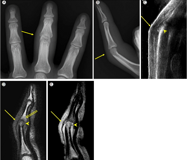
Septic arthritis (A) AP view and (B) lateral radiographs of the 3rd finger of a hand show symmetric joint space narrowing, periarticular soft tissue swelling and bone erosion (arrow) of the PIP joint. (C) Ultrasonography of the dorsal portion of the PIP joint of the finger shows cortical erosion (arrowhead) and periarticular soft tissue inflammation (arrows). (D, E) On MRI of the finger, the proximal phalangeal head, mid phalangeal base and periarticular soft tissue of the PIP joint show low signal intensity on T1-weighted image (D). After contrast infusion, fat suppressed T1-weighted image shows signal enhancement of the proximal phalangeal head and periarticular soft tissue sparing mid phalangeal base. The area of low signal intensity of the mid phalangeal base suggests reactive bone marrow edema not inflammation.

Tuberculous dactylitis. (A) AP view and (B) lateral radiographs of the 3rd finger of a hand show osteomyelitis with cold abscess of the proximal phalanx. The joints space of the PIP joint of the finger is relatively preserved (arrow) (C) Ultrasonography of the volar portion of the PIP joint shows effusion (arrow) in the joint and large erosion of the proximal phalangeal head (arrowhead).
류마티스 관절염
염증성 관절염이 두 개 이상의 관절에 있을 때 류마티스 관절염의 가능성과 음성혈청반응 척추관절증(seronegative spondyloarthropathy)을 생각해야 한다. 손과 발에 관절염이 있을 때 방사선 검사에서 염증성 관절염의 소견이 여러 개의 관절에서 양측성 그리고 대칭적으로 근위부(proximal)에 분포하고 골 증식이 없으면 류마티스 관절염일 가능성이 높다(Fig. 5). MRI는 조기의 류마티스 관절염을 평가하고, 동반된 건염 여부를 확인하고, 활액막의 두께와 골수와 연부조직 염증 정도를 기준으로 치료반응을 관찰하는 데 유용하다(Fig. 6) [6]. 만성기에는 관절주위 골다공증, 균질한 관절강협착, 골 미란, 연부조직 부종 외에 관절의 아 탈구와 연골하낭종이 동반되기도 한다. 류마티스 관절염은 활액막의 전반적으로 염증을 일으키는 질환이기 때문에 retrocalcaneal bursa같은 윤활낭(bursa)과 건초(tendon sheath)에도 염증을 일으킨다. 손과 발 외에도 무릎 관절, 고관절, 천장관절, 견관절에도 관절염이 있을 수 있으며, 척추에서 C1-2에 윤활관절에 염증성 관절염이 생겨서 odontoid process에 골 미란이 생기고(Fig. 7), 윤활관절에 인접한 C1-2를 연결하는 인대 손상으로 atalantodental space가 3 mm 이상 넓어지는 atlantoaxial subluxation이 생기기도 한다(Fig. 8). 이때 방사선 검사에서 경추를 굴곡한 자세에서만 atlantoaxial subluxation이 나타나는 경우도 있다.

Rheumatoid arthritis of hand and wrist. Radiograph of a hand shows joint space narrowing, bone erosion (arrow heads) and periarticular soft tissue swelling (arrow) in pancompartment of the wrist and metacarphophalangeal and proximal interphalangeal joints of the hand.
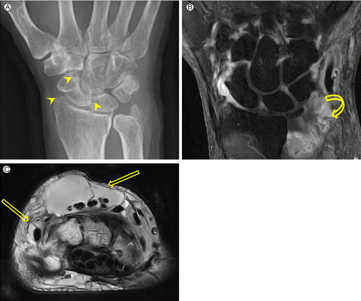
Rheumatoid arthritis of wrist. (A) Radiograph shows marginal erosions (arrow heads) in midcarpal compartment and radiocarpal compartment. (B) On fat suppressed T1-weighted MR coronal scan of the wrist, pannus formation (curved arrow). is noted in distal radioulnar compartment with effusion. (C) On T2-weighted MR axial scan shows tenosynovitis of extensor tendons (open arrows).
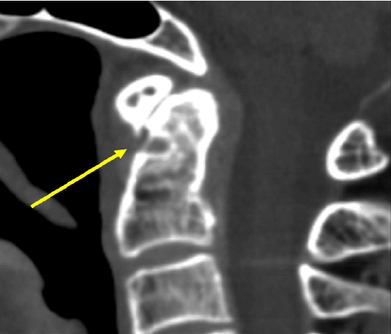
Rheumatoid arthritis of C1-2. Sagittal CT scan of C-spine shows a bone erosion of the anterior portion of odontoid process of C2 (arrow).
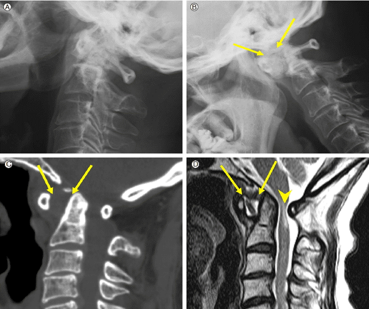
Rheumatoid arthritis of C1-2 with subluxation. (A, B) Lateral views of C-spine show C1-2 subluxation (arrows) not with neck extension (A) but with neck flexion (B). (C, D) Sagittal scan of CT (C) and MR T2-weighted image (D) of C-spine show C1-2 subluxtion (arrows) with narrowing of spinal canal. Spinal cord shows high signal intensity on MR T2-weighted image suggesting compressive myelopathy (arrowhead).
음성혈청반응 척추관절증(seronegative spondyloarthropathy)
염증성 관절염이 여러 관절에 생기며, 손과 발에 생기는 경우에 원위부 관절을 침범하고 골 증식의 소견을 보이면 음성혈청반응 척추관절증을 먼저 의심해야 한다. 강직성 척추염, Psoriatic arthritis, Reiter syndrome (Reactive arthritis) 등이 이에 해당하고 Psoriatic arthritis와 Reiter syndrome은 비교적 드물다.
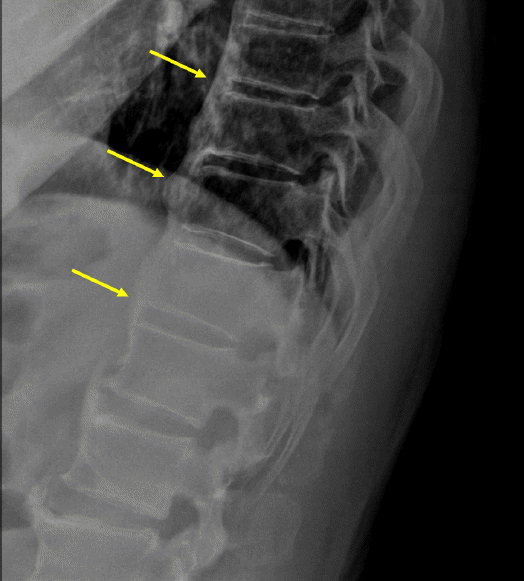
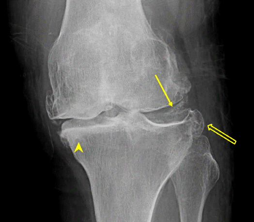
강직성 척추염은 axial skeleton을 주로 침범하고 peripheral joint를 침범하기도 한다(Fig. 9). 척추에서는 골염(osteitis), syndesmophyte 형성, 척추후관절(facet joint) 염증, 그리고 척추제 융합이 생긴다(Fig. 10). 천장관절에서는 양측성 대칭적으로 염증이 생기며 대체로 척추의 염증보다 먼저 생긴다. 처음에는 관절면의 연골하 골 피질의 하얀 선이 끊어지거나 희미해지는 것으로 시작하여 골 미란의 크기가 커지게 된다(Fig. 11). 골 미란 주변에는 골 경화가 보일 수 있고 관절 내의 염증이 진행되면서 결국 관절강이 좁아지고 골 융합이 생긴다(Fig. 12). 단순 방사선 검사에서 천장관절이 정상으로 보이거나 이상소견이 뚜렷하지 않을 때 MRI로 관절내부 삼출액과 관절주변 골수부종이 있으면 진단할 수 있다(Fig. 13). 또한 단순 방사선 검사에서 이상 소견이 보이지만 염증성 관절염인지 퇴행성 관절염인지 구분하기 힘들 때는 CT로 감별할 수 있다. 척추를 침범하는 경우에는 초기에 추간판과 척추체 인접부(Discovertebral junction)에 전방 가장자리에 골 미란이 있다가 골 경화가 심해지는데 이를 “shiny corner sign”이라고 한다(Fig. 10). 척추체 전면과 측면에 골 증식이 일어나면 “squared” 모양을 보이며, annulus fibrosus의 외측에 골화가 진행되면 syndesmophyte가 나타나는데, 이때 척추의 전면에 출렁거리는 듯한 전종인대의 골화를 보이는 diffuse idiopathic skeletal hyperostosis (DISH)의 경우와 감별해야한다(Fig. 14). Syndesmophyte가 두꺼워지고 연결되면 bamboo spine의 소견이 보이고 비교적 척추 골절이 쉽게 일어난다(Fig. 10). 후관절(facet joint)의 염증은 진행하여 관절의 융합이 생기고, 척추의 AP view에서 후방 극간인대(posterior interspinous ligament)의 골화로 “dagger sign”이 보이며 후관절(facet joint)의 융합과 극간 인대(interspinous ligament)의 골화는 “trolleytrack sign”으로 보인다(Fig. 12). Peripheral joint를 침범하는 경우에는 양측성으로 균일한 관절강 협착을 보이고 고관절의 경우에서는 acetabular protrusion과 연골하 낭종이 생기고 대퇴골 경부에 골 증식이 보인다(Fig. 12).

Ankylosing spondylitis of peripheral joints. (A, B) Radiographs of a foot (A) and a knee (B) show marginal bone erosions (arrowheads) and bony proliferations (arrows).
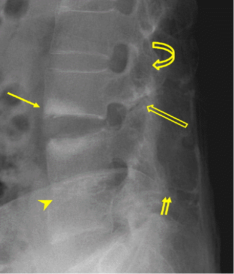
Ankylosing spondylitis of L-spine. Lateral radiograph of L-spine shows syndesmophyte formation (arrowhead), ankylosis of facet joints (curved arrow), ossification of posterior interspinous ligament (double arrows), osteitis of anterior corner of discovertebral junction (shiny-corner sign) (arrow), and a distractive fracture of L2-3 (open arrow).
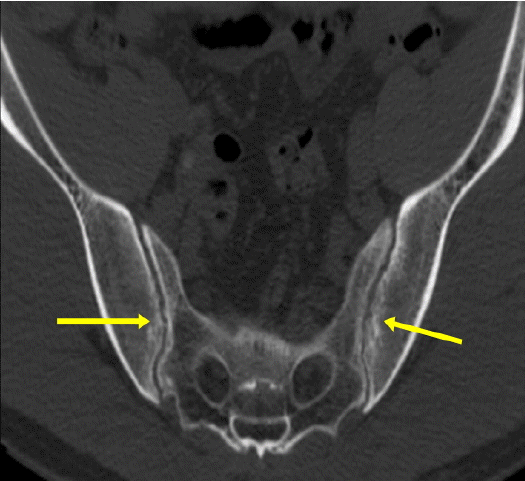
Ankylosing spondylitis of SI joint. Axial scan of CT of pelvis shows bone erosions of both SI joints (arrows).
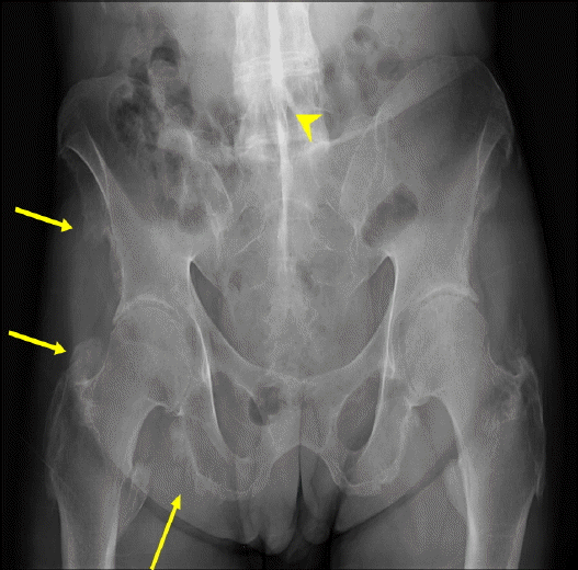
Ankylosing spondylitis of SI joint. Radiograph of pelvis shows bony ankylosis of both SI joints, Symmetric joint space narrowing of both hip joints, bony proliferations (arrows) of both pelvic bones and both proximal femurs. There is a white vertical line at the midline of lumbosacral spine; “dagger sign” (arrowhead).
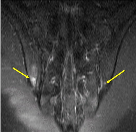
Ankylosing spondylitis of SI joint. On MR oblique coronal scan of pelvis, fat suppressed contrast enhancement T1-weighted image shows signal enhancement (arrows) in joint spaces and periarticular bones of the lower portion of both SI
DISH (Diffuse idiopathic skeletal hyperostosis) of thoracolumbar spine. Lateral radiograph of thoracolumbar spine shows flowing ossification (arrows) of anterior longitudinal ligament at the anterior portion of the thoracolumbar spine.
퇴행성 관절염
퇴행성 관절염은 비대칭성 관절강 협착, 골 경과와 골극, 염증성 관절염에서 보이는 골 미란이 없는 연골하 낭종이 특징적이 소견이다. 가장자리 골극이 퇴행성 관절염을 진단하는데 도움을 주는 소견이라고 하면, 관절강 협착, 골 경화, 연골하 낭종은 중증 정도를 평가하는 데 사용된다. 퇴행성 관절염이 영상소견에서 보일 때 침범된 관절의 위치, 환자의 나이와 기타 영상 소견으로 퇴행성변화를 동반하는 다른 종류의 관절염의 가능성을 생각해야 한다.
골관절염(Typical osteoarthritis)
관절의 퇴행성 변화는 일상생활에서 생기는 미세하고 반복적인 외상으로 인해 관절연골이 손상되고 닳고 파괴되는것으로서 환자의 자세의 습관과 행동에 따라 퇴행성 변화의 차이가 생긴다. 퇴행성 변화는 주로 40대 전후에 생기기 시작하며 무릎의 경우 관절강 협착이 내측에 비 대칭성으로 나타나고 고관절의 경우 상방에 관절강 협착이 오면서 대퇴 골두가 상방으로 이동한다. 손에서는 사용하는 동작과 정도에 따라서 first carpometacarpal joint 또는 interphalangeal joint에 퇴행성 변화가 생긴다(Figs. 2 and 15).
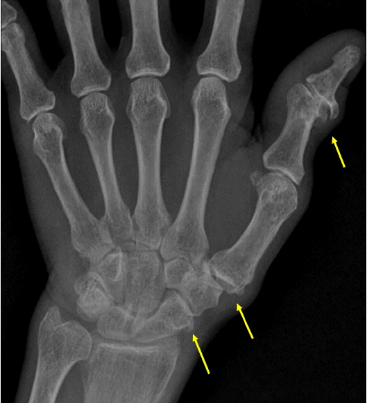
Osteoarthritis of hand. Radiograph shows spurs (arrows) and sclerosis of the 1st interphalangeal joint, the 1st carpometacarpal joint, and trapezioscaphoid joint.
이차성 골관절염(Secondary osteoarthritis)
퇴행성 변화가 흔히 발생하는 위치가 아닌 곳에 생기거나, 중증 정도가 심하고, 나이도 비교적 어린 경우에는 다른 원인에 의한 퇴행성 변화가 있는지 살펴보아야 한다.
외상은 가장 흔한 이차성 골관절염의 원인으로 연골손상을 동반하는 외상이 있었거나 운동선수의 경우나 직업적으로 과도하게 관절연골에 손상이 생기는 경우에 퇴행성 변화가 일찍 오고 좌, 우 한 쪽에만 나타난다.
CPPD는 연골, 활액막, 힘줄과 인대에 석회질이 침착되는 질환으로 결정 침작이 관절연골을 손상시켜서 퇴행성 변화를 나타나게 한다(Fig. 16). 무릎은 CPPD가 가장 흔히 발견되는 곳으로 외측 무릎관절 또는 무릎뼈 대퇴골 관절에만 변화가 있으면 골관절염과 감별이 가능하다. 분포 양상은 일반적으로 양측성이다. 영상소견이 일부분 골관절염과 유사하나 다음의 몇 가지의 차이점을 보인다[7].
CPPD of knee. Radiograph shows chondral calcinosis in lateral meniscus (arrow) with large spurs (open arrow) and severe subchondral sclerosis (arrow head).
침범하는 관절부위의 차이
관절질환이 몸무게가 실리는 관절에 발생하기도 하지만 퇴행성 관절질환이 흔히 나타나지 않는 손목, 팔꿈치, 어깨 관절에서도 발견된다.
관절내부에서 침범하는 위치의 차이
손목에서 radiocarpal에만 퇴행성변화가 단독으로 생기거나 또는 무릎에서 patellofemoral compartment의 병변이 단독적으로 생기거나 또한 심하게 손상된 경우에는 CPPD결절 침착 질환을 의심할 수 있다(Fig. 17).
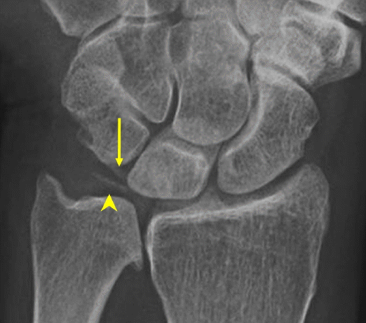
CPPD of wrist. Radiograph shows chondral calcinosis in trigangular fibrocartilage (arrow head) and ligament calcification in lunotriquetral ligament (arrow).
크기가 큰 연골하 낭종
심한 골 파괴성 변화
다양한 골극 형성
Neuropathic joint는 초기에 골관절염과 유사하나 진행할수록 골화, 골분절(fragmentation), 아 탈구(subluxation)가 점점 뚜렷해진다. 초기의 neuropathic joint와 골관절염과의 감별에 도움이 되는 소견은 신경손상이 있는 위치에 관절변화가 생기는 것이다. 그러므로 환자의 병력이 영상 검사 진단에 필요하다. 예를 들어 DM foot에서는 midfoot에 neuropathic joint가 생기는 특징이다(Fig. 18).
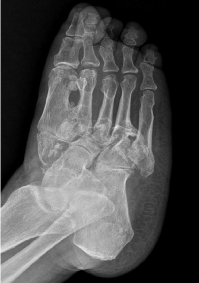
Neuropathic joint. Oblique radiograph of a foot of a DM patient shows fragmentation, disorganization and sclerosis of mid foot.
기타
통풍(Gout)은 관절강 협착이나 골다공증을 동반하지 않는 monosodium urate cystal 침착에 의한 것으로 통풍에서 보이는 골 미란은 특징적으로 관절면 가장자리에서 벗어나서 경계가 잘 그려지는 punched-out appearance를 보인다. monosodium urate crystal이 방사선 검사에서 고음영으로 보이지 않는 경우가 많기 때문에 결정 침착이 있는 곳은 연부조직의 심한 부종으로만 보이고 이때 초음파검사에서 결정 침착이 고음영으로 보여 진단에 유용하며 MRI에서는 모든 스캔에서 저 신호강도로 보인다(Fig. 19). 가장 흔히 침착되는 곳은 first metatarsophalangeal joint이다. 초기에는 한 개의 관절에서 시작하나 후기에는 여러 관절을 침범한다. 손과 발의 다른 관절에 침착되는 경우가 드물지 않고 팔꿈치의 olecranon bursa에 침착되기도 한다.
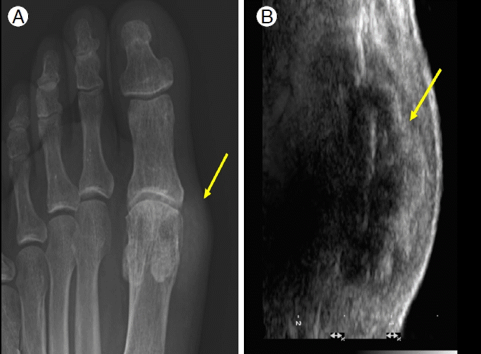
Gout. (A) Radiograph shows soft tissue swelling (arrow) of the medial portion of the 1st metatarsal head with adjacent bone erosion. (B) US of the medial portion of the 1st MTP joint shows echogenic crystal deposition in the soft tissue swelling.
SLE (systemic lupus erythematosus)는 방사선 검사에서 관절면 협착이 흔하지 않으며 주로 metacarpophalangeal joint의 subluxation 생기며 AP view에서는 잘 보이지 않고 oblique view를 찍어야만 알 수 있다. 관절주변 골다공증과 연부조직 부종은 보일 수 있다.
결 론
관절염이 의심될 때 관절강 협착이 있는지 먼저 보고 골 미란이 있는지 골극이 있는지 살펴보아야 한다. 관절강의 협착이 있을 때 골미란이 있으면 염증성 관절염, 골극이 있으면 퇴행성 관절염을 고려하여야 하며 염증성 관절염이 하나의 관절에 생길 때는 가장 먼저 세균성 관절염의 가능성을 확인하는 것이 중요하다. 여러개의 관절에 생길 때는 관절염의 분포와 골 증식이 있는지를 평가하여 류마티스 관절염과 음성혈청반응 척추관절증(seronegative spondyloarthropathy)을 구분한다. 퇴행성 변화가 있는 관절에서는 침범된 관절의 위치와 진행 정도, 나이 등을 고려하여 이차성 관절염이 의심되면 원인을 찾아보아야 한다. 관절강 협착이 없고 경계가 명확한 관절주위 골미란이 있으며 종괴모양의 연부조직부종이 있을 때는 통풍을 고려하여야 한다. 가장 기본적인 영상 검사인 단순 방사선 검사에서는 관절염이 진행되어 관절 주변 뼈에 변화가 있을 때 이상소견이 나타나고, CT는 단순 방사선 검사보다 예민하게 이상소견을 발견하는 데 도움이 되며 관절구조가 복잡할 때 유리하다. US는 관절내부 삼출액과 관절주변 연부조직의 변화를 잘 볼 수 있고 단순 방사선 검사에서 잘 나타나지 않는 석회질 침착을 예민하게 볼 수 있는 장점이 있으나 연골과 골 파괴를 평가하는 데 제한이 있고 다른 검사에 비해서 검사결과가 검사자에 좌우되고 재현성이 떨어진다. MRI는 검사에 포함된 관절의 관절연골, 골수의 변화, 관절주위 연부조직 변화를 평가하는 데 유용하며 치료경과 관찰에 도움을 준다. 이때 조영증강 검사를 같이 실시하여야 활액막과 삼출액을 구분하여 활액막의 비후 정도를 평가할 수 있고 골수와 연부조직의 염증범위를 정확하게 평가할 수 있으며 감영성 관절염인 경우 골수 내부 또는 주변조직에 농양이 동반되었는지 확인할 수 있다.