만성내장질환 환자에서 발생한 십이지장 및 공장의 허혈성 장염 1예
A Case of Ischemic Duodenal and Jejunal Enteritis in a Patient with Chronic Splanchnic Disease
Article information
Abstract
위장관 허혈은 내장동맥 협착, 혈전 및 저혈류 상태의 혈관수축에 의해 발생한다. 하지만 상부위장관에 동맥협착이 있다고 해도 풍부한 측부순환으로 허혈성 손상은 드물다. 저자들은 구토, 상복통 및 토혈을 주소로 내원한 92세 여자 환자를 경험하였다. 심전도에서 발작성 심방세동을 가지고 있었다. 환자는 상부위장관 내시경에서 십이지장 제2부에 미만성 분절형태의 점막 부종, 홍반 및 출혈을 보였다. 복부 혈관조영 전산화 단층촬영에서 복강동맥과 상장간막동맥의 협착이 있었고 십이지장 제2부의 근위부부터 공장의 근위부까지 분절형 동심성 비대가 보였다. 환자는 일주일 동안 양전자 펌프 억제제 및 수액치료를 받았다. 추적 상부위장관 내시경에서 이전 검사에 비해 호전된 점막을 관찰할 수 있었다. 결론적으로 십이지장 및 근위부 공장은 혈액순환이 매우 풍부하여서 허혈성 손상이 발생하는 경우는 매우 드물지만 만성내장질환을 가지고 있는 환자에서 유발인자로 인해 발생할 수 있다는 사실에 주의해야 한다.
Trans Abstract
Gastrointestinal ischemia happens by splanchnic artery stenosis, thrombus, or physiological vasoconstriction during a low-blood-stream state. However, even if arterial stenosis exists in the upper gastrointestinal tract, ischemic injury is very rare due to rich collateral circulation. The authors experienced 92-year-old female patient with vomiting, epigastric pain, and hematemesis. An electrocardiogram showed paroxysmal atrial fibrillation. The patient had diffuse and segmental mucosal edema, erythema, and hemorrhage in the second part of the duodenum on esophagogastroduodenoscopy (EGD). On abdomen computed-tomography angiography, stenosis of the celiac and superior mesenteric arteries was observed, and segmental concentric wall thickness was seen from the proximal second portion of the duodenum to the proximal jejunum. The patient was treated with PPI and fluid therapy for one week. At follow-up EGD, the mucosa had improved compared with the previous EGD examination. In conclusion, ischemic injury rarely affects the duodenum and jejunum; however, it can develop in the presence of inducing factors. (Korean J Med 2012;82:704-708)
서 론
위장관 허혈은 주로 심폐기능질환을 가지는 고령의 환자에서 발생하며 위험인자로는 50세 이상, 울혈성 심부전, 부정맥, 최근의 급성심근경색, 혈량저하증, 저혈압 또는 패혈증, 이전에 동맥색전증을 앓았던 경우, 혈관염, 심부정맥 혈전증, 과응고상태(단백 C와 S 부족증, 항트롬빈 III 부족증, 활성형단백저항증) 등이 있다[1].
상부위장관의 경우는 동맥의 협착이 있다고 해도 측부순환이 풍부해서 실제로 허혈성 위염, 십이지장염 및 공장염을일으키는 경우는 매우 드물다. 그러나 상부위장관에 측부순환이 풍부하더라도 혈전에 의한 급성색전증이 발생하거나 만성내장동맥질환(chronic splanchnic disease)이 있는 환자에서 위험인자가 중복되어 작용하는 경우에는 상부위장관에 허혈성 손상을 유발할 수 있다. 십이지장에 허혈성 장염이 발생한 경우는 세계적으로 3예에 불과하며 국내에서도 장간막정맥 혈전증에 의한 십이지장 허혈성 장염이 1예가 보고되었을 뿐 발생의 보고가 매우 드물다[2-5].
저자들은 구토 및 토혈을 주소로 내원한 92세 여자 환자에서 상부위장관 내시경을 실시하였고, 십이지장에 중증의 허혈성 손상을 보였다. 혈관조영단층촬영에서 복강동맥(celiac artery) 및 상장간막동맥(superior mesenteric artery)의 협착을 보였고, 십이지장뿐만 아니라 근위부 공장까지 장벽의 분절성 비대를 보여서 허혈성 십이지장염 및 공장염을 진단하였으며, 대증적 치료로 허혈성 장염의 호전을 관찰하였기에 문헌고찰과 함께 보고한다.
증 례
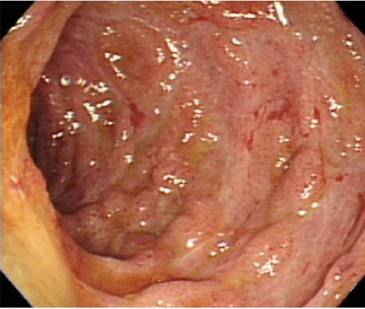
92세 여자 환자가 내원 2일 전부터 상복부 통증이 있었고, 내원 전일부터 구토 및 토혈을 주소로 내원하였다. 과거력에서 15년 전에 고혈압을 진단받았고, 3년 전에 좌측 고관절 인공관절 치환술을 시행 받았다. 가족력에서 특이소견은 없었고 흡연 및 한약의 복용력은 없었으며 고혈압으로 투약중이었으나 아스피린을 포함한 비스테로이드성소염제 및 항응고제는 복용하고 있지 않았다. 환자는 상복부에 칼로 베이는 듯한 지속적인 통증을 보였으며 동반된 증상으로 오심 및 구토, 토혈을 호소하고 있었다. 내원 시 혈압 140/80 mmHg, 맥박 89회/분, 호흡수 20회/분, 체온은 36.3oC였다. 진찰 소견에서 상복부에 압통을 호소하였으나 반발통은 없었으며 그 외에 특이소견은 관찰되지 않았다. 검사실 소견으로 혈액 검사에서 백혈구 9,250/mm3, 혈색소 14.0 g/dL, 혈소판 272,000/mm3이었다. 생화학 검사에서 총 단백 5.93 g/dL, 알부민 3.13 g/dL으로 감소되어 있는 소견 외에 이상 소견은 보이지 않았다. 또한 과응고 검사 및 혈관염 검사는 정상이었다. 심전도에서는 발작성 심방세동이 관찰되었고 상부위장관 내시경에서 위 및 십이지장의 구부는 정상이었고, 십이지장 제2부위의 근위부에서부터 내시경이 진입하여 도달할 수 있는 제3부위 근위부까지 미만성으로 중증의 점막의 부종, 발적소견을 보였으며, 삼출물이 다수 관찰되었고 부분적으로 다발성 궤양 및 출혈 소견을 보였다(Fig. 1). 환자는 전신상태가 양호하지 못하였고 고령의 환자여서 소장내시경 및 대장내시경은 실시하지 못하였다. 복부 혈관조영단층촬영과 소장조영술을 실시하여 복강동맥과 상장간막동맥에 의미 있는 협착이 관찰되었고 근위부 십이지장부터 근위부 공장까지 장관벽의 분절성, 동심성 비후를 관찰할 수 있었다(Figs. 2 and 3). 조직검사를 실시하였고 전반적인 염증세포의 침윤과 함께 국소적인 섬유화가 관찰되었으나 혈관염의 증거는 없었다(Fig. 4). 환자는 복부전산화단층촬영에서 혈전의 증후는 없었고, 심방세동은 발작성이어서 추가적인 항응고치료는 시행하지 않았으며, 양전자펌프억제제와 수액치료를 일주일간 시행한 후에 추적 상부위장관 내시경을 실시하였고, 경도의 점막 부종 및 발적 소견이 있었으나 궤양 및 출혈 등의 소견은 보이지 않아 이전 검사에 비해 호전되었다고 판단되었다(Fig. 5).

Esophagogastroduodenoscopy of the duodenum. Circumferential and segmental mucosal edema and erythema from the proximal second portion to the third portion of the duodenum. Multiple scattered ulcers, exudates, and hemorrhage are seen.
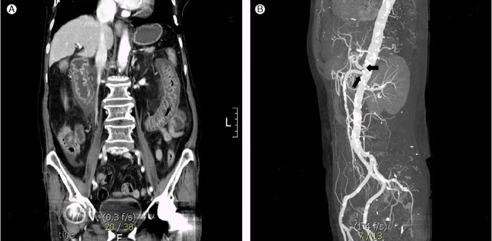
Contrast-enhanced abdominal CT and CT angiography. (A) Segmental concentric bowel-wall thickening from the duodenal second portion to the proximal jejunum. (B) Celiac axis and orifice of the superior mesenteric artery showing a narrowed portion (arrow), but the gastric artery, gastroepiploic artery, and pancreaticoduodenal arcade are relatively well traced. There was no evidence of definite occlusive vascular lesions.
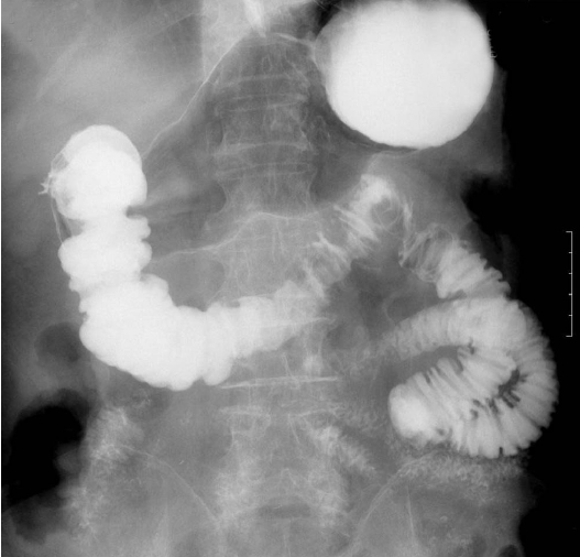
Small-bowel series finding. Irregular segmental fold thickening in the duodenum and proximal jejunum.

Microscopic findings of ischemic duodenitis. The duodenal biopsy showed focal fibrosis and infiltration with polymorphonuclear leukocytes and lymphocytes (H&E stain, × 200).
Esophagogastroduodenoscopic finding 7 days after the previous examination. Circumferential and segmental mild mucosal edema and erythema remain. However, the degree of mucosal injury is much improved compared with the initial endoscopy. There was no evidence of ulcer exudate or hemorrhage.
고 찰
위장관 또는 내장의 허혈은 장관 내 동맥에 여러 가지 원인으로 폐쇄가 발생하거나 저혈류 상태에서 생리적인 혈관수축에 의해 발생한다. 실제로 내장동맥의 협착 빈도는 높은 것으로 알려져 있으며, 평균나이 69세인 980명에 대한 후향적 연구에서 다양한 원인으로 인해 혈관 조영술을 시행하였을 때 8%에서 적어도 하나 이상의 내장동맥에 의미 있는 협착이 있었다[6]. 즉, 고령, 당뇨, 고혈압 등 내장동맥 협착의 위험인자를 가지고 있는 환자일수록 위장관 허혈의 위험은 증가한다.
위, 소장 및 대장에 공급되는 혈액은 크게 세 가지 주요 동맥으로 이루어진다. 첫째 가지는 복강동맥으로 위, 근위부 십이지장, 간 및 비장에 혈액을 공급한다. 둘째 가지는 상장간막동맥으로 원위부 십이지장, 소장 및 근위부 대장에 혈액을 공급한다. 셋째 가지는 하장간막동맥으로 원위부 대장 및 직장에 혈액을 공급한다[7]. 십이지장은 복강동맥의 위십이지장동맥(gastroduodenal artery) 분지와 상장간막동맥의 췌십이지장동맥(pancreaticoduodenal artery) 분지에 의한 이중 혈류공급을 받는 장기이기 때문에 허혈성 장염의 빈도는 매우 드물다. 하지만 세 가지 주요 내장동맥 중에 두 가지 이상이 폐쇄되거나 심한 협착을 보일 경우에는 위나 십이지장에 혈액공급이 감소되어서 정상 위 및 십이지장에 점막 방어가 파괴되고 염증 및 궤양을 유발할 수 있다[1]. 본 증례의 경우에도 복강동맥과 상장간막동맥에 의미 있는 협착이 존재하였던 경우에 허혈성 손상이 발생한 경우이다.
위장관 허혈의 특징적인 증상은 없으며 일반적으로 복통 및 혈성설사, 구토 등의 비특이적인 증상을 보인다. 국내의 한 연구에 의하면 허혈성 대장염의 경우 복통이 85%, 혈성설사가 65%, 복부팽만감이 30%, 오심 및 구토증세를 보이는 경우가 17%로 보고되었다[8]. 상부위장관의 경우에는 현재까지 자세한 임상양상에 대한 연구는 없는 상태이나 그동안 보고되었던 증례를 살펴보면 상복부 동통, 구토, 식후 발생되는 통증 등이 비교적 흔한 증상이며 그 외에도 체중감소, 토혈, 혈변 등의 소견을 보였다[2-5].
위장관에 발생하는 허혈성 손상은 급성과 만성으로 나눌 수 있다. 급성으로는 급성내장증후군(acute splanchnic syndrome)으로 내장순환의 단절로 인한 급작스런 복통을 특징으로 하며 색전 및 말기동맥혈전폐쇄(end stage arterial thrombotic occlusions), 비폐쇄장간막허혈 등에 의한 급성 위장관 손상이 이에 속한다. 만성으로는 만성내장질환(chronic splanchnic disease)과 만성내장증후군(chronic splanchnic syndrome)으로 나눌 수 있는데 만성내장질환은 내장 동맥에 의미 있는 협착은 있지만 풍부한 측부순환으로 증상은 없는 경우이고 만성내장증후군은 만성내장질환에서 허헐성 증상을 가지는 경우이다[7]. 허혈성 손상이 발생하는 기전은 일반적으로 허혈기와 재관류기로 구분될 수 있다. 허혈기에는 에너지를 함유하는 삼인산아데노신(ATP)이 고갈되면서 폐쇄막(tight junctions)의 분열을 유도하여 장관 내의 상피방어 기능을 감소시키며[9], 재관류기에는 혈액이 허혈 조직으로 유입되면서 산소는 활성산소종(reactive oxygen species)으로 전환되고 단백질과 DNA에 독성 작용을 하여 손상된 조직을 더욱 악화시킨다. 따라서 허혈성 단계에서의 손상보다는 재관류가 일어났을 때 더 심한 점막 손상을 받는다[10,11]. 최근에는 이러한 기전에 근거해서 항산화제를 사용하여 허혈/재관류 위장관 손상에 대한 치료 연구가 활발히 진행되고 있다[12].
위장관의 허혈성 손상에 대한 진단은 임상적 증상을 통해서는 어려우므로 다양한 진단적 방법들을 동원해야 할 경우가 많다. 선택적 장간막혈관조영술은 높은 진단율과 중재적 시술을 할 수 있기 때문에 증상이 있는 내장동맥 경색이 강하게 의심되는 환자에서 표준 진단법이다. 그러나 제한점이 있는데 심한 중환자에서 혈관조영술을 실시하기 어렵고 음성으로 결과가 나오는 경우가 많으며, 수술적 처치가 필요한 환자에게 치료 지연을 일으킬 수 있다[1]. 단순 내장동맥 협착이나 일시적 허혈이 의심되는 환자들은 최근에는 혈관조영자기공명영상이나 혈관조영단층촬영으로 내장동맥의 협착 여부를 진단하고 있다. 혈관조영단층촬영의 장점으로는 급성 증상을 가지는 환자에서 동맥 혹은 정맥의 폐쇄, 장관벽의 확장이나 조영증강 등을 진단할 수 있다. 또한 비침습적이고 비용면에서도 혈관조영술과 비교해서 경쟁력을 가질 수 있다[13]. 혈관조영자기공명영상은 내장 및 문맥순환계의 실제적인 혈액 유량을 측정할 수 있어서 식후에 혈액유량의 증강을 이용해서 혈관 협착이 있는 환자와 정상인 사이에서의미 있는 차이를 진단할 수 있다[14]. 특징적인 내시경적 소견에 대한 연구는 현재까지 없는 상태이지만 그동안의 증례를 참고해 보면, 십이지장의 제2부분에서 다수의 미란이 동반된 부종성, 출혈성 점막 소견이 있었고, 십이지장의 하행부위에 염증 및 종주형 궤양의 형태를 보인 경우도 있었다[3,4]. 조직학적으로는 고유판의 부종, 다형핵 백혈구의 침착 등의 급성 염증성 변화를 보이거나 단지 점막에 부종성 변화만 보이는 경우와 섬유화가 동반된 점막 또는 점막하의 허혈성 변화를 보이는 경우도 있었다[2-4].
내장동맥 허혈의 진단 및 치료에는 임상적인 증상에 기초하여 즉각적이고 순차적인 접근이 필요하다. 급성동맥폐쇄나 혈전증과 같은 급성내장증후군의 경우에는 초기에 내시경 및 혈관조영단층촬영, 혈관조영자기공명영상, 혈관조영술 등으로 빠르게 진단하여서 수술적 치료 혹은 풍선확장 및 스텐트 삽입술, 혈전용해제 치료 등을 고려해야 생존율을 향상시킬 수 있으며, 일시적인 저혈류에 의한 경우에는 수액요법 및 금식 등의 대증적 치료로 호전을 기대할 수 있다.