영구형 인공심박동기 삽입술 후 발생한 스트레스성 심장 근육병증 1예
Stress-Induced Cardiomyopathy as a Complication of Permanent Pacemaker Implantation
Article information
Abstract
스트레스성 심장 근육병증은 감정적 혹은 육체적 스트레스를 받는 상황에서 생길 수 있으며, 급성 관상동맥증후군과 임상양상이 유사하므로 감별진단이 중요하다. 영구형 인공심박동기 삽입술 후 합병증으로 스트레스성 심장 근육병증이 발생할 수 있으며 시술 후 심전도와 심초음파를 통한 경과관찰이 필요하다. 저자들은 영구형 인공심박동기 삽입술 후 발생한 스트레스성 심장 근육병증 증례 1예를 경험하였기에 문헌고찰과 함께 보고하는 바이다.
Trans Abstract
Stress-induced cardiomyopathy is a disease characterized by acute transient left ventricular dysfunction following exposure to stressful situations. We encountered an 80-year-old woman with complete atrioventricular block and normal LV systolic function. After permanent pacemaker implantation, electrocardiogram showed inverted T-waves in precordial leads. Follow-up echocardiographic findings indicated dyskinesia of the apical wall. Final diagnosis was stress-induced cardiomyopathy associated with a physically stressful condition (i.e., pacemaker implantation). (Korean J Med 2012;82:609-613)
서 론
스트레스성 심장 근육병증은 일시적인 급성 좌심실 기능의 저하를 특징으로 하는 질환으로 심전도의 이상 소견 및 혈액검사에서 심근효소의 증가를 보이며 관상동맥 조영술은 정상 소견을 보인다[1]. 스트레스성 심장 근육병증은 감정적 혹은 육체적 스트레스를 받는 상황에서 생길 수 있으며 좌심실의 기능저하가 일시적이므로 일반적으로 예후는 좋은 것으로 알려져 있다. 스트레스성 심장 근육병증은 급성 관상동맥증후군과 임상양상이 유사하므로 감별진단이 중요하다. 여러 원인이 스트레스성 심장 근육병증의 원인으로 보고되고 있으나 영구형 인공심박동기 삽입술 후 발생한 스트레스성 심장 근육병증의 증례는 드물다. 이에 저자들은 영구형 인공심박동기 삽입술 후 발생한 스트레스성 심장 근육병증 1예를 경험하였기에 문헌고찰과 함께 보고하는 바이다.
증 례
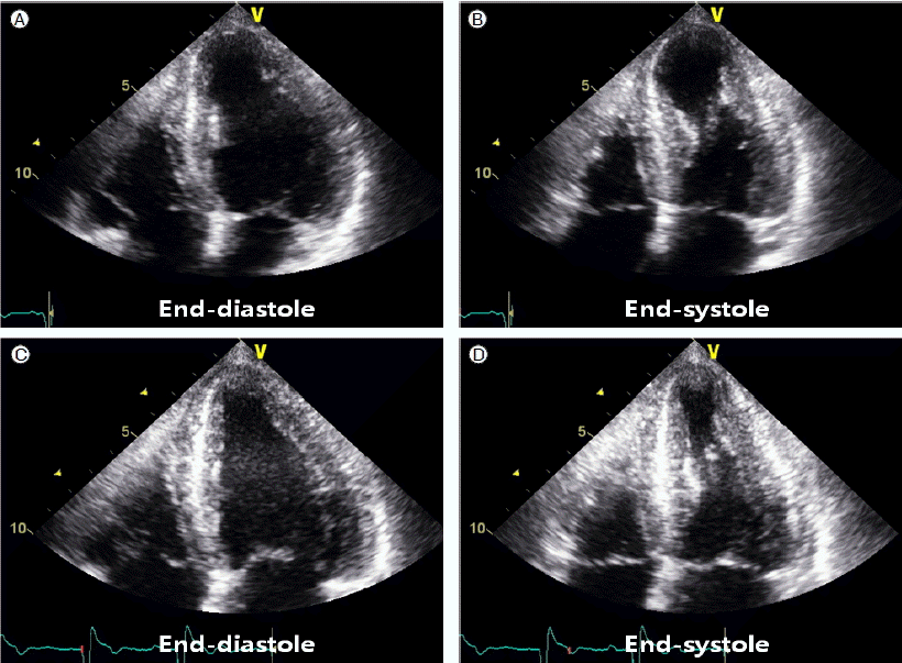
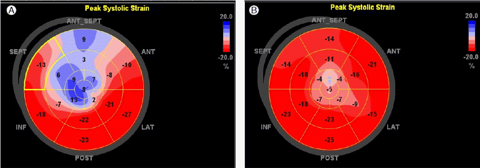
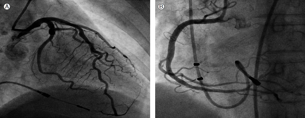
80세 여자 환자가 내원 당일 갑자기 발생한 호흡곤란을 주소로 내원하였다. 흉통, 어지러움, 실신 등의 동반 증상은 없었다. 환자는 과거력 및 가족력에서 특이사항은 없었고 복용하는 약물은 없었다. 생체활력징후는 혈압 130/60 mmHg, 심박수 40회/분, 호흡수 20회/분, 체온은 37.0℃였다. 신체 이학적 검사에서 심박동은 규칙적이었으나 서맥이 관찰되었고 흉부 청진에서 심잡음이나 수포음, 천명음은 관찰되지 않았다. 내원 초기 환자의 단순흉부방사선촬영에서 폐부종이나 심비대 소견은 없었고 초기 심전도에서 완전방실차단 소견이 관찰되었다. 혈액검사에서 이상 소견이 없었으며 경흉부심초음파에서 좌심실의 기능은 구혈률 60%로 정상범위였고 심실벽운동은 정상이었다. 내원 후 24시간 심전도 검사를 시행하여 지속적인 완전방실차단 소견을 확인할 수 있었고, 3일간 심전도 감시를 하였으나 정상 박동으로 회복되지 않아 내원 4일째 VDD (ventricular pacing, dual chamber sensing, dual chamber function) 유형의 영구형 인공심박동기 삽입술을 시행하였다. 인공심박동기 전극의 위치는 우심실 첨부에 위치되었고 심박동기는 잘 작동하였다. 내원 5일째 환자는 특이 증상을 호소하지 않았으나 심전도에서 이전에 보이지 않던 T파의 전위가 전흉부 V3에서 V6까지 관찰되었다(Fig. 1). 당시 시행한 환자의 혈액검사 결과 CK-MB 11.7 ng/mL (정상: 0.6-6.3 ng/mL), troponin I 1.36 ng/mL (정상: 0.0-0.04 ng/mL)로 심근효소의 증가소견이 관찰되었다. 경흉부심초음파의 맨눈 소견에서 심첨부의 dyskinesia가 관찰되었다(Fig. 2). Automated Function Imaging 방법으로 이면성 longitudinal strain 값을 측정했을 때 기저부 및 중간부 전중격(anterospetum), 중간부 하중격(inferoseptum)과 전체 심첨부의 dyskinesia를 확인할 수 있었다(Fig. 3). 내원 6일째 환자는 여전히 증상은 없었으며 심전도에서 T파의 전위 및 심근효소의 증가가 지속되었다. 내원 7일째 급성 심근경색을 배제하기 위하여 관상동맥 조영술을 시행하였고 결과는 정상이었다(Fig. 4). 내원 10일째 경흉부심초음파를 다시 시행하였다. 좌심실 심첨부와 심실 중격의 dyskinesia가 운동저하(hypokinesia) 수준으로 좌심실 벽운동 장애가 호전된 것을 확인할 수 있었다(Figs. 2 and 3). 최종적으로 영구형 인공심박동기 삽입술 후 발생한 스트레스성 심장 근육병증으로 진단하였고 환자는 호전된 상태로 퇴원하여 외래에서 경과관찰 중이다.

Electrocardiography performed immediately after dual-chamber pacemaker implantation (A) showed a well functioning pacing rhythm. Follow-up electrocardiography performed the day after pacemaker implantation (B) showed inverted T-waves in the precordial leads (V3-V6).

Echocardiography performed on admission showed dyskinetic motion at the apical segments of the left ventricle in the apical four-chamber view (A: end-diastole; B: end-systole). Follow-up echocardiography revealed improvement of the dyskinetic motion of the apical segments (C: end-diastole; D: end-systole).

(A) Global longitudinal strain assessed by 2-dimensional strain echocardiography showed dyskinetic motion of the basal to mid-anteroseptum, mid-inferoseptum, and whole apical segments on bull’s eye display. (B) The global strain image showed that the wall motion abnormality at apical and septal walls was improved from dyskinesia to hypokinesia on day 5 after presentation.
고 찰
스트레스성 심장 근육병증은 1990년대 일본에서 처음으로 takotsubo 심장 근육병증으로 명명되어 보고된 질환으로[2], transient left ventricular apical ballooning syndrome 혹은 broken heart syndrome으로 불리기도 한다. 최근까지 스트레스성 심장 근육병증의 여러 가지 원인과 기전이 보고되고 있다. 그중 수술과 침습적인 시술은 잘 알려진 원인들이다. 그러나 영구형 인공심박동기 삽입술 후 발생한 스트레스성 심장 근육병증의 증례는 드물며[3-5], 국내에서는 아직 보고된 바가 없다.
스트레스성 심장 근육병증은 허혈성 흉통, 호흡곤란 등의 증상과 ST 분절의 상승, T파의 전위 등의 심전도변화 및 심근효소의 증가를 특징으로 하는 질환으로 급성 관상동맥증후군과 유사한 양상을 보이므로 이에 대한 감별진단은 임상적으로 중요하다[2]. 관상동맥의 연축, 카테콜아민과 연관된 독성, 자율신경과 성호르몬의 불균형 등이 스트레스성 심장 근육병증의 병인으로 제시되고 있다[6-8]. 최근 Mayo Clinic에서 스트레스성 심장 근육병증에 대한 다음과 같은 수정된 진단기준을 제안하였다. 일시적인 좌심실벽운동 이상이 있고, 관상동맥 조영술에서 관상동맥 협착 혹은 급성 혈관 내 동맥경화반 파열의 증거가 없어야 하며, 새로운 심전도의 이상 혹은 troponin 상승 소견이 있어야 한다[9]. 스트레스성 심장 근육병증의 예후는 일반적으로 좋은 것으로 알려져 있으나 심부전, 심인성 쇼크, 급성 승모판 역류, 심실벽내 혈전 형성, 좌심실벽의 파열을 초래할 수 있다[5]. 사망률은 다양하게 보고되고 있으나 단일 3차 의료기관의 한 보고에 의하면 사망률 16%까지 높게 나타나고 있다[9]. 그러므로 스트레스성 심장 근육병증의 조기 진단과 집중 관찰 및 적절한 보존적인 치료가 반드시 필요하다.
본 증례의 환자는 호흡곤란이나 흉통 등의 증상을 보이지 않았으나 영구형 인공심박동기 삽입술 후 시행한 심전도에서 T파의 전위가 보이는 변화가 있었고 경흉부심초음파에서 좌심실의 심실벽운동 이상이 새롭게 관찰되어 스트레스성 심장 근육병증의 가능성을 생각할 수 있었다. 이후 급성 심근경색을 감별하기 위해 시행한 관상동맥 조영술 결과에서 이상소견이 없어 스트레스성 심장 근육병증을 확진할 수 있었다. 스트레스성 심장 근육병증의 유발인자로 증상을 동반한 발작성 상심실빈맥과 서맥 등과 같은 부정맥이 보고된 바가 있다[10]. 본 증례는 완전방실차단이 있었으나 영구형 인공심박동기 삽입술을 전후하여 심전도와 심근효소 수치 및 심초음파 소견이 변한 것이 확인되었으므로 이 환자는 영구형 인공심박동기 삽입술에 합병된 스트레스성 심장 근육병증이라고 할 수 있다. 본 증례의 환자는 시술 후 절대안정 상태였기 때문에 호흡곤란이나 흉통 등의 증상이 나타나지 않았던 것으로 생각한다. 이 환자의 스트레스성 심장 근육병증의 직접적인 원인으로 통증이나 출혈을 생각할 수 있고, 감정적인 stress도 가능한 원인으로 생각할 수 있다.
본 증례의 환자와 같이 증상이 없는 경우 스트레스성 심장 근육병증의 초기 진단은 어려울 수 있다. 또한 스트레스성 심장 근육병증에서 나타나는 심전도 변화 및 심초음파에서의 심실벽운동 이상소견은 영구형 인공심박동기 삽입과 연관되어 발생할 경우 우심실 심첨부의 pacing으로 발생하는 심실비동기화(dyssynchrony) 소견으로 혼동될 수 있다. 따라서 본 증례를 통하여 증상이 없다하더라도 영구형 인공심박동기 삽입술 후 심전도 및 심초음파를 이용한 집중적인 경과관찰이 필요하다는 것을 알 수 있었다.
