결석을 동반한 담낭염 환자에게서 발견된 이중관 총담관 기형 1예
Case Report of a Double Common Bile Duct with Gallstone Cholecystitis
Article information
Abstract
간담도계는 해부학적 기형과 변형이 비교적 흔한 편이나 총담관의 기형은 매우 드물다. 총담관의 기형은 수술 시 담관의 손상을 일으킬 수 있으며, 상부위장관의 종양을 비롯하여 담도결석 등 여러 가지 합병증을 일으킬 수 있어서 임상적으로 매우 중요하다. 저자들은 결석을 동반한 담낭염 환자에게서 이중관 총담관 기형 중 type Vb를 발견하여 이에 문헌고찰과 함께 보고한다.
Trans Abstract
Anatomical variation in the bile duct system is relatively common. Nevertheless, a double common bile duct is an extremely rare asymptomatic variant. Recognition of this anomaly is important clinically, because it can lead to complications, including choledocholithiasis, cholangitis, pancreatitis, and upper gastrointestinal malignancies. A correct diagnosis of this rare anomaly is also important because complications can occur in surgery if the anomaly is not recognized preoperatively. Recently, we encountered a very rare case of a double common bile duct associated with gallstone cholecystitis. A 33-year-old female was admitted to our hospital complaining of epigastric pain after meals. She had single biliary drainage from double common bile ducts with communicating channels. We report the case and review the literature on double common bile ducts. (Korean J Med 2011;80:208-211)
서 론
간담도계는 발생학적 특징으로 인하여 해부학적인 기형이 흔한 장기 중 하나이다[1]. 담도계 기형의 분류는 보고자에 따라 다르게 분류하고 있는데, 1958년 Hayes 등[2]은 400예의 담도계 수술 중 189예의 기형을 발견하였으며 총담관의 기형은 2예, 0.5%로 간관이나 담낭 그리고 담낭관의 기형에 비해 흔하지 않으며 이중관 총담관[3], 총담관낭종[4], 게실[5], 소판막 그리고 담췌관 합류이상[6,7] 등이 성인에서 보고되고 있다.
이중관 총담관의 기형은 1543~1986년까지 24예가 보고되었을 만큼 매우 드물며[3], 대부분 증상을 일으키지 않으나 상부위장관 종양을 비롯한 담도 결석, 담관염, 췌장염, 총담관낭종, 췌담도 연결부위의 기형 등을 일으킬 수 있다. 따라서 이러한 합병증으로 인하여 외과적 치료가 필요하게 될 시에 담도계 손상을 유발할 수 있으며, 이에 대한 이해가 담관계 기형의 예후에 큰 영향을 줄 수 있으므로 임상적으로 매우 중요하다[8]. 저자들은 담낭염 치료 도중 발견된 이중관 총담관 기형을 문헌고찰과 함께 보고한다.
증 례
33세 여자가 10일 전부터 시작된 식후에 발생하는 심와부 통증이 있어 내원하였다. 과거력에서 특이 병력은 없었으며 가족력, 사회력에서도 특이 사항은 없었다. 환자의 활력 징후는 혈압 110/70 mmHg, 맥박 72회/분, 호흡 18회/분, 체온 37℃였다. 신체검사에서 공막에 황달 소견을 보였고, 복부진찰 소견에서 심와부 압통이 있었으나 반발통은 없었으며, 머피씨 징후가 보였다. 복부에 종괴는 촉지되지 않았다.
입원 당시 검사 소견에서 혈색소 11.5 g/dL, 백혈구 5,500/mm3, 혈소판 304,000/mm3였으며, 생화학 검사에서 총 빌리루빈 3.9 mg/dL, 직접빌리루빈 1.8 mg/dL, γ-GT 223 IU/L, AST 84 IU/L, ALT 342 IU/L, 아밀라아제 37 U/L, 알칼라인 포스파타아제 599 IU/L이었다.
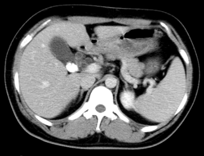
복부전산화 단층촬영에서 담낭에 결석이 보이며, 두 개의 분리된 간외담관이 보였으며, 후방외측에 있는 담관에 결석이 보였다. 또한 간내담관과 간외담관의 확장 소견이 있었다(그림 1).

Contrast-enhanced axial computed tomography (CT) shows two separate extrahepatic bile ducts, one of which was filled with stones
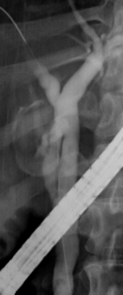
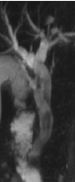
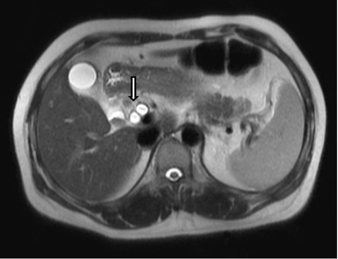
제 2병일째에 내시경적 역행성 담췌관조영술을 시행하였다. 내시경적 역행성 담췌관조영술에서 간내담관과 간외담관의 경도의 확장소견이 있었으며 간외담관에 다수의 비정형의 충만결손이 보였다. 또한 두 개의 총담관이 관찰되었으며 원위부와 근위부에서 만나 융합하여 서로 교통이 있었고, 십이지장에서 하나로 개구하였다. 내시경적 유두 풍선 확장술 시행 후 결석 제거용 풍선으로 결석을 제거하였고, 담낭에는 다수의 충만결손이 관찰되었다(그림 2). 시술 후 시행한 자기공명췌담도조영술에서 두 개의 간외담관이 덩굴처럼 서로 감겨 근위부와 원위부에서 만나는 것을 확인하였으며, 결석은 제거되었으나 담즙 찌꺼기가 남아있는 상태였다(그림 3, 그림 4). 제6병일째 담낭제거 수술을 시행하였으며 수술 중에 담관조영술을 시행하였다. 담관조영술에서 내시경적 역행성 담췌관조영술과 자기공명췌담도조영술에서 보였던 것과 같이 두 개의 분리된 간외담도가 췌장두부에서 만나 총담관을 이루며 근위부에 교통가지가 있었다. 담낭절제술 후 제9병일째 합병증 없이 퇴원하였으며 담낭의 병리소견에서 만성담낭염, 콜레스테롤 폴립, 콜레스테롤증이 있었다.

Endoscopic retrograde cholangiopancreatography (ERCP) with contrast injection shows the proximal and distal unions of the double common bile duct and another stone-filled extrahepatic bile duct.
고 찰
간담도계는 해부학적 기형과 변형이 비교적 흔한 장기로 간외담관의 변이는 여러 형태로 나타날 수 있고 대개 발생학적 기형이나 회전이상, 태아 간게실(hepatic diverticulum)의 비적절한 분리와 융합에 의하여 발생한다[1]. 1972년 Goor와 Ebert [9]는 발생학적 특징과 해부학적 모양에 따라 담도계 기형을 1) 이소성 담낭(ectopic gallbladder) 2) 담낭관 기형(anomalies of the cystic duct) 3) 중복 담낭(duplication of gallbladder) 4) 총담관기형(anomalies of the common hepatic duct) 5) 부담관(accessory bile duct) 6) 교통성 부담관(communicating accessory bile duct) 7) 총담관낭종(cysts of the biliary tree) 8) 십이지장으로의 중복담도 배출(double drainage to the duodenum) 9) 부간(accessory liver) 등 아홉 가지로 분류하였다.
간담도계 기형 중 이중관 총담관 기형은 매우 드물다. 1543년 Vesalius 등[10]에 의해 처음 보고되었으며 Teilum [3]에 따르면 1986년까지 24예가 보고되었다. 우리나라에서는 1989년 Kae 등[11]에 의해 발견된 type II 이중관 총담관이 보고되었고, 2005년 이중관 총담관에 발생한 바터씨 팽대부암이 보고된 바 있다[12]. 이러한 담관계 기형은 대부분 무증상 상태로 잘 발견되지 않으나 담관계 외과적 치료 시에 담관계 기형에 대한 해부학적 이해부족으로 인한 담관계 손상을 유발시켜 합병증을 유발할 수 있으므로 이에 대한 해부학적인 인지가 중요하다고 하겠다.
Saito 등[13]의 분류에 기초하여 Choi 등[14]은 이중관 총담관의 부총담관의 개구위치와 관계없이 형태학적인 분류를 다음과 같이 하였다. Type I, 총담관에 격막이 존재; type II, 총담관이 원위부에서 분지를 내면서 각각의 개구가 존재; type III, 총담관이 두 개이나 간외 교통하지 않음; type IV, 각각의 개구를 가진 두 개의 총담관의 간외 연결통로가 한 개 이상 존재; type V, 두 개의 총담관이 만나 개구는 하나이며 근위부에 연결통로가 없으면 Va, 있으면 Vb라 한다(그림 5).
또한 이중관 총담관의 개구 위치에 따라 십이지장으로 정상 개구하는 경우와 십이지장 이외의 장기로 개구하는 경우로 분류할 수 있다. Yamashita 등[8]은 이중관 총담관의 형태학적인 분류보다는 개구 위치에 따른 임상적 중요성에 주목하였는데 이중관 총담관의 개구 위치로 위의 소만부(16예), 십이지장의 제1부위(8예), 십이지장의 제2부위(9예), 췌관(7예), 격막(4예)을 보고하였다. 또한 이중관 총담관의 개구 위치에 따라 발생할 수 있는 심각한 합병증으로 담도계를 포함하는 상부 소화기계의 종양 발생, 담도 결석, 총담관 낭종, 췌담도 연결 부위의 기형 등이 있다고 보고하였다. 이처럼 이중관 총담관의 개구위치는 담관계 기형의 예후에 영향을 줄 수 있으므로 임상적으로 매우 중요하다.
이번 증례는 근위부에서 교통하는 두 개의 총담관이 만나 십이지장으로 하나의 개구를 하는 type Vb이며, 결석을 동반한 담낭염을 주소로 내원하였다가 발견하였다. 복부전산화 단층촬영상에서 이중관 총담관임을 발견하였으며, 내시경적 역행성 담췌관조영술과 자기공명췌담도조영술, 수술 중 담관조영술상에서도 확인할 수 있었다. type Vb는 매우 드물어 2008년 이전에는 보고된 바 없었다[15].


