위창자간막동맥의 단독 벽내 혈종
Isolated intramural hematoma of the superior mesenteric artery
Article information
A 46-year-old man was admitted to our hospital because of abrupt-onset epigastric and back pain, which was aggravated following meals. He avidly exercised strenuously, including running marathons and weight lifting. The physical examination was unremarkable except for mild direct tenderness in the epigastric area. Ultrasonography of the upper and lower abdomen was normal esophagogastroduodenoscopy showed minimal reflux esophagitis, and his pain was not relieved by a number of medications. Computed tomography angiography (CTA) revealed an intramural hematoma without dissection obstructing the true lumen of the proximal to middle portion of the superior mesenteric artery (SMA) (Fig. 1, arrows). Low-molecular-weight heparin was administered, followed by warfarin, and the patient improved after 1 week of treatment. Follow-up CTA at 4 months indicated complete resorption of the intramural hematoma and restoration of the true lumen of the SMA (Fig. 2, arrows). One year later, the patient remains asymptomatic without further treatment. There are rare reports of isolated dissection of the SMA, especially in Asian populations. The causes are unknown and the treatment options vary, and include surgery, endovascular treatment, or conservative management dependent on the extent of dissection and the associated complications1-3).
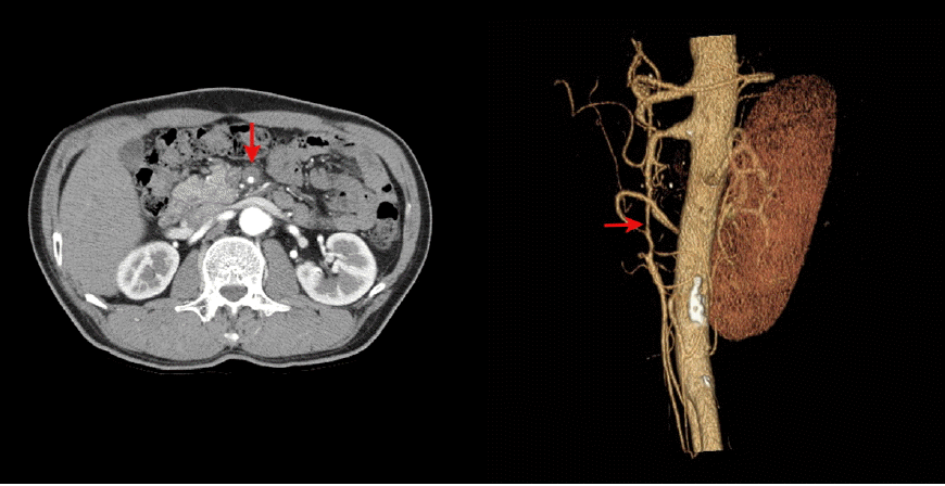
Computed tomography angiography shows an intramural hematoma without dissection obstructing the true lumen of the proximal to middle portion of the superior mesenteric artery (arrows).
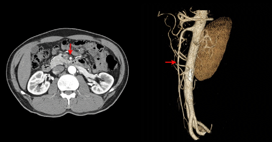
Computed tomography angiography after 4 months shows complete resorption of the intramural hematoma and restoration of the true lumen of the superior mesenteric artery (arrows).