췌장미부 종양으로 오인된 부비장 결핵 1예
A case of accessory splenic tuberculosis mimicking a distal pancreatic tumor
Article information
Abstract
저자들은 33세 남자 환자에서 비장 경색을 동반한 췌장미부 종양으로 오인된 부비장 및 비장 결핵을 경험하였기에 문헌고찰과 함께 보고하고자 한다.
Trans Abstract
Abdominal tuberculosis usually affects the gastrointestinal tract, peritoneum, lymph nodes, liver or spleen. Tuberculosis of the spleen is uncommon, except when associated with miliary dissemination. We report a case of a 33-year-old man with tuberculosis of the accessory spleen, which was originally suspected to be a distal pancreatic tumor. He was admitted with a history of left upper quadrant abdominal pain for 3 months. Computed tomography imaging of the abdomen revealed a 4.5 cm sized poorly defined hypodense mass in the distal pancreas and showed multiple focal hypodense lesions in the enlarged spleen. We performed distal pancreatectomy and splenectomy under the preoperative expectation of a distal pancreatic tumor. Microscopic examination of the specimens revealed accessory splenic tuberculosis associated with splenic tuberculosis. Following this, he was treated with appropriate antituberculosis drugs and was discharged without any complications. (Korean J Med 79:53-56, 2010)
서 론
결핵은 전신적 질환이며, 인체의 모든 장기에 침범할 수 있다. 이 중 복부 결핵은 주로 림프절, 소화관, 장간막에서 발생할 수 있으며 다른 장기를 침범하는 경우는 드물다. 특히, 비장을 침범하는 경우는 매우 드문 것으로 알려져 있다1,2).
저자들은 비장 경색을 동반한 췌장미부 종양으로 오인하여 수술을 통해 진단한 부비장 및 비장 결핵 1예를 경험하였기에 문헌고찰과 더불어 보고하는 바이다.
증 례
33세 남성이 3개월 전부터 시작된 좌측 복부 동통과 피로를 주소로 내원하였다. 나이지리아 국적의 외국인 노동자로 1주 전부터 상기 증상이 심해졌다고 하였다.
5년 전 말라리아로 치료받고 완치된 것 외 특이병력은 없었고, 입원 당시 혈압 130/80 mmHg, 맥박 80회/분, 체온은 36.5℃였다. 이학적 검사상 만성 병색을 보였고, 좌측상부복부에 압통이 있었으며 종괴가 촉지되었다. 심폐 청진시에는 특이소견 보이지 않았다. 입원 당시 말초혈액검사에서 백혈구 3,910/L, 혈색소 13.2 g/dL, 헤마토크리트 39.6%, 혈소판 178,000/L, MCV 72.8 fL, MCH 24.2 pg, MCHC 33.2 g/dL이었고, 혈청생화학검사에서 총 단백질 7.1 g/dL, 알부민 3.4 g/dL, 총 빌리루빈 0.89 mg/dL, AST 31 U/L, ALT 38 U/L, ALP 124 IU/L, r-GTP 22 IU/L, FBS 105 mg/dL, LDH 512 IU/mL, Na 142 mmol/L, K 4.8 mmol/L, Cl 98 mmol/L, BUN 11.3 mg/dL, Creatinine 1.1 mg/dL, C-reactive protein 4.21 mg/dL이었다. 일반면역혈청검사에서 HBs Ag, HBs Ab, anti-HCV Ab, anti-HIV Ab 모두 음성이었으며 CEA 1.64 ng/mL, CA 19-9 47.14 U/mL이었다.
단순 흉부 X-선에서 특이소견 보이지 않았으며 복부 초음파촬영술에서 비장 크기가 20 cm 이상으로 커져 있고 다발성의 저에코 부위가 관찰되었다. 복부 전산화단층촬영상 췌장미부에 4.5×4.5 cm 가량의 췌장에서 발생한 것으로 보이는 경계가 불명확하고 불균질한 저밀도를 보이는 종괴와 다발성 저음영 병변을 동반한 20 cm 가량의 비장종대가 관찰되었다(그림 1). 비장 경색을 동반한 췌장 미부 종양으로 생각되어 수술을 시행하였다. 수술시 췌장 미부에 4×4 cm 크기의 부드러운 종괴를 볼 수 있었고, 종대된 비장에서 황백색의 다발성 결절들을 관찰할 수 있었다. 또한, 후복막부에 다발성의 림프절 종대가 있어 췌장 미부에 발생한 신생물 의심하에 원위부 췌장 절제술, 비장 절제술, 림프절 곽청술을 시행하였다.
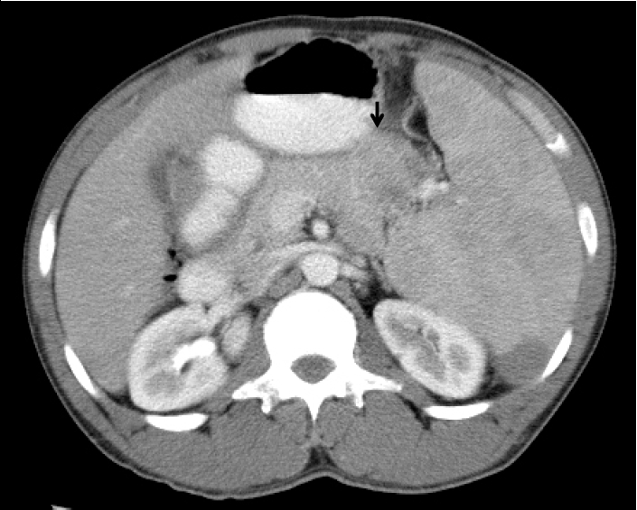
Abdomen CT shows a 4.5 cm sized poorly defined hypodense mass (arrow) in the distal pancreas and multiple focal hypodense lesions in the enlarged spleen.
절제된 검체의 육안적 소견상 췌장미부 종괴는 4.5×4.5cm 크기의 어두운 분홍색조를 보이며 절개시 췌장조직에 유착된 것으로 보이는 종괴에 황색조의 경계가 뚜렷한 다발성 결절들이 관찰되었다(그림. 2A). 비장은 19.0×14.0×9.0 cm 크기로 종대되어 있고, 황색조의 경계가 뚜렷한 다양한 크기의 다발성 결절들이 관찰되었다(그림. 2B). 병리조직학적 검사 상 췌장 미부 종괴로 의심된 부위는 정상 췌장 조직에 유착된 부비장이었으며, 부비장 조직 내에서 건락성 괴사를 보이는 만성 육아종성 병변이 관찰 되었다(그림. 2C). 비장 내 결절에서도 동일한 만성 육아종성 병변이 관찰되었다(그림. 2D). 또한, 종대된 후복막 림프절에서도 만성 육아종성 병변을 보여 부비장과 비장 및 후복막 림프절에 발생한 결핵으로 진단하였다. 수술 1주 후 시행한 추적 복부 전산화단층촬영상 수술로 인한 복강기종 외 특이소견 보이지 않았다. 폐렴구균에 대한 예방 접종과 항결핵제 투여하면서 외래에서 경과관찰 중이다.
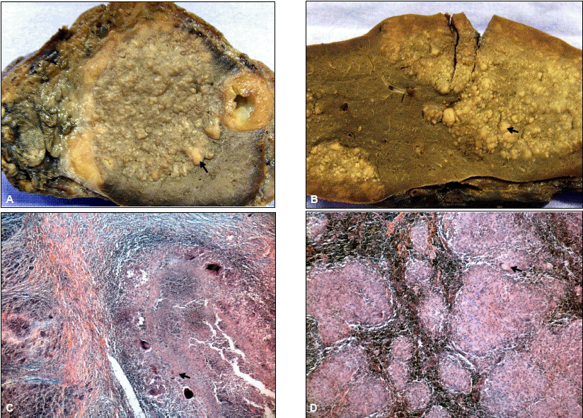
(A) Gross findings of the resected distal pancreatic mass. The mass measured 4.5×4.5 cm. The cut surface appeared multiple yellowish-white nodules (arrow). (B) Gross findings of the resected spleen show multiple nodules coalescing to form large yellowish white mass of firm consistency (arrow). (C) Microscopic findings of the presumed distal pancreatic mass which in fact was the accessory spleen adherent to the pancreatic tail and showing chronic granulomatous inflammation with Langhans' giant cells (arrow) and caseous necrosis, consistent with accessory splenic tuberculosis (H&E stain, ×40). (D) Microscopic examination shows chronic granulomatous inflammation with Langhans' giant cells (arrow) and caseous necrosis, consistent with splenic tuberculosis (H&E stain, x40)
고 찰
결핵은 공중 보건의 개선과 진단, 치료의 발달로 인해 선진국에서는 유병율이 감소하였으나 후진국 및 개발도상국에서는 여전히 상당한 유병율을 보인다. 2009년 보고된 WHO의 통계에 따르면 2007년 세계적으로 인구 10만명당 139명의 새로운 결핵 환자들이 발생하고 있으며3), 질병관리본부가 발표한 2007년도 결핵환자 신고현황 연보에 따르면 국내에서도 2007년도에 인구 10만명당 71.6명의 새로운 환자가 발생하였다4). 폐외 결핵은 전체 결핵환자의 11~12% 가량에서 보고되고 있으나 이 중 비장결핵은 매우 드물다1,2).
비장 결핵은 일반적으로 비특이적인 임상증상을 보이며 주로 발열, 오한, 체중 감소, 만성피로, 좌상복부 동통 및 압통 등을 보일 수 있다. 비장 종대를 동반하기도 하며 불명열의 양상을 보이기도 한다5). 진단은 초음파촬영술에서 저에코 음영이나 조영 증강된 복부 전산화단층촬영에서 저밀도 음영으로 관찰되는 것이 도움이 될 수 있으나 진단적 가치를 보이는 특이적 소견은 아니며6,7) 림프종, 전이암, 비장낭종, 농양, 비장경색, 혈관종 또는 과오종 등과 감별이 필요하다8). 확진을 위해서는 미세침흡인생검술, 비장 생검 또는 비장 절제술 후 검체 등에 대한 조직학적 검사를 요한다9,10). 치료는 대부분의 환자들이 6~9개월 간의 항결핵요법에 잘 반응하며 비장 절제술이 필요한 경우는 드물다11). 그러나 항결핵요법에 잘 반응하지 않거나 약제 투여가 불가할 시에는 비장절제술을 시행하기도 한다.
부비장 결핵은 일반적으로 10~30% 빈도를 보이는 비교적 흔한 소견이다12,13). 복강내 어디서든 발견할 수 있으며, 주로 비장문에서 발견된다14,16). 대부분 2 cm 미만의 크기를 보이며 초음파에서 고진폭 경계를 갖는 구형 혹은 난원형의 에코발생 종괴로 관찰되고, 색채 초음파에서 혈관문을 관찰하면 90%의 민감도를 보인다15). 복부 전산화단층촬영에서는 뚜렷한 경계를 가지고 주위 췌장조직보다는 높으며 비장과 같은 밀도를 보이는 균질한 조영 소견을 관찰 할 수 있다16).
문헌에서는 비장 결핵을 초음파, 복부 전산화단층촬영 소견을 바탕으로 미세침흡인생검 또는 비장 생검만으로 진단 가능한 것으로 보고하고 있으나9,10,17), 본 례에서는 복부 전산화단층촬영상 부비장이 크기가 4.5 cm 가량으로 증가되어 있고, 췌장과의 경계가 불분명하며 불균일한 저밀도의 조영 증강 보임으로써 췌장 미부에 발생한 종양으로 오인하여 절제 후 검체의 조직 검사를 통해 진단하였다.
만성 피로, 좌측 복부 동통, 비장 종대를 통해 비장결핵을 의심하여 조직 생검을 고려할 수 있었으나 비장 경색을 동반한 췌장 미부 종양시 유사한 증상 보일 수 있고, 비장 종대는 췌장 미부에 동반된 부비장 결핵을 종양으로 오인함으로써 간과하였다.
최근 HIV 감염, 장기이식으로 인한 면역억제제 사용, 항암요법 등의 빈도가 증가하면서 선진국에서도 결핵이 증가하는 양상을 보이며 비장 결핵 역시 빈도가 늘어날 것으로 예상된다. 비장 결핵은 특이증상이 없는 경우가 많고 방사선 소견만으로 진단하기 어렵기 때문에 적절한 치료 시기를 놓치기 쉽다. 특히, 부비장 결핵이 동반된 경우 더욱 진단이 어려울 수 있다. 이에 저자들은 비장경색과 췌장미부에 발생한 종양으로 오인된 비장 및 부비장의 결핵을 보고함으로써 유사한 환자에서 감별 진단에 도움을 주고자 한다.