위 정맥류를 동반한 간문맥의 해면상 변형

Cavernous Transformation of the Portal Vein with Gastric Varix

Article information

Korean J Med. 2010;79(1):11-12
Publication date (electronic) : 2010 July 1
Department of Internal Medicine, Seoul National University Hospital Gangnam Healthcare Center, Seoul, Korea
양종인, 강정묵, 김동희, 박민정, 조상헌
서울대학교병원 강남센터 내과

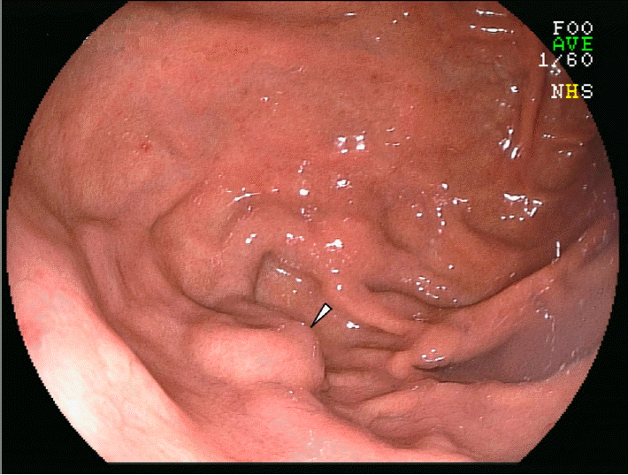
66세 남자가 건강 검진을 위하여 내원하였다. 환자는 고혈압과 당뇨병을 꾸준히 관리해 오고 있는 것 외에는 다른 병력은 없었고, 토혈, 흑혈변, 복부 팽만감 등의 호소도 없었으며 다른 불편감의 호소도 없었다. 일반혈액검사는 백혈구 4,500/μL, 혈소판 169,000/μL, 혈색소 14.6 g/dL로 정상범위였고, 프로트롬빈시간도 103% (0.98 INR)로 정상범위였다. 간기능검사치는 ALT 48 IU/l(정상치: <40), gamma glutamyl transferase 69 IU/l (정상치: 11~63) 외에는 콜레스테롤 131 mg/dL, 알부민 4.3 g/dL, 빌리루빈 1.3 mg/dL, 알칼리성 포스파타제 38 IU/l 및 AST 36 IU/l로 정상범위였다. 혈청검사에서 HBsAg과 anti-HCV는 음성이었다. 종양표지자 검사에서 aFP 1.2 ng/mL와 CA 19-9 13.3 U/mL로 모두 정상범위였다. 복부 평가를 위하여 시행한 초음파 검사에서 간문(porta hepatis)과 췌두부에 구불구불하게 확장된 정맥들과 비장비대가 관찰되었으나, 간은 비교적 정상 음영을 보였다(Fig. 1). 복부 CT에서도 현저하게 구불구불한 문맥과 비장정맥 및 문맥 주위로 잘 발달된 측부혈관들이 뚜렷하게 관찰되었다(Fig. 2, Fig. 3). 위내시경에서는 궁륭부에 국한된 정맥류가 관찰되었다(그림 4).
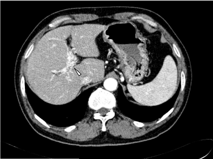
Figure 1.

Abdominal ultrasonography shows the cavernous transformation of the portal vein (arrowhead).

Figure 2.

Abdominal computed tomography shows the cavernous transformation of the portal vein with many collateral vessels in the liver (arrowhead).
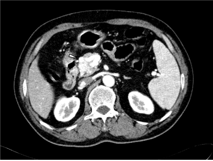
Figure 3.

Abdominal computed tomographyshows the cavernous transformation of the portal vein with many collateral vessels in the pancreatic head (arrowhead).

Figure 4.

Gastroscopy shows a dilated submucosal vein in the fundus (arrowhead isolated gastric varix 1).

간문맥의 해면상 변형은 다양한 원인들에 의하여 간문맥이 폐쇄되고 이 주변으로 많은 측부 혈관들이 발달하여 발생하는 질환으로 그 빈도가 매우 드물다1,2). 소아 및 청소년에서는 주로 선천적 기형에 의하여 발생되며 수술적으로 문맥압을 교정하는 치료를 고려할 수 있다3). 성인에서는 빈도가 더욱 드문 것으로 알려져 있으며 유발 원인질환도 다양하므로 원인질환이 있는지에 대한 평가도 필요하다. 간문맥의 해면상 변형의 주 증상은 위장관 출혈으로 알려져 있다. 실제 본 증례도 위의 궁륭부에 정맥류가 있었던 점을 고려하면, 그 빈도가 드물기는 해도 간문맥의 해면상 변형이 의심되는 환자에서는 내시경 시술에서 점막하 종양이 의심될 경우에도 정맥류의 가능성을 고려하여 조직검사 등에 유의하여야 하겠고, 위장관 출혈을 유발할 수 있는 약물을 사용할 때에도 주의가 필요하다고 판단된다4).

References

1. Zhang LJ, Yang GF, Jiang B, Wen LQ, Shen W, Qi J. Cavernous transformation of portal vein: 16-slice CT portography and correlation with surgical procedure of orthotopic liver transplantation. Abdom Imaging 33:529–535. 2008;
2. 송 상훈, 서 기현, 김 재한, 오 명진, 황 헌규, 김 완섭, 김 영민, 문 종호, 조 영덕, 이 문성, 허 경렬, 문 철, 구 동억, 이 혜경, 심 찬섭. 간문맥의 해면상 변형에 의해 발생된 폐쇄성 황달 1예. 대한소화기내시경학회지 19:999–1004. 1999;
3. Zhang H, Zhang N, Li M, Jin W, Pan S. Surgical treatment of portal vein cavernous transformation. World J Surg 28:708–711. 2004;
4. 장 재영. 식도 및 위정맥류의 예방과 치료. 대한내과학회지 75:6–14. 2008;

Article information Continued

Figure 1.

Abdominal ultrasonography shows the cavernous transformation of the portal vein (arrowhead).

Figure 2.

Abdominal computed tomography shows the cavernous transformation of the portal vein with many collateral vessels in the liver (arrowhead).

Figure 3.

Abdominal computed tomographyshows the cavernous transformation of the portal vein with many collateral vessels in the pancreatic head (arrowhead).

Figure 4.

Gastroscopy shows a dilated submucosal vein in the fundus (arrowhead isolated gastric varix 1).