


ьДЬ ыба
ъ░ДьЭШ эФ╝ызЙэХШ эШИьвЕ(subcapsular hematoma)ьЭ┤ыЮА Glisson capsule ы░СьЧР эШИьХбьЭ┤ ъ│аьЭ┤ыКФ ъ▓ГьЭД ызРэХЬыЛд[1]. ъ░ДьЭШ эФ╝ызЙ ы░СьЧР эШИьХбьЭ┤ ъ│аьЭ┤ыКФ ъ▓╜ьЪ░ыКФ ъ░ДэЧРьаБьЬ╝ыбЬ ы│┤ъ│аыРШыКФыН░, ъ░ДэЧРьаБьЬ╝ыбЬ ы│┤ъ│аыРШыКФ ьжЭыбАыУдьЭШ ъ▓╜ьЪ░ ыМАы╢Аы╢Д ьЩ╕ьГБ, ьЭШэХЩьаБ ь▓Шь╣Ш, ъ░ДьвЕъ┤┤ьЭШ эММэШИ ыШРыКФ эШИьХбьЭСъ│а ьЮеьХа ыУ▒ ь╢ЬэШИьЭШ ьЫРьЭ╕ьЭ┤ ыРа ьЭ┤ьЬаыУдьЭ┤ эЩХьЭ╕ыРШый░, ыЪЬыа╖эХЬ ьЫРьЭ╕ьЭ┤ ы░Ьъ▓мыРШьзА ьХКыКФ ъ▓╜ьЪ░ыКФ ыНФьЪ▒ ыУЬым╝ыЛд[1]. ыШРэХЬ эФ╝ызЙэХШ ь╢ЬэШИьЭА ыМАы╢Аы╢Д ьЭ╝эЪМьД▒ьЭШ ьЮДьГБ ъ▓╜ъ│╝ые╝ ы│┤ьЭ┤ыКФ ъ▓ГьЬ╝ыбЬ ы│┤ъ│аыРШъ│а ьЮИьЬ╝ый░, ы░Шы│╡ьаБьЬ╝ыбЬ ьЮмы░ЬэХШыКФ ьЮДьГБ ъ▓╜ъ│╝ые╝ ы│┤ьЭ┤ыКФ ъ▓╜ьЪ░ыКФ ыНФьЪ▒ ыУЬым╝ыЛд. ьаАьЮРыУдьЭА эК╣ы│ДэХЬ ьЫРьЭ╕ьЭД эЩХьЭ╕эХа ьИШ ьЧЖьЧИьЬ╝ый░, ы░Шы│╡ьаБьЬ╝ыбЬ ъ░ДьЭШ эФ╝ызЙэХШ эШИьвЕьЭД ы│┤ьШАыНШ ьжЭыбАые╝ ъ▓╜эЧШэХШьЧм ым╕эЧМъ│аь░░ъ│╝ эХиъ╗Ш ы│┤ъ│аэХШъ│аьЮР эХЬыЛд.
ьжЭ ыбА
эЩШ ьЮР: 52ьД╕ ьЧмьЮР
ьг╝ ьЖМ: ы░Шы│╡ыРШыКФ ьЪ░ьГБы│╡ы╢А эЖ╡ьжЭ
эШДы│Сыае: 4ъ░ЬьЫФ ьаД ьЪ░ьГБы│╡ы╢А эЖ╡ьжЭьЬ╝ыбЬ ьЩ╕ы╢Аы│СьЫР ьЭСъ╕ЙьЛдьЭД ы░йым╕эХШьШАьЬ╝ый░, ьШБьГБ ъ▓АьВмьГБ S8 ъ╡мьЧньЧР ьХ╜ 13 cm эБмъ╕░ьЭШ эФ╝ызЙэХШ эШИьвЕьЭ┤ эЩХьЭ╕ыРШьЧИыЛд. ыЛ╣ьЛЬ эК╣ы│ДэХЬ ьЫРьЭ╕ьЭ┤ ы░Ьъ▓мыРШьзА ьХКьХШьЬ╝ый░, ьГЭь▓┤ ьзХэЫД ьХИьаХьаБьЬ╝ыбЬ ы│┤ьб┤ьаБ ь╣ШыгМые╝ ьЛЬэЦЙ эЫД эЪМы│╡эХШьЧм эЗ┤ьЫРэХШьШАыЛд. ьЭ┤эЫД ъ▓╜ъ│╝ ъ┤Аь░░ ьдС 2ъ░ЬьЫФ ьаД ьЪ░ьГБы│╡ы╢А эЖ╡ьжЭьЭ┤ ьЮмы░ЬэХШьЧм ьЩ╕ы╢Аы│СьЫР ьЭСъ╕ЙьЛдьЧРьДЬ ыЛдьЛЬ эЩХьЭ╕эХЬ ъ▓░ъ│╝, ъ│╝ъ▒░ эФ╝ызЙэХШ эШИьвЕьЭА эБмъ╕░ъ░А ъ░РьЖМэХШьШАьЬ╝ыВШ, S6 ъ╡мьЧньЧРьДЬ ьГИыбЬьЪ┤ эФ╝ызЙэХШ эШИьвЕьЭ┤ эЩХьЭ╕ыРШьЧИыЛд. ь╢ЬэШИ ьзАьЖН ьЧмы╢А эЩХьЭ╕ьЭД ьЬДэХ┤ эШИъ┤Аьб░ьШБьИаьЭД ьЛЬэЦЙэХШьШАьЬ╝ыВШ эЩЬыПЩьД▒ьЭШ ь╢ЬэШИ ы╢АьЬДыКФ ьЧЖьЧИьЬ╝ый░ ь╢Фъ░А ьГЭь▓┤ьзХэЫД ыУ▒ьЭА ьХИьаХьаБьЭ┤ьЦ┤ьДЬ ы│┤ьб┤ьаБ ь╣ШыгМые╝ ьЛЬэЦЙэХШьШАыЛд. ьЭ┤эЫД ы│╡эЖ╡ьЭА эШ╕ьаДыРШьЧИъ│а, ьХИьаХьаБ ьГБэГЬыбЬ эЗ┤ьЫРэХШьШАьЬ╝ыВШ, ьЭ╝эЪМьД▒ьЭ┤ ьХДыЛМ ы░Шы│╡ьД▒ьЭШ ъ▓╜ъ│╝ые╝ ы│┤ьЧм, ьЫРьЭ╕ ы░П ь╣ШыгМьЧР ыМАэХ┤ ьЮмьГБыЛ┤ьЭД эЭмызЭэХШьЧм ы│╕ьЫРьЧР ыВ┤ьЫРэХШьШАыЛд.
ъ│╝ъ▒░ыае: ъ╕░ьаА ъ│аэШИьХХьЭ┤ ьЮИьЦ┤ amlodipine 5 mg 1ьЭ╝ 1эЪМ ы│╡ьЪйьдСьЧР ьЮИьЬ╝ый░, aspirin ыШРыКФ warfarinьЭД эПмэХиэХЬ эХнэШИьЖМэМРьаЬ ы░П эХньЭСъ│аьаЬьЭШ ы│╡ьЪй ьЭ┤ыаеьЭА ьЧЖьЧИыЛд. 4ыЕД ьаД ьвМь╕б ьЬаы░йьХФ ьзДыЛи эЫД ьЬаы░йьаИьаЬьИаьЭД ьЛЬэЦЙы░ЫьХШъ│а, ьЭ┤эЫД ьЧРьКдэК╕ыбЬъ▓Р ьИШьЪйь▓┤ ьЦСьД▒ ьЖМъ▓мьЬ╝ыбЬ эГАыкйьЛЬэОЬ эИмьХ╜ьЭД ьЛЬьЮСэХШьШАьЬ╝ый░ ыВ┤ьЫР ьЛЬъ╣МьзА 20 mg 1эЪМ/ьЭ╝ьЭД эИмьХ╜эХШъ│а ьЮИьЧИыЛд.
ьВмэЪМыае ы░П ъ░Аьб▒ыае: эК╣ьЭ┤ ы│СыаеьЭА ьЧЖьЧИыЛд.
ьЛаь▓┤ ьзДь░░ ьЖМъ▓м: эШИьХХ 162/84 mmHg, ызеы░Х 90эЪМ/ы╢Д, эШ╕эЭб ьИШ 20эЪМ/ы╢Д, ь▓┤ьШи 36.7тДГьШАьЬ╝ый░ ьЪ░ьГБы│╡ы╢А ь┤ЙьзДьЛЬ ъ▓╜ып╕эХЬ ьХХэЖ╡ьЭ┤ ьЮИьЧИьЬ╝ыВШ ы░Шы░Ь ьХХэЖ╡ьЭА ьЧЖьЧИыЛд. ьЮеъ╕░ ы╣ДыМА ьЖМъ▓мьЭА ьЧЖьЧИъ│а, ьвЕъ┤┤ыКФ ь┤ЙьзДыРШьзА ьХКьХШыЛд.
ъ▓АьВмьЛд ьЖМъ▓м: ызРь┤ИэШИьХб ъ▓АьВмьЧРьДЬ ы░▒эШИъ╡м 5,070/mm3 (эШ╕ьдСъ╡м 51%, эШ╕ьВ░ъ╡м 2.8%, эШ╕ьЧ╝ъ╡м 0.8%, ыж╝эФДъ╡м 36.1%), эШИьГЙьЖМ 13.5 g/dL ы░П эШИьЖМэМР 162,000/mm3ыбЬ ьаХьГБ ы▓ФьЬДьШАыЛд. ъ░ДьЭШ эФ╝ызЙэХШ эШИьвЕ ы░ЬьГЭ ьЭ┤ьаД эШИьЖМэМРьЭШ ьИШь╣ШыКФ ьЭ┤ьаД 3ыЕДъ░Д 150,000-200,000/mm3ьЭШ ы▓ФьЬДыбЬ ьХИьаХьаБьЬ╝ыбЬ ьЬаьзА ьдСьЭ┤ьЧИыЛд. ьГЭэЩФэХЩ ъ▓АьВмыКФ ыкиыСР ьаХьГБ ы▓ФьЬДьШАыЛд. эШИьХб ьЭСъ│а ъ▓АьВмыКФ prothrombin time 13.3 sec, international normalized ratio 1.01, activated partial thromboplastin time 32.4 sec, fibrinogen 281 mg/dL, platelet function testьЩА aggregation test ьаХьГБ ьЖМъ▓мьЭ┤ьЧИьЬ╝ый░ ь╢Фъ░АыбЬ ьЛЬэЦЙэХЬ von Willebrand factor ы░П factor VIII ъ▓АьВм ыкиыСР ьаХьГБ ьЖМъ▓мьЭ┤ьЧИыЛд. ьвЕьЦС эСЬьзАьЮР ъ▓АьВмыКФ carcinoembryonic antigen, cancer antigen 19-9, alpha-fetoproteinые╝ ьЛЬэЦЙэХШьШАьЬ╝ый░ ыкиыСР ьаХьГБ ы▓ФьЬД ыВ┤ьЧР ьЮИьЧИыЛд. BэШХ ъ░ДьЧ╝ ы░ФьЭ┤ыЯмьКд ъ┤Аыаи ъ▓АьВмыКФ hepatitis B (HB) virus surface antigen(-), anti-HBs Ab(+), anti-HBc IgG(-)ьШАьЬ╝ый░ CэШХ ъ░ДьЧ╝ ы░ФьЭ┤ыЯмьКд ъ┤Аыаи ъ▓АьВмьЧРьДЬ anti-hepatitis C virus Ab(-)ьШАыЛд. ьЭ╕ь▓┤ ый┤ьЧн ъ▓░эХН ы░ФьЭ┤ыЯмьКд(human immunodeficiency virus) ъ▓АьВмыПД ьЭМьД▒ьЭ┤ьЧИыЛд. Enzyme-linked immunosorbent assay ъ╕░ы░Ш ъ╕░ьГЭь╢й эХнь▓┤ ъ▓АьВмьЧРьДЬ ъ░ДэЭбь╢й(Clonorchis sinensis) эХнь▓┤ьЧР ыМАэХ┤ 0.055ыбЬ ьЭМьД▒ьЭ┤ьЧИьЬ╝ый░(ьЦСьД▒ cutoff: 0.234), эПРэЭбь╢й(Paragonimus westermani)ьЧР ыМАэХ┤ьДЬыКФ 0.278ыбЬ ьЦСьД▒ ьЖМъ▓мьЭ┤ьЧИыЛд(ьЦСьД▒ cutoff: 0.232). ъ░ЬэЪМь╢й(Toxocara canis) эХнь▓┤ ъ▓АьВмыКФ ьЭМьД▒ьЭ┤ьЧИыЛд. ыМАы│А ъ▓АьВмьЧРьДЬ ъ╕░ьГЭь╢йьЭА эЩХьЭ╕ыРШьзА ьХКьХШыЛд.
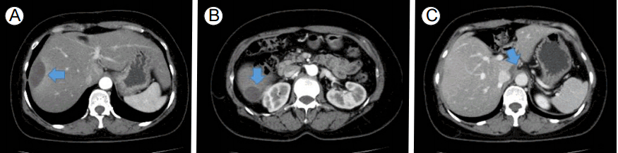
ьШБьГБ ъ▓АьВм ьЖМъ▓м: 4ъ░ЬьЫФ ьаД ьЪ░ьГБы│╡ы╢А эЖ╡ьжЭьЭ┤ ь▓ШьЭМ ы░ЬьГЭэХШьШАьЭД ыЛ╣ьЛЬ ьЛЬэЦЙэХЬ ьЩ╕ы╢А ы│╡ы╢А ьШБьГБьЧРьДЬ ьЪ░ь╕б ъ░ДьЭШ S8ьЧР 13 cmьЭШ ыаМьжИыкиьЦС(lentiform)ьЭШ эФ╝ызЙэХШ эШИьвЕьЭ┤ эЩХьЭ╕ыРШьЧИьЬ╝ый░, ьЭ┤ ы│Сы│А ьЭ┤ьЩ╕ьЧР ыЛдые╕ ы│Сы│АьЭА эЩХьЭ╕ыРШьзА ьХКьХШыЛд(Fig. 1). 2ъ░ЬьЫФ ьаД ьЪ░ьГБы│╡ы╢А эЖ╡ьжЭ ьЮмы░ЬьЛЬ ьЛЬэЦЙэХЬ ьЩ╕ы╢А ьШБьГБ ъ▓АьВмьЧРьДЬыКФ ьЭ┤ьаДьЧР эЩХьЭ╕ыРЬ S8 ъ╡мэЪНьЭШ ы│Сы│АьЭА ъ░РьЖМэХЬ ьЦСьД▒ьЭ┤ьЧИьЬ╝ыВШ, S6 ьШБьЧньЧР 8.8 cmьЭШ ьГИыбЬьЪ┤ эФ╝ызЙэХШ эШИьвЕьЭ┤ эЩХьЭ╕ыРШьЧИыЛд(Fig. 2). ы│╕ьЫРьЧР ыВ┤ьЫРэХШьЧм ьЮДьГБ ъ▓╜ъ│╝ые╝ эЩХьЭ╕эХШъ╕░ ьЬДэХ┤ ыЛдьЛЬ ь┤мьШБэХЬ ы│╡ы╢А ьШБьГБ ъ▓АьВмьЧРьДЬ S8 ы░П S6 ъ╡мьЧньЭШ эШИьвЕьЭА эБмъ╕░ъ░А ъ░РьЖМэХШьШАьЬ╝ыВШ S1 ьШБьЧньЧР 1.7 cm эБмъ╕░ьЭШ ьГИыбЬьЪ┤ эФ╝ызЙэХШ эШИьвЕьЭ┤ ъ┤Аь░░ыРШьЧИыЛд(Fig. 3).
ь╣ШыгМ ы░П ъ▓╜ъ│╝: эЩШьЮРыКФ ыВ┤ьЫР ыЛ╣ьЛЬ ым┤ьжЭьГБьЭ╕ ьГБэГЬьШАыЛд. ъ╕░ьГЭь╢й ы░ШьЭС ъ▓АьВмьЧРьДЬ эЩХьЭ╕ыРЬ эПРэЭбь╢й ьЦСьД▒ ьЖМъ▓мьЧР ыМАэХ┤ praziquantelьЭД ы│╡ьЪйэХШьШАьЬ╝ый░, эШИьХбьвЕьЦСыВ┤ъ│╝ьЩА ьГБьЭШ эЫД эГАыкйьЛЬэОЬ ы│╡ьЪйьЭД ьдСьзАэХШьШАыЛд. ьЭ┤эЫД эШДьЮмъ╣МьзА 4ъ░ЬьЫФъ░Д ьжЭьГБьЭШ ьЮмы░Ь ьЧЖьЭ┤ ь╢ФьаБ ъ┤Аь░░ ьдСьЧР ьЮИыЛд.
ъ│а ь░░
ъ░ДьЭШ эФ╝ызЙэХШ эШИьвЕьЭА Glisson's capsuleьЭ┤ ъ░ДьЭШ ьЛдьзИ ьВмьЭ┤ьЧР эШИьХбьЭ┤ ь╢ХьаБыРШыКФ ъ▓ГьЭД ьЭШып╕эХШый░, ьЭ╝ы░ШьаБьЬ╝ыбЬ ьЪ░ь╕б ъ░ДьЧРьДЬ ы░ЬьГЭ ы╣ДьЬиьЭ┤ ьХ╜ 75% ьаХыПДьЭ┤ыЛд[1]. ъ░ДьЭШ эФ╝ызЙэХШ эШИьвЕьЭА ьШБьГБ ъ▓АьВмые╝ эЖ╡эХ┤ ьзДыЛиэХа ьИШ ьЮИыЛд. ь╗┤эУиэД░ыЛиь╕╡ь┤мьШБ(computed tomography, CT)ьЭД ьЛЬэЦЙэХШый┤, эШИьвЕьЭА ы╣Дьб░ьШБьжЭъ░Х CTьЧРьДЬ ъ░ДьЛдьзИы│┤ыЛд ъ│аьЭМьШБьЭД ы│┤ьЭ┤ъ▓М ыРШый░, ьЭСъ│аыРШьзА ьХКыКФ ъ▓╜ьЪ░ыЭ╝ый┤ ьб░ьШБьжЭъ░Х эЫДьЧРыКФ ъ░ДьЛдьзИы│┤ыЛд ьаАьЭМьШБьЬ╝ыбЬ ы│┤ьЭ┤ъ▓М ыРЬыЛд. ьЮРъ╕░ъ│╡ыкЕьШБьГБ(magnetic resonance imaging)ьЧРьДЬыКФ T1, T2 ъ░Хьб░ ьШБьГБ ыкиыСРьЧРьДЬ ъ░ДьЛдьзИы│┤ыЛд ьаАъ░ХыПД ьЛаэШ╕ые╝ ы│┤ьЭ┤ый░, T2 ъ░Хьб░ ьШБьГБьЧРьДЬ ыНФ ыЪЬыа╖эХЬ эК╣ьзХьЭД ы│┤ьЭ╕ыЛд[2]. ьШБьГБ ъ▓АьВмыКФ ьаАыеШыРЬ ьХбь▓┤ьЭШ эК╣ьД▒ы┐Р ьХДыЛИыЭ╝, ъ░ДьЛдьзИыВ┤ ы│Сы│АьЭ┤ ьЮИыКФьзАыПД ь╢Фъ░АыбЬ эЩХьЭ╕эХа ьИШ ьЮИыЛд.
ым╕эЧМьЧР ы│┤ъ│аыРШъ│а ьЮИыКФ ъ░Д эФ╝ызЙэХШ эШИьвЕьЭШ ьЫРьЭ╕ыУдыбЬыКФ эБмъ▓М ьЮДьЛаъ│╝ ьЧ░ъ┤АыРЬ ъ▓╜ьЪ░[3], ьЭШэХЩьаБ ьЛЬьИаъ│╝ ьЧ░ъ┤АыРЬ ъ▓╜ьЪ░[4,5], ъ░ДыВ┤ ьвЕьЦСьЭ┤ эММэШИыРЬ ъ▓╜ьЪ░[6,7] ыУ▒ъ│╝ ьЧ░ъ┤АыРЬ ы│┤ъ│аыУдьЭ┤ ы╣Дъ╡РьаБ эЭФэХШыЛд. ьЭ┤ ьЩ╕ьЧРыПД ъ░Д ыЖНьЦС[8], ъ╕░ьГЭь╢й ъ░РьЧ╝[9], ъ░ДьЮРы░Шы│С(peliosis hepatitis) [10]ьЭ┤ ьЫРьЭ╕ьЭ┤ый░, ьЭ╝ы╢А ьЫРьЭ╕ ып╕ьГБьЭ╕ ъ▓╜ьЪ░ыПД ы│┤ъ│аыРШъ│а ьЮИыЛд[1]. эШДьЮмъ╣МьзА ы│┤ъ│аыРЬ ъ░ДьЭШ эФ╝ызЙэХШ эШИьвЕьЭШ ьЫРьЭ╕ыУдьЭА эСЬ 1ьЧР ьаХыжмэХШьШАыЛд.
ы│╕ ьжЭыбАыПД ы╣Дъ╡РьаБ ьЮШ ьХМыадьзД ьЮДьЛа, ьЭШэХЩьаБ ьЛЬьИа ыШРыКФ ъ░ДыВ┤ ьвЕьЦС ыУ▒ ьЫРьЭ╕ьЭ┤ ьЧЖьЧИыНШ эЩШьЮРыбЬ, ьжЭьГБьЭ┤ ыкЗ ъ░ЬьЫФ ъ░Дъ▓йьЭД ыСРъ│а ьЮмы░ЬьД▒ ъ▓╜ъ│╝ые╝ ы│┤ьЭ┤ъ│а ьЮИьЧИьЬ╝ый░, ъ▓АьВм ьЛЬызИыЛд ъ░ДыВ┤ эФ╝ызЙэХШ эШИьвЕ ы░ЬьГЭ ы╢АьЬДъ░А ыЛмыЮРыЛдыКФ ьаРьЧРьДЬ эШДьЮм ьзДэЦЙыРШыКФ ьЫРьЭ╕ьЭ┤ ьЮИыКФ ъ▓ГьЬ╝ыбЬ эМРыЛиэХШьШАыЛд. ьЮмы░ЬьД▒ ъ▓╜ъ│╝ые╝ ы│┤ьЭ┤ъ│а ьЮИъ│а, эФ╝ызЙэХШ эШИьвЕъ│╝ ъ╕░ьГЭь╢йъ│╝ ьЧ░ъ┤АьД▒ыПД ы│┤ъ│аыРЬ ы░Ф ьЮИьЬ╝ый░, ъ╕░ьГЭь╢йьЭ┤ ьЭ┤ыПЩэХШый┤ьДЬ ы░Шы│╡ьаБьЬ╝ыбЬ эФ╝ызЙэХШ эШИьвЕьЭД ьЬаы░ЬэХШьШАьЭД ъ░АыКеьД▒ьЭ┤ ы░░ьаЬыРШьзА ьХКьХД, ъ░АыКеэХЬ ъ╕░ьГЭь╢й ъ┤Аыаи ъ▓АьВмые╝ ьЛЬэЦЙэХШьШАыЛд. ъ▓АьВм ъ▓░ъ│╝ эПРэЭбь╢й ы░ШьЭСьЭ┤ ьЦСьД▒ ьЖМъ▓мьЭД ы│┤ьШАьЬ╝ыВШ, ым╕эЧМьГБ эПРэЭбь╕╡ьЭ┤ ьЫРьЭ╕ьЬ╝ыбЬ ы│┤ъ│аыРЬ ы░Ф ьЧЖьЬ╝ый░, эХнь▓┤ ы░ШьЭСызМьЬ╝ыбЬ эПРэЭбь╢йьЭ┤ эФ╝ызЙэХШ ь╢ЬэШИьЭШ ьЫРьЭ╕ьЬ╝ыбЬ ыЛиьаХэХШъ╕░ ьЦ┤ыадьЫаыЛд. Fasciolaъ░А ьЫРьЭ╕ьЬ╝ыбЬ ы│┤ъ│аыРШьЧИьЬ╝ыВШ ы│╕ эЩШьЮРьЧРьДЬыКФ ьЬаь╢йьЭД эЩХьЭ╕эХа ьИШыКФ ьЧЖьЧИыЛд. ъ╕░ьГЭь╢йьЭ┤ эФ╝ызЙэХШ ь╢ЬэШИьЭШ ьЫРьЭ╕ьЬ╝ыбЬ эЩХьзДэХа ьИШ ьЧЖьЧИьЬ╝ыВШ, ьЬДэЧШ-ьЭ┤ыУЭ ъ│аыадэХШьЧм эПРэЭбь╢й ы░ШьЭС ьЦСьД▒ьЧР ыМАэХ┤ьДЬыКФ эЩШьЮРьЩА ьГБьЭШ эЫД ъ╡мь╢йьаЬые╝ ы│╡ьЪйэХШъ╕░ыбЬ эХШьШАыЛд. ыШРэХЬ 4ыЕД ыПЩьХИ ы│╡ьЪйэХЬ эГАыкйьЛЬэОЬьЭ┤ эШДьЮм ьжЭьГБьЭД ьЬаы░ЬэХШьШАьЭД ъ░АыКеьД▒ ыШРэХЬ ьЩДьаДэЮИ ы░░ьаЬэХа ьИШ ьЧЖьЧИыЛд. эК╣эЮИ эГАыкйьЛЬэОЬьЭ┤ ъ░ДьЮРьГЙы░Шы│СьЭШ ьЬаы░Ь ьЫРьЭ╕ьЬ╝ыбЬ ы│┤ъ│аыРЬ ы░Ф ьЮИъ│а, ъ░ДьЮРьГЙы░Шы│С ыШРэХЬ эФ╝ызЙэХШ эШИьвЕьЭШ ьЫРьЭ╕ьЬ╝ыбЬ ы│┤ъ│аыРШъ│а ьЮИьЦ┤, эШИьХбьвЕьЦСыВ┤ъ│╝ьЩА ьГБьЭШ эЫД эГАыкйьЛЬэОЬыПД ьдСыЛиьЭД ьЛЬэЦЙэХШыКФ ъ▓ГьЭ┤ ьЬДэЧШ-ьЭ┤ыУЭ ый┤ьЧРьДЬ ыНФ ьЪ░ьЫФэХа ъ▓ГьЬ╝ыбЬ эМРыЛиыРШьЦ┤, эГАыкйьЛЬэОЬ ы│╡ьЪйыПД ьдСыЛиэХШыПДыбЭ ъ╢Мъ│аэХШьШАыЛд.
ъ░ДьЭШ эФ╝ызЙэХШ эШИьвЕьЭ┤ ьзДыЛиыРЬ ъ▓╜ьЪ░, ы│┤ьб┤ьаБ ь╣ШыгМ ыШРыКФ ьИШьИаьаБ ь╣ШыгМые╝ ъ▓░ьаХэХШыКФ ыН░ ьЮИьЦ┤ ьЧ░ьЖНьаБьЭ╕ ьШБьГБ ъ▓АьВм ы░П эШИьГЙьЖМ ыУ▒ьЭШ эШИьХб ъ▓АьВм ыУ▒ьЭД ьЛЬэЦЙэХШый░ эШИьвЕьЭ┤ ьзДэЦЙэХШьзА ьХКыКФьзА ьЧмы╢Аые╝ ыкиыЛИэД░ызБэХШыКФ ъ▓ГьЭ┤ ьдСьЪФэХШыЛд. ьЭ╝ы░ШьаБьЬ╝ыбЬ эММьЧ┤ыРШьзА ьХКыКФ эФ╝ызЙэХШ эШИьвЕьЭА ы│┤ьб┤ьаБ ь╣ШыгМые╝ ьЛЬэЦЙэХШый░, ьШИэЫДыКФ ьЦСэШ╕эХШыЛд[1]. ьИШьИаьаБ ь╣ШыгМыКФ эФ╝ызЙэХШ эШИьвЕьЭШ эММьЧ┤ ыШРыКФ эММьЧ┤ьЭ┤ ьЮДы░ХэХЬ ъ▓╜ьЪ░ьЧР ыМАэХ┤ ьаБьЪйэХШыКФ ъ▓ГьЭ┤ ь╢Фь▓ЬыРЬыЛд. ьЭ╝ы░ШьаБьЬ╝ыбЬ эФ╝ызЙ эММьЧ┤ьЛЬ 75%ъ░А ьВмызЭэХЬыЛд[1]. ъ╕░ьб┤ьЧР ы│┤ъ│аыРЬ ьжЭыбАыУд ьдС ьЮДьЛа, ьЭШэХЩьаБ ь▓Шь╣ШыВШ ьЩ╕ьГБ, ъ░ДыВ┤ ьвЕьЦСьЭШ эММьЧ┤ъ│╝ ьЧ░ъ┤АыРЬ ьжЭыбАые╝ ьаЬьЩ╕эХЬ ыЛдые╕ ьЫРьЭ╕ьЧР ьЭШэХЬ ь╣ШыгМ ы░П ьЮДьГБ ъ▓╜ъ│╝ыКФ эСЬ 2ьЧР ьаХыжмэХШьШАыЛд.
ьЪФ ьХ╜
ьЩ╕ьГБ ыШРыКФ ьЭСъ│а ьЮеьХа ыУ▒ьЭ┤ ыПЩы░ШыРШьзА ьХКыКФ ьГБэГЬьЧРьДЬ ьЮРы░ЬьаБьЭ╕ эФ╝ызЙэХШ эШИьвЕьЭШ ы░ЬьГЭьЭА ыУЬым╕ ъ▓╜ьЪ░ьЭ┤ыЛд. ыУЬым╕ ьЫРьЭ╕ьЭ┤ьзАызМ ьжЭыбАыбЬ ы│┤ъ│аыРЬ ъ▓╜ьЪ░ыКФ ьЮДьЛаъ│╝ ьЧ░ъ┤АыРЬ ьЮРъ░ДьаДьжЭ ыШРыКФ ьаДьЮРъ░ДьжЭ ы░П hemolysis, elevated liver enzymes, low platelets (HELLP)ьжЭэЫДъ╡░ ыУ▒ьЭ┤ ьЮИьЬ╝ый░ ъ░ДьД╕эПмьХФьвЕ, ъ░ДьГШьвЕ ы░П ъ░ДьЭШ ьХЕьД▒ ы░П ьЦСьД▒ ьвЕьЦСьЭШ эММьЧ┤ъ│╝ьЭШ ьЧ░ъ┤АьД▒ьЭ┤ ы│┤ъ│аыРЬ ы░Фъ░А ьЮИыЛд. ыШРыКФ ьЭШэХЩьаБ ьЛЬьИа, ъ░РьЧ╝, ьХ╜ым╝ьЧР ьЭШэХЬ ъ░ДьЮРьГЙы░Шы│СыПД ьЭ╝ы╢А ы│┤ъ│аыРШьЦ┤ ьЮИыЛд.
ы│╕ ьжЭыбАыКФ ьЩ╕ьГБьЭ┤ыВШ ьЭСъ│а ьЮеьХа ыУ▒ьЭ┤ ыПЩы░ШыРШьзА ьХКыКФ ьГБэГЬьЧРьДЬ ы░Шы│╡ьаБьЬ╝ыбЬ эФ╝ызЙэХШ эШИьвЕьЭ┤ ы░ЬьГЭэХШьШАьЬ╝ый░, ьЧмыЯм ым╕эЧМъ│аь░░ьЭД эЖ╡эХ┤ эЩХьЭ╕ыРЬ ъ░АыКеэХЬ ьЫРьЭ╕ыУдьЧР ыМАэХ┤ ъ░Ры│ДэХШьШАьЬ╝ыВШ ыЪЬыа╖эХЬ ьЫРьЭ╕ьЭД ы░ЭэШАыВ╝ ьИШыКФ ьЧЖьЧИыЛд. ыЛдызМ, ъ░АыКеьД▒ьЭД ьЩДьаДэЮИ ы░░ьаЬэХШъ╕░ ьЦ┤ыадьЫаыНШ ъ╕░ьГЭь╢й ъ░РьЧ╝ьЧР ыМАэХ┤ эХнъ╡мь╢йьаЬ ь╣ШыгМ ы░П эГАыкйьЛЬэОЬ ы│╡ьЪйъ│╝ ьЧ░ъ┤АыРЬ ъ░ДьЮРьГЙы░Шы│СьЭШ ъ░АыКеьД▒ьЧР ыМАэХ┤ эГАыкйьЛЬэОЬ ьдСыЛи ыУ▒ьЭШ ьб░ь╣Шые╝ ь╖иэХШьШАьЬ╝ый░, ьЭ┤эЫД ы░Шы│╡ьаБьЬ╝ыбЬ ы░ЬьГЭэХШыНШ ъ▓╜ъ│╝ъ░А ьЮДьГБьаБьЬ╝ыбЬ эШ╕ьаДыРШыКФ ьЦСьГБьЭД ы│┤ьШАыЛд. ьЭ┤ьЧР ы╣ДыбЭ ъ┤Аь░░ ъ╕░ъ░ДьЭ┤ ь╢йы╢ДэХШъ▓М ъ╕╕ьзА ьХКьзАызМ, ым╕эЧМъ│аь░░ъ│╝ эХиъ╗Ш ы│┤ъ│аэХШыКФ ы░ФьЭ┤ыЛд.


 PDF Links
 PubReader
 ePub Link
 Full text via DOI
 Download Citation
 Print
Print


