동기능부전증후군으로 발생한 폐고혈압 1예
Reversible Pulmonary Hypertension due to Sick Sinus Syndrome
Article information
Abstract
폐고혈압은 흔하지 않으나 치명적인 질환으로 적합한 원인을 찾아 교정하는 것이 중요하다. 이에 저자들은 동기능부전 및 동정지로 인한 좌심실 질환으로 폐고혈압의 발생을 심도자술로 증명하였고, 동기능부전 및 동정지를 치료함으로써 폐고혈압의 호전을 보인 1예가 있어 보고하는 바이다.
Trans Abstract
A 60-year-old man visited the hospital after experiencing dyspnea after exertion for 2 weeks. An electrocardiogram showed sinus arrest with junctional escape rhythm at 40 beats/min. Transthoracic echocardiography showed that the right ventricular systolic pressure (RVSP) was approximately 71 mmHg and that the left ventricular ejection fraction was preserved. The ratio of peak early diastolic transmitral inflow velocity to early diastolic peak mitral annular velocity (E/E’) was 29. Cardiac catheterization revealed a systolic pulmonary artery pressure (SPAP) of 63 mmHg, a mean pulmonary artery pressure of 27 mmHg, and a pulmonary capillary wedge pressure of 22 mmHg with a rhythm of 40 beats/min. The patient was diagnosed with pulmonary hypertension (group 2) due to sick sinus syndrome. SPAP decreased to 48 mmHg during atrial pacing at 60 beats/min. After permanent pacemaker insertion, RVSP decreased from 71 mmHg to 44 mmHg. In this case, passive group 2 pulmonary hypertension occurred due to sick sinus syndrome.
서 론
폐고혈압증의 원인은 다양하며, 그 치료 또한 원인이 되는 질환에 대한 교정이 중요하다. 폐고혈압증의 원인 중에는 좌심실 질환에 의해 발생할 수 있으며, 특히 심실의 이완 기능 부전에 따른 폐고혈압이 발생할 수 있다[1,2]. 이에 저자들은 동기능부전 및 접합부 이탈 박동으로 인한 심실이완기능부전이 발생하고, 이로 인해 발생한 폐고혈압을 심도자술로 증명하였고, 심장박동조율기 삽입 후 폐고혈압의 호전을 보인 증례를 경험하였기에 보고하는 바이다.
증 례
환 자: 60세 남자
주 소: 운동시 호흡곤란
현병력: 60세 남자 환자가 내원 2주 전부터 운동시 발생하는 호흡곤란으로 입원하였다.
과거력: 환자는 20년 정도 되는 고혈압 및 당뇨병 이환력이 있으며, 2년 전 신장 이식 및 1년 전 경피경관 관상동맥성형술을 시행받았고, 3개월 전 심방빈맥으로 우심방의 분계능선(crista terminalis) 및 Cavo-tricuspid 부위에 고주파절제를 받았다. 내원 6개월 전 시행한 심초음파에서 우심실의 크기는 정상이었고, 최대 삼첨판역류 유속은 2.6 m/sec으로 우심방압을 5 mmHg로 추정하였을 때, 우심실 수축기압은 31 mmHg로 정상 소견이었다. 또한 초기 이완기 경승모판 최대 혈류속도(E)와 이완기 후기 심방수축에 의한 혈류속도(A)의 비(E/A)는 0.7이었고, 감속 시간(deceleration time)은 308 msec, 초기 이완기 경승모판 최대 혈류속도(E)와 초기 이완기 승모판륜의 최대 운동속도(e’)의 비(E/e’)는 14로 경도의 이완 기능 장애 소견을 보였다. 좌심방 부피 지수는 43.8 mL/m2였다.
가족력 및 사회력: 특이사항은 없었다.
이학적 소견: 내원 당시 급성 병색을 보였으며, 혈압 160/60 mmHg, 맥박 40회/분, 호흡수 20회/분, 체온은 36.5°C, 산소포화도 99%였다. 두경부 진찰에서 특이 소견은 없었다. 흉부 청진에서 심음은 규칙적이었으며 수축기 심잡음이 청진되었고, 양측 폐 하엽에서 수포음이 청진되었다. 복부는 편평했으며, 압통 및 반발 압통은 없었고, 간 비 종대도 없었다. 양 하지에서는 2+의 함요 부종이 관찰되었다.
검사 소견: 내원 당시 말초 혈액 검사에서 혈색소 10.6 g/dL였고, 생화학 검사에서 혈액요소질소 28.2 mg/dL, 크레아티닌 1.44 mg/dL, 디 이중체(D-dimer) 415 ng/dL로 증가되어 있었다. 그 외 백혈구, 혈소판, 아스파르테이트아미노전달효소, 알라닌아미노전달효소, 나트륨, 칼륨, 프로트롬빈시간, 활성화부분트롬보플라스틴시간은 정상 수치내 있었다.
영상 소견: 흉부 단순 방사선 사진에서 심장비대 및 폐울혈, 양측 흉수 소견이 관찰되었다(Fig. 1). 흉부 컴퓨터 방사선단층촬영에서 폐혈전색전증 소견은 없었다.

Chest radiography. The chest radiograph shows bilateral pleural effusion and central pruning of the pulmonary vessel, indicating pulmonary edema and cardiomegaly.
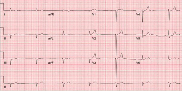
심전도: 분당 40회의 동정지 및 접합부 이탈 박동이 관찰되었다(Fig. 2).

Electrocardiography (ECG). The ECG reveals sinus arrest with junctional escape rhythm at 40 beats/min and left axis deviation.
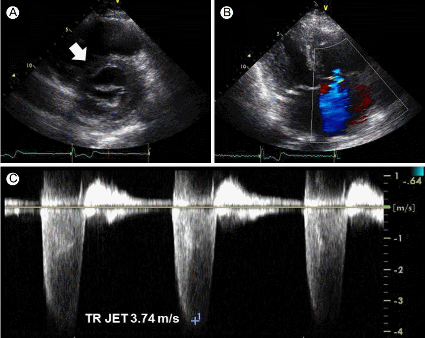
심초음파: 내원하여 시행한 심초음파에서 좌심실 박출 계수는 72%로 정상 범위였으나, 이완기 D-형 좌심실 소견이 관찰되었다. e’은 5 cm/sec로 감소되어 있었으며, E/e’ 비율은 29로 증가되어 있었다. 좌심방 부피 지수는 60.9 ml/m2로 이전 검사에 비해 증가한 소견이었다. 경도의 우심실 확장이 관찰되었으나 우심실 수축 기능은 정상이었다. 중등도의 삼첨판 역류 소견이 관찰되었고, 최대 삼첨판역류 유속은 3.74 m/sec로 우심방압을 15 mm로 추정하였을 때 우심실 수축기압은 71 mmHg로 중증 폐고혈압에 합당한 소견을 보였다(Fig. 3, Table 1).

Transthoracic echocardiography. (A) In the parasternal short axis view, interventricular septal flattening, known as the “D-shape sign” (arrow), was observed. (B) Color Doppler imaging of the tricuspid valve revealed moderate tricuspid regurgitation. (C) The maximal velocity of tricuspid regurgitation jet (TR JET) was 3.74 m/s.
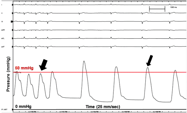
심도자 소견: 분당 40회의 서맥에서 시행한 심도자 검사상 수축기 및 평균 폐동맥압은 63 mmHg, 27 mmHg이었고, 폐모세혈관쐐기압은 22 mmHg로 폐고혈압으로 확진할 수 있었다(Fig. 4, Table 1). 심장전기생리 검사용 카테터를 우심방에 위치시킨 후, 우심방을 분당 60회로 조율하며 폐동맥압 및 폐모세혈관쐐기압을 측정하였다. 수축기 폐동맥압은 48 mmHg, 폐모세혈관쐐기압은 16 mmHg으로 호전되었다(Fig. 4, Table 1).

Changes in pulmonary artery pressure during atrial pacing and sinus arrest with junctional escape rhythm. During atrial pacing at 60 beats/min, systolic pulmonary artery pressure decreased to 48 mmHg (big arrow) from 68 mmHg (small arrow).
치료 및 경과: 운동시 호흡곤란 및 양하지 함요 부종을 조절하기 위해 이뇨제 치료를 시작하였으나 증상의 호전은 뚜렷하지 않았으며, 동정지 소견은 지속되었다. 최저 맥박수는 분당 40회 최대 맥박수는 분당 45회였으며 평균 맥박수는 분당 42회였다. 입원 7병일째 영구형 심장박동조율기 삽입술을 시행하였다. 환자에게 영구형 심장박동조율기 삽입 후 맥박수를 분당 60회로 유지하였다. 입원 8병일째 측정한 심초음파에서 경도의 우심실 확장 및 중등도의 삼첨판역류는 변화가 없었으나, 최대 삼첨판역류 유속은 3.14 m/s로 감소되었고, 우심실 수축기혈압이 44 mmHg로 호전되었다. E/e’은 20으로 감소되고, e’은 6.7 cm/sec로 상승되었다(Table 1). 환자는 전반적인 상태 호전 및 운동시 호흡곤란 및 양하지 함요 부종이 호전되어 퇴원하였다.
고 찰
폐고혈압은 안정시 심도자술로 측정한 평균 폐동맥압 ≥ 25 mmHg로 정의한다[1,3]. 좌심실 질환에 의한 폐고혈압(2군)에서는 폐모세혈관쐐기압 > 15 mmHg이며, 경폐압력 차이(transpulmonary pressure gradient)가 12-15 mmHg 이하일 때 수동성 폐고혈압(passive pulmonary hypertension), 경폐압력 차이가 12-15 mmHg 이상일 때 반응성 폐고혈압(reactive pulmonary hypertension)으로 나눌 수 있다[1,3]. 좌심실 질환으로 인한 폐고혈압증의 병태 생리는 우선적으로 좌심실의 수축 또는 이완 기능부전으로 인해 좌심실의 충만압이 상승하게 되고, 이로 인해 폐모세혈관쐐기압이 증가하면서 결과적으로 폐고혈압증이 발생하게 되는데[3], 저자들은 서맥으로 인하여 수동성 폐고혈압이 발생하였다가 심박동 조율 후 폐고혈압이 호전되는 증례를 확인하여 이를 보고하였다.
본 저자의 환자는 평균 폐동맥압이 27 mmHg, 폐모세혈관쐐기압이 22 mmHg, 경폐압력 차이가 5 mmHg로 좌심실 질환에 의한 폐고혈압이며, 수동성 폐고혈압에 해당한다. 환자는 내원 당시 동기능부전 및 분당 40회의 서맥이 관찰되었는데, 내원 1년 전 심방빈맥으로 고주파절제를 받기 전에도 서맥이 일시적으로 있었으며, 이후 고주파절제술이 동기능부전을 더 악화시켰을 가능성도 있겠다. 환자는 운동시 호흡곤란, 양 하지의 함요 부종 및 양측 흉수 소견으로 심부전의 증상 및 징후를 보였고, 심초음파상 좌심실 박출 계수는 72%로 정상소견으로 서맥에 의한 심부전으로 평가하였다[4,5]. 본 환자에서의 폐고혈압 발생의 병태 생리는 우선적으로 서맥으로 인한 정상적인 구혈이 일어나지 않으면서 좌심실의 충만압이 상승하게 되고, 그로 인해 후향적으로 폐동맥압이 상승하였을 것으로 판단된다[3]. 또한 환자는 입원 기간 중 지속적인 동정지 및 접합부 이탈 박동이 관찰되었고, 심초음파상 좌심방의 수축은 관찰되지 않았는데[6,7], 이러한 좌심방의 수축 기능의 상실로 후향적으로 폐동맥압의 상승에 영향을 주었을 가능성이 있겠다. 이에 저자들은 심도자술에서 서맥이 회복되면서 수축기 폐동맥혈압의 감소를 보인 것을 확인할 수 있었다(Fig. 4). 환자는 영구형 심장박동조율기 삽입 후 서맥에서 회복되었고, 심초음파상 폐고혈압증은 호전 양상을 보였다.
