


ņä£ ļĪĀ
Ļ░Ģņ¦üņä▒ ņ▓ÖņČöņŚ╝(ankylosing spondylitis)ņØĆ ņ▓ÖņČöĻ┤ĆņĀłņŚ╝(spondyloarthritis) ņ¦łĒÖśĻĄ░ ņżæņŚÉņä£ Ļ░Ćņן ņŻ╝ļÉ£ ņ¦łĒÖśņ£╝ļĪ£ ņČĢņä▒(axial) Ļ┤ĆņĀłņØś ņÜ┤ļÅÖ ņןņĢĀ ļ░Å Ļ▓Įņ¦üņØä ĒŖ╣ņ¦Ģņ£╝ļĪ£ ĒĢ£ļŗż[1]. ņĄ£ĻĘ╝ Ļ┤ĆņĀłņŚ╝ ņ¦łĒÖśņØś ņĪ░ĻĖ░ ņ¦äļŗ© ļ░Å ņ╣śļŻīņŚÉ ļīĆĒĢ£ Ļ┤Ćņŗ¼ņØ┤ ļåÆņĢäņ¦ÉņŚÉ ļö░ļØ╝ Ļ▓ĮņČö-ņÜöņČö Ļ┤ĆņĀłņØ┤ ņ£ĄĒĢ®ļÉśņ¢┤ ļ¬®ņØä ņĀ£ļīĆļĪ£ Ļ░Ćļłäņ¦Ć ļ¬╗ĒĢśļŖö ņŗ¼ĒĢ£ ņāüĒā£ļĪ£ ļ│æņøÉņØä ņ▓śņØī ļ░®ļ¼ĖĒĢśļŖö ņé¼ļĪĆļŖö ņĀüņ¢┤ņĪīļŗż. ĒĢśņ¦Ćļ¦ī ņĪ░ņĪ░Ļ▓Įņ¦üņØ┤ ļÅÖļ░śļÉ£ Ļ░Ģņ¦üņä▒ ņ▓ÖņČöņŚ╝ņØä ņČöĻ░äĒīÉ ĒāłņČ£ņ”Ø, ņÜöņČö ļČĆņ£äņØś ņŚ╝ņóī ļō▒ ĻĖ░Ļ│äņĀü ņÜöĒåĄ(mechanical pain)ņ£╝ļĪ£ ļ©╝ņĀĆ ņśżņØĖĒĢśļŖö Ļ▓ĮņÜ░Ļ░Ć ņĀüņ¦Ć ņĢŖļŗż. ļśÉĒĢ£ Ļ░Ģņ¦üņä▒ ņ▓ÖņČöņŚ╝ņŚÉ ņØśĒĢ┤ ļ░£ņāØĒĢ£ ļ¦Éņ┤ł Ļ┤ĆņĀłņŚ╝ņØ┤ļéś ļŗżņ¢æĒĢ£ Ļ┤ĆņĀł ņÖĖ ņ×äņāüņāüņØ┤ Ļ░äĒś╣ ĒāĆņ¦łĒÖśņ£╝ļĪ£ Ļ░äņŻ╝ļÉśĻ│Ā ņ׳ļŗż. ņØ┤ ĻĖ░ĒÜīļź╝ ĒåĄĒĢśņŚ¼ Ļ░Ģņ¦üņä▒ ņ▓ÖņČöņŚ╝ ļĢī ļ│┤ņØ┤ļŖö ņŚ╝ņ”Øņä▒ ņÜöĒåĄ(inflammatory back pain)ņØś ņŻ╝ņÜö ĒŖ╣ņä▒ņØä ņé┤ĒÄ┤ļ│┤Ļ│Ā ņØ┤ ņ¦łĒÖśņØś ņ¦äļŗ©ņŚÉ ņ×ÉĻĖ░Ļ│Ąļ¬ģņśüņāüņØ┤ ņ¢┤ļ¢ż ļÅäņøĆņØä ņżä ņłś ņ׳ļŖöņ¦Ć ņé┤ĒÄ┤ļ│┤Ļ│Āņ×É ĒĢ£ļŗż.
ļ│Ė ļĪĀ
Ļ░Ģņ¦üņä▒ ņ▓ÖņČöņŚ╝ņØś ņ×äņāüņāü
Ļ░Ģņ¦üņä▒ ņ▓ÖņČöņŚ╝ņØś ņ×äņāüņāüņØĆ Ēü¼Ļ▓ī Ļ┤ĆņĀł ļ░Å Ļ┤ĆņĀł ņÖĖ ņ×äņāüņāüņ£╝ļĪ£ ĻĄ¼ļČäļÉ£ļŗż. Ļ┤ĆņĀłņØĆ ņČĢņä▒ Ļ┤ĆņĀł ņÖĖņŚÉļÅä ļ¦Éņ┤ł Ļ┤ĆņĀłņØ┤ ņ╣©ļ▓öļÉĀ ņłś ņ׳ņ£╝ļ®░ ņČĢņä▒ Ļ┤ĆņĀł ņ╣©ļ▓ö ļĢī ļéśĒāĆļéśļŖö ņŻ╝ņÜö ņ”Øņāü ņżæ ĒĢśļéśļŖö ņŚ╝ņ”Øņä▒ ņÜöĒåĄņØ┤ļŗż[2] (Table 1). ņ▓ÖņČöĻ┤ĆņĀłņŚ╝ ĒÖśņ×ÉņØś 75%ņŚÉņä£ ņ▓½ ņ”ØņāüņØ┤ ņŚ╝ņ”Øņä▒ ņÜöĒåĄņ£╝ļĪ£ ļéśĒāĆļéśļ®░ ļīĆĻ░£ ņÜöņČöļéś ņÜöņ▓£ņČö ļČĆņ£äņŚÉņä£ļČĆĒä░ ņŗ£ņ×æļÉ£ļŗż[1]. ņŖżĒŖĖļĀłņ╣ŁņØ┤ļéś ļ╣äņŖżĒģīļĪ£ņØ┤ļō£ņä▒ ĒĢŁņŚ╝ņĀ£ļź╝ ļ│ĄņÜ®ĒĢ£ Ēøä ņ”ØņāüņØ┤ ļéśņĢäņ¦Ćļ®░ Ē£┤ņŗØ ĒøäņŚÉļŖö ņśżĒ׳ļĀż ņĢģĒÖöļÉśļŖö Ļ▓ĮĒ¢źņØ┤ ņ׳ļŗż. ļ│┤ĒåĄ ņĪ░ņĪ░Ļ▓Įņ¦üņØ┤ 30ļČä ņØ┤ņāü ļÅÖļ░śļÉśļ®░ ĒåĄņ”Ø ļĢīļ¼ĖņŚÉ ņāłļ▓ĮņŚÉ ņ×ĀņØä ņäżņ╣Ā ņłś ņ׳ļŗż. ņŚ╝ņ”Øņä▒ ņÜöĒåĄņØĆ ļĢīļĪ£ļŖö Ļ▓Įļ»ĖĒĢśĻ▓ī ļéśĒāĆļéśĻĖ░ ļĢīļ¼ĖņŚÉ ļ¬©ļź┤Ļ│Ā ņ¦ĆļéśĻ░ł ņłś ņ׳ņ£╝ļ®░ ļīĆĻ░£ ļ░śļ│ĄļÉśļŖö ņ”Øņāü ĒśĖņĀä ļ░Å ņĢģĒÖöņØś Ļ▓ĮĻ│╝ļź╝ ļ│┤ņØĖļŗż. ņ▓ÖņČöĻ┤ĆņĀł Ļ░ä ņ£ĄĒĢ®ņ£╝ļĪ£ Ļ░Ģņ¦üņØ┤ ļ░£ņāØļÉśļ®┤ ņŚ╝ņ”Øņä▒ ņÜöĒåĄņØĆ Ļ░ÉņåīĒĢśļ®┤ņä£ Ļ┤ĆņĀłņÜ┤ļÅÖ ņĀ£ĒĢ£ņŚÉņä£ ņśżļŖö ĻĖ░ļŖź ņןņĢĀĻ░Ć ļ░£ņāØĒĢ£ļŗż. ņ”ē ņÜ┤ļÅÖ Ēøä ņ”ØņāüņØ┤ ļéśļ╣Āņ¦ĆļŖö ĒŖ╣ņ¦ĢņØä Ļ░Ćņ¦ĆļŖö ĻĖ░Ļ│äņĀü ņÜöĒåĄņØ┤ ņŚ╝ņ”Øņä▒ ņÜöĒåĄĻ│╝ ļÅÖļ░śļÉĀ ņłś ņ׳ļŗż. ļéśņĢäĻ░Ć ņ▓ÖņČö Ļ░ä ņØĖļīĆņÖĆ ļŖæĻ│©ņ▓ÖņČö(costovertebral) Ļ┤ĆņĀł, ĒØēļŖæ(sternocostal) Ļ┤ĆņĀłņØś Ļ│©ĒÖöļĪ£ ņÜöņČöņĀäļ¦īņ”Ø, ĒØēņČöĒøäļ¦īņ”ØņØ┤ ļ░£ņāØĒĢśĻ│Ā ļ¬®ņØ┤ ĻĄ¼ļČĆņĀĢĒĢ┤ņ¦äļŗż. Ļ░Ģņ¦üņØ┤ ņŗ¼ĒĢśņ¦Ć ņĢŖļŹöļØ╝ļÅä Ļ┤ĆņĀł ņÜ┤ļÅÖ ņןņĢĀĻ░Ć ņŗ¼ĒĢĀ ņłś ņ׳ļŖöļŹ░, ņØ┤ļŖö ņØ┤ņ░©ņĀüņØĖ ĻĘ╝ņ£Ī ņŚ░ņČĢ(spasm) ļĢīļ¼ĖņØ┤ļŗż. Ļ░Ģņ¦üņä▒ ņ▓ÖņČöņŚ╝ ĒÖśņ×ÉņØś 1/3ņŚÉņä£ Ļ│ĀĻ┤ĆņĀł, ņ¢┤Ļ╣©Ļ┤ĆņĀłņØä ņ╣©ļ▓öĒĢśļŖö ļ░ö, Ļ│ĀĻ┤ĆņĀłņØś Ļ▓ĮņÜ░ ņ¢æņĖĪņ£╝ļĪ£ ņś¼ ņłś ņ׳Ļ│Ā Ļ┤ĆņĀłĒīīĻ┤┤ ļ░Å ņŗ¼ĒĢ£ Ļ┤ĆņĀł ļ│ĆĒśĢņØä ņĢ╝ĻĖ░ĒĢĀ ņłś ņ׳ļŗż[3].
ļ¦Éņ┤łĻ┤ĆņĀł ņ╣©ļ▓öņØĆ ļźśļ¦łĒŗ░ņŖż Ļ┤ĆņĀłņŚ╝Ļ│╝ļŖö ļŗ¼ļ”¼ ņŻ╝ļĪ£ ĒĢśņ¦Ć Ļ┤ĆņĀłņŚÉ ļ░£ņāØĒĢśļŖö ļ╣äļīĆņ╣Łņä▒ ņåīņłśņä▒ Ļ┤ĆņĀłņŚ╝(oligoarthritis)ņ£╝ļĪ£ ļéśĒāĆļé£ļŗż. ļĢīļĪ£ļŖö ņåÉĻ░ĆļØĮņØ┤ļéś ļ░£Ļ░ĆļØĮņØś Ļ┤ĆņĀłĻ│╝ ņŻ╝ņ£ä ņŚ░ļČĆņĪ░ņ¦üĻ╣īņ¦Ć ņóģņ░ĮļÉśļŖö ņ¦ĆņŚ╝(dactylitis)ņØ┤ ļ░£ņāØĒĢśĻĖ░ļÅä ĒĢ£ļŗż.
Ļ▒┤, ņØĖļīĆ, ĻĘ╝ļ¦ē, Ļ┤ĆņĀł Ēö╝ļ¦ēņØ┤ ļ╝łņÖĆ ļČĆņ░®ļÉśļŖö ļČĆņ£äņŚÉ ņŚ╝ņ”ØņØ┤ ļ░£ņāØĒĢśļŖö ļČĆņ░®ļČĆņŚ╝(enthesitis)ņØĆ Ļ░Ģņ¦üņä▒ ņ▓ÖņČöņŚ╝ņØä ļ╣äļĪ»ĒĢ£ ņ▓ÖņČöĻ┤ĆņĀłņŚ╝ņŚÉņä£ ļ│┤ņØ┤ļŖö ņŻ╝ņÜö ņ×äņāüņāüņØ┤ļŗż. ļČĆņ░®ļČĆņŚ╝ņØĆ ņĢäĒé¼ļĀłņŖżĻ▒┤ņØ┤ļéś ņĪ▒ņĀĆĻĘ╝ļ¦ēņØ┤ ļČĆņ░®ĒĢśļŖö ļ░£ļÆżĻ┐łņ╣śņŚÉ ņל ļ░£ņāØĒĢśļŖöļŹ░ ņĢäņ╣©ņŚÉ ņØ╝ņ¢┤ļéśņä£ ļ░£ļÆżĻ┐łņ╣śĻ░Ć ļĢģņŚÉ ļŗ┐ņØä ļĢī ņŗ¼ĒĢśĻ▓ī ņĢäĒöäĻ│Ā Ļ▒Ėņ£╝ļ®┤ņä£ ĒśĖņĀäļÉ£ļŗż. ļČĆņ░®ļČĆņŚ╝ņØĆ ņןĻ│©ļŖźņäĀņØ┤ļéś Ļ▓ĮĻ│©ņĪ░ļ®┤(tibial tuberosity), Ļ░ĆņŖ┤ ņĀäļ®┤ņŚÉļÅä ļ░£ņāØĒĢĀ ņłś ņ׳ļŗż. ļČĆņ░®ļČĆņŚ╝ ņÖĖņŚÉļÅä ĒØēņćäĻ│©(sternoclavicular) Ļ┤ĆņĀł, ĒØēĻ│©ļ│æņŚ░Ļ│©(manubriosternal) Ļ┤ĆņĀł, ĒØēļŖæĻ┤ĆņĀłņØś Ļ┤ĆņĀłņŚ╝ņ£╝ļĪ£ Ļ░ĆņŖ┤ ņĀäļ®┤ņØ┤ ņĢäĒöī ņłś ņ׳ņ£╝ļ®░, ņŗ¼ĒĢ┤ņ¦Ćļ®┤ ĒØēĻ│ĮņØś ĒīĮņ░ĮņØä ņĀĆĒĢ┤ĒĢśĻĖ░ļÅä ĒĢ£ļŗż.
Ļ░Ģņ¦üņä▒ ņ▓ÖņČöņŚ╝ņŚÉņä£ ļÅÖļ░śļÉśļŖö Ļ┤ĆņĀł ņÖĖ ņ×äņāüņāüņ£╝ļĪ£ ĒżļÅäļ¦ēņŚ╝(uveitis), Ļ▒┤ņäĀ(psoriasis), ņŚ╝ņ”Øņä▒ ņןņ¦łĒÖś(inflammatory bowel disease) ļō▒ņØ┤ ņ׳ļŗż. ņØ┤ ņżæ ĒżļÅäļ¦ēņŚ╝ņØ┤ Ļ░Ćņן ĒØöĒĢśļ®░(ĒÖśņ×ÉņØś 20- 30%) ļīĆĻ░£ ņĀäļ░®ņŚÉ ļ░£ņāØĒĢ£ļŗż[4]. HLA-B27 ņ£ĀņĀäņ×É ņ¢æņä▒ņØĖ ĒÖśņ×ÉņŚÉņä£ ļŹö ņל ņāØĻĖ░Ļ│Ā ĒĢ£ņ¬Į ļłłņØś ĒåĄņ”Ø, ļ░£ņĀü, ļłłļČĆņŗ¼Ļ│╝ ļłłļ¼╝ ņ”ØĻ░ĆĻ░Ć ļéśĒāĆļé£ļŗż. ĒżļÅäļ¦ēņŚ╝ņØ┤ ņØśņŗ¼ļÉĀ Ļ▓ĮņÜ░ ļ░öļĪ£ ņĢłĻ│╝Ļ▓Ćņ¦äņØ┤ ĒĢäņÜöĒĢśļ®░ ņ╣śļŻīĻ░Ć ļŖ”ņ¢┤ņ¦Ćļ®┤ ņ£Āņ░®ņØ┤ļéś ņŗ£ļĀźĻ░Éņåī ļō▒ ĒĢ®ļ│æņ”ØņØ┤ ļ░£ņāØĒĢ£ļŗż. Ļ░Ģņ¦üņä▒ ņ▓ÖņČöņŚ╝ ĒÖśņ×ÉņŚÉņä£ Ļ▒┤ņäĀņØ┤ ļÅÖļ░śļÉĀ Ļ▓ĮņÜ░, ņČĢņä▒ Ļ┤ĆņĀłņØä ņ╣©ļ▓öĒĢ£ Ļ▒┤ņäĀ Ļ┤ĆņĀłņŚ╝ņØś Ļ░ĆļŖźņä▒ņØ┤ ņ׳ļŖöņ¦ĆļÅä ņé┤ĒÄ┤ļ│┤ļŖö Ļ▓āņØ┤ ĒĢäņÜöĒĢśļŗż. ņŚ╝ņ”Øņä▒ ņןņ¦łĒÖśĻ│╝ Ļ░Ģņ¦üņä▒ ņ▓ÖņČöņŚ╝ņØ┤ ĒĢ©Ļ╗ś ļ░£Ļ▓¼ĒĢśļŖö Ļ▓ĮņÜ░Ļ░Ć ņ׳ļŖöļŹ░[5], ĒĢ£ ļ│┤Ļ│ĀņŚÉņä£ļŖö ņןņ”ØņāüņØ┤ ņŚåļŖö Ļ░Ģņ¦üņä▒ ņ▓ÖņČöņŚ╝ ĒÖśņ×ÉņØś 20-70%ņŚÉņä£ ļīĆņן ņĀÉļ¦ēņŚÉ ņŚ╝ņ”ØņØ┤ ņĪ┤ņ×¼ĒĢśņśĆļŗż. ļ░śļīĆļĪ£ ņŚ╝ņ”Øņä▒ ņןņ¦łĒÖś ĒÖśņ×ÉņØś 28-35%ņŚÉņä£ ņ▓£ņןĻ┤ĆņĀłņŚ╝(sacroiliitis)ņØä ļ╣äļĪ»ĒĢ£ ņČĢņä▒ ņ▓ÖņČöĻ┤ĆņĀłņŚ╝ņØ┤ ņ׳ļŖö Ļ▓āņ£╝ļĪ£ ļ░£Ēæ£ļÉśņŚłļŗż[6]. ņØ┤ņÖĖļÅä ļō£ļ¼╝ņ¦Ćļ¦ī ņŗ¼ņןņ░©ļŗ©, ļīĆļÅÖļ¦ēĒīÉļ¦ēļČĆņĀä, Ļ░äņ¦łņä▒ ĒÅÉļĀ┤, IgA ņŗĀņןļ│æņ”ØņØ┤ Ļ░Ģņ¦üņä▒ ņ▓ÖņČöņŚ╝Ļ│╝ ļÅÖļ░śļÉĀ ņłś ņ׳ļŗż.
Ļ░Ģņ¦üņä▒ ņ▓ÖņČöņŚ╝ņØś ņ¦äļŗ©ņØä ņ£äĒĢ£ Ļ▓Ćņé¼
ĒśłņĢĪĻ▓Ćņé¼
ņĢäņ¦üĻ╣īņ¦Ć Ļ░Ģņ¦üņä▒ ņ▓ÖņČöņŚ╝ņØä ĒÖĢņ¦äĒĢĀ ņłś ņ׳ļŖö ĒśłņĢĪĻ▓Ćņé¼ļŖö ņŚåļŗż. ņĀüĒśłĻĄ¼ ņ╣©Ļ░Ģņ¦Ćņłś(erythrocyte sedimentation rate)ņÖĆ C-ļ░śņØæ ļŗ©ļ░▒(C-reactive protein, CRP) ņāüņŖ╣ņØĆ ņĢĮ 40%ņØś ĒÖśņ×ÉņŚÉņä£ ļ░£Ļ▓¼ļÉ£ļŗż. CRPĻ░Ć ņ¦äļŗ©ņØä ņ£äĒĢ£ ĒĢäņłś Ļ▓Ćņé¼ļŖö ņĢäļŗłņ¦Ćļ¦ī CRPĻ░Ć ļåÆņØĆ ĒÖśņ×ÉĻĄ░ņŚÉņä£ TNF-╬▒ ĻĖĖĒĢŁņĀ£ņŚÉ ļ░śņØæņØ┤ ļŹö ņóŗņ£╝ļéś ņŗĀņāØĻ│© ĒśĢņä▒(new bone formation)ņØ┤ ļŹö ņל ņØ╝ņ¢┤ļéĀ ņłś ņ׳ļŖö Ļ▓āņ£╝ļĪ£ ņĢīļĀżņĀĖ ņ׳ļŗż[7,8]. Ļ░Ģņ¦üņä▒ ņ▓ÖņČöņŚ╝ ĒÖśņ×ÉņØś ņĢĮ 90%ņŚÉņä£ HLA-B27 ņ£ĀņĀäņ×É Ļ▓Ćņé¼Ļ░Ć ņ¢æņä▒ņØ┤ļéś[9] ņØ┤ Ļ▓Ćņé¼ ļŗ©ļÅģņ£╝ļĪ£ Ļ░Ģņ¦üņä▒ ņ▓ÖņČöņŚ╝ņØä ņ¦äļŗ© ļé┤ļ”┤ ņłśļŖö ņŚåļŗż. ĻĄŁļé┤ ņŚ░ĻĄ¼ņŚÉņä£ļÅä ļ│┤ņśĆļō»ņØ┤ 1,700ļ¬ģņØś Ļ▒┤Ļ░ĢļīĆņĪ░ĻĄ░ ņżæ 100ļ¬ģ(5.9%)ņŚÉņä£ HLA-B27ņ£ĀņĀäņ×ÉĻ░Ć ņ¢æņä▒ņØ┤ĻĖ░ ļĢīļ¼ĖņØ┤ļŗż[10]. HLA-B27ņ£ĀņĀäņ×É Ļ▓Ćņé¼ļŖö ņ▓ÖņČöĻ┤ĆņĀłņŚ╝ņØ┤ ņØśņŗ¼ļÉśņ¦Ćļ¦ī ņśüņāüņåīĻ▓¼ņØ┤ ņØ┤ņŚÉ ĒĢ®ļŗ╣ĒĢśņ¦Ć ņĢŖņØĆ Ļ▓ĮņÜ░ņŚÉļ¦ī ļÅäņøĆņØ┤ ļÉĀ ņłś ņ׳ļŗż.
ņ▓ÖņČöĻ┤ĆņĀłņØś ņśüņāüĻ▓Ćņé¼
Ļ░Ģņ¦üņä▒ ņ▓ÖņČöņŚ╝ņØś ņ¦äļŗ©ņØä ņ£äĒĢ£ ĒĢäņłśņĀüņØĖ Ļ▓Ćņé¼ļĪ£ ņ▓£ņןĻ┤ĆņĀłņØ┤ ĒżĒĢ©ļÉ£ Ļ│©ļ░śņØś ļŗ©ņł£ X-ņäĀ Ļ▓Ćņé¼Ļ░Ć ĻĘ╝Ļ░äņØ┤ ļÉśļŖö ņśüņāüņØ┤ļŗż. ĻĘ╝ļלņŚÉļŖö ņ▓ÖņČöĻ┤ĆņĀłņŚ╝ņØś ņĪ░ĻĖ░ ņ¦äļŗ©ņØ┤ļéś ņĀĢļ░ĆĒĢ£ ņ▓ÖņČöĻ┤ĆņĀł ĒÅēĻ░Ćļź╝ ņ£äĒĢśņŚ¼ ņ×ÉĻĖ░Ļ│Ąļ¬ģņśüņāüņØ┤ ļČĆĻ░üļÉśĻ│Ā ņ׳ļŗż.
ļŗ©ņł£ X-ņäĀ Ļ▓Ćņé¼
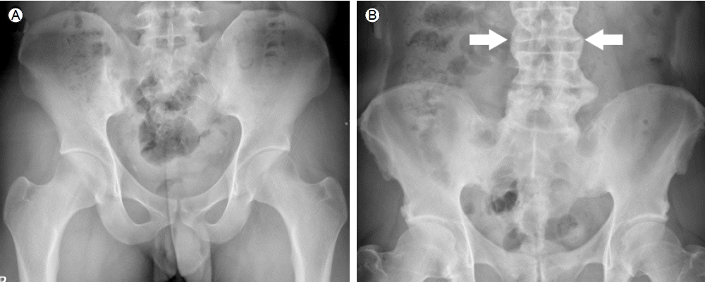
Ļ░Ģņ¦üņä▒ ņ▓ÖņČöņŚ╝ ĒÖśņ×ÉņØś ņ▓ÖņČöĻ┤ĆņĀł ņ╣©ļ▓öņØĆ ļīĆĻ░£ ņ▓£ņןĻ┤ĆņĀłļČĆĒä░ ņŗ£ņ×æļÉ£ļŗż. Ļ│©ļ░ś ņĀäĒøäņØś ļŗ©ņł£ X-ņäĀ Ļ▓Ćņé¼ņŚÉņä£ņä£ ņ▓£ņןĻ┤ĆņĀłņŚ╝ņØś ļŗ©Ļ│äļź╝ ĒÅēĻ░ĆĒĢĀ ņłś ņ׳ļŗż[11] (Table 2, Fig. 1). ļŗ©ņł£ X-ņäĀ Ļ▓Ćņé¼ļŖö Ļ░Ģņ¦üņä▒ ņ▓ÖņČöņŚ╝ ĒÖśņ×ÉņØś ņ▓ÖņČöņ▓┤(vertebral body) ļ│ĆĒÖö-ņØĖļīĆĻ▓░ĒĢ®(syndesmophyte), ņ▓ÖņČöņ▓┤ņØś ņé¼Ļ░üĒÖö(squaring), ņ£ĄĒĢ®(fusion)-ļź╝ ļ│┤ļŖö ļŹ░ļÅä ņé¼ņÜ®ļÉ£ļŗż. ņ▓ÖņČöņ▓┤ ļ│ĆĒÖöĻ░Ć ņ׳ņØä ļĢī Ļ░Éļ│äĒĢ┤ņĢ╝ ĒĢĀ ņ¦łĒÖśņ£╝ļĪ£ ļ»Ėļ¦īņä▒ ņøÉļ░£ņä▒ ļ╝łļīĆĻ│╝Ļ│©ņ”Ø(diffuse idiopathic skeletal hyperostosis, DISH)ņØä ļōż ņłś ņ׳ļŗż. ļæÉ ņ¦łĒÖś ļ¬©ļæÉ ņ▓ÖņČöņØś ņÜ┤ļÅÖņĀ£ĒĢ£, ņ▓ÖņČöņØś ļ╝łļÅīĻĖ░ Ļ│╝ĒśĢņä▒ņØ┤ ļÅÖļ░śļÉĀ ņłś ņ׳ņ¦Ćļ¦ī DISHņŚÉņä£ļŖö ņ▓ÖņČöņØś Ēć┤Ē¢ēņä▒ ļ│ĆĒÖöĻ░Ć Ļ┤Ćņ░░ļÉśĻ│Ā ņ▓£ņןĻ┤ĆņĀłņØś ņ╣©ļ▓öņØ┤ ļō£ļ¼╝ļŗżļŖö ņ░©ņØ┤ņĀÉņØ┤ ņ׳ļŗż. ņØ┤ ņÖĖņŚÉļÅä ņ▓£ņןĻ┤ĆņĀłņØś Ļ│©Ļ┤ĆņĀłņŚ╝, ņČ£ņé░Ļ│╝ ņŚ░Ļ┤ĆļÉ£ ņ▓£ņןĻ┤ĆņĀł ļ│ĆĒÖö(osteitis condensans ilii) ļō▒ņØ┤ Ļ░Éļ│äņ¦äļŗ©ņŚÉ ĒżĒĢ©ļÉ£ļŗż. ļŗ©ņł£ X-ņäĀ Ļ▓Ćņé¼ļŖö Ļ▓Ćņé¼ ļ╣äņÜ®ņØ┤ ņĀĆļĀ┤ĒĢśĻ│Ā ĒīÉļÅģņØ┤ ņÜ®ņØ┤ĒĢśļéś Ļ│©ļ│ĆĒÖöĻ░Ć ļ░£ņāØĒĢśĻĖ░ ņĀäņŚÉļŖö ņ¦äļŗ©ņØä ļé┤ļ”┤ ņłś ņŚåļŗżļŖö ļŗ©ņĀÉņØ┤ ņ׳ļŗż.
ņ×ÉĻĖ░Ļ│Ąļ¬ģņśüņāü(magnetic resonance imaging, MRI)
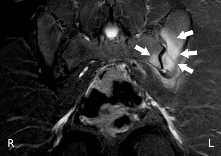
ņ▓£ņןĻ┤ĆņĀł MRIĻ░Ć ņĀ£Ļ│ĄĒĢ┤ņżä ņłś ņ׳ļŖö ņŻ╝ņÜö ņĀĢļ│┤ļŖö Ļ┤ĆņĀł Ēś╣ņØĆ Ļ┤ĆņĀł ņŻ╝ņ£äņØś ŌĆśņŚ╝ņ”ØŌĆÖ ņåīĻ▓¼ņØ┤ļŗż[12]. MRIņØś T2 fat suppression ņśüņāüņØä ĒåĄĒĢśņŚ¼ Ļ┤ĆņĀł ņŻ╝ņ£äņØś Ļ│©ņłś ļČĆņóģ(bone marrow edema)ņØä ļ│┤ņŚ¼ņżīņ£╝ļĪ£ņŹ© ļŗ©ņł£ X-ņäĀ Ļ▓Ćņé¼ļ│┤ļŗż ņĪ░ĻĖ░ņŚÉ ņ▓£ņןĻ┤ĆņĀłņŚ╝ņØä ņ¦äļŗ©ĒĢĀ ņłś ņ׳ļŗż(Fig. 2). ņĪ░ņśüņĀ£ļź╝ ņé¼ņÜ®ĒĢ£ T1 Ļ░ĢņĪ░ņśüņāüļÅä ņ▓£ņןĻ┤ĆņĀł ņŻ╝ņ£äņØś Ļ│©ņŚ╝(osteitis)ņØä ņ¦äļŗ©ĒĢśļŖö ļŹ░ ļÅäņøĆņØä ņżä ņłś ņ׳ļŗż. ņØ┤ļĪ£ņŹ© MRIļĪ£ ļŗ©ņł£ X-ņäĀ Ļ▓Ćņé¼ ņåīĻ▓¼ļ¦īņ£╝ļĪ£ļŖö ļČłļČäļ¬ģĒĢ£ ŌĆśļ╣äļ░®ņé¼ņäĀ ņ▓£ņןĻ┤ĆņĀłņŚ╝(non-radiographic sacroiliitis)ŌĆÖņØä ņ¦äļŗ©ĒĢĀ ņłś ņ׳Ļ▓ī ļÉśņŚłļŗż[12]. ĒĢśņ¦Ćļ¦ī MRIļŖö Ļ│ĀĻ░ĆņØś Ļ▓Ćņé¼ņØ┤Ļ│Ā Ļ░Ģņ¦üņä▒ ņ▓ÖņČöņŚ╝ ĒÖśņ×ÉņØ┤Ļ▒░ļéś ĻĘĖ ņ¦äļŗ©ņØ┤ ņØśņŗ¼ļÉśļŖö Ļ▓ĮņÜ░ 1ĒÜīņŚÉ ĒĢ£ĒĢśņŚ¼ ļ│┤ĒŚśĻĖēņŚ¼Ļ░Ć ņØĖņĀĢļÉśĻ│Ā ņ׳ļŗż. ļĢīļĪ£ļŖö MRIņŚÉņä£ ņŚ╝ņ”ØņØ┤ļØ╝Ļ│Ā ņāØĻ░üļÉśļŖö ņåīĻ▓¼ņØ┤ ņ¦Ćļ░®ļ│Ćņä▒(fat degeneration)Ļ│╝ Ļ░Éļ│äņØ┤ ņ¢┤ļĀżņÜĖ ņłś ņ׳ļŗż[13]. ņ▓ÖņČö MRIņŚÉņä£ ņ▓ÖņČöņ▓┤ņØś ļ¬©ņä£ļ”¼ņŚÉ ļ│┤ņØ┤ļŖö ņŚ╝ņ”Øļ│æļ│ĆņØ┤ ņŗĀņāØĻ│©ņØś ņĀäĻĄ¼ļ│æļ│ĆņØ┤ļØ╝ļŖö Ļ░ĆņäżņØ┤ ļīĆļæÉļÉśņŚłņ¦Ćļ¦ī ņØ┤ļ¤¼ĒĢ£ ļ│æļ│Ć ņżæ ņØ╝ļČĆņŚÉņä£ļ¦ī ņØĖļīĆĻ▓░ĒĢ®, ņŗĀņāØĻ│© ĒśĢņä▒ņ£╝ļĪ£ ņ¦äĒ¢ēĒĢśļŖö Ļ▓āņ£╝ļĪ£ ļ░ØĒśĆņĪīļŗż[14]. ņĄ£ĻĘ╝ ņŚ░ĻĄ¼ņŚÉņä£ļŖö ņŚ╝ņ”Øņä▒ ļ│æļ│ĆĻ│╝ ņ¦Ćļ░®ļ│Ćņä▒ņØ┤ ļÅÖņŗ£ņŚÉ ņĪ┤ņ×¼ĒĢśļŖö ņ▓ÖņČöņ▓┤ ļ¬©ņä£ļ”¼ņŚÉņä£ ņŗĀņāØĻ│©ņØ┤ ļ¦ÄņØ┤ ĒśĢņä▒ļÉ£ļŗżļŖö ņŻ╝ņןņØ┤ ņĀ£ĻĖ░ļÉśņŚłļŗż[15].
ņ╗┤Ēō©Ēä░ ļŗ©ņĖĄ ņ┤¼ņśü
ņ▓£ņןĻ┤ĆņĀłņØś ņ╗┤Ēō©Ēä░ ļŗ©ņĖĄ ņ┤¼ņśüņØĆ Ļ│©ļ»Ėļ×Ć(bone erosion), Ļ│©Ļ▓ĮĒÖö(bone sclerosis) ļō▒ņØä Ļ┤Ćņ░░ĒĢśļŖö ļŹ░ ņ£ĀņÜ®ĒĢśļéś ņ┤łĻĖ░ ņŚ╝ņ”Ø ļ│ĆĒÖöļź╝ Ļ┤Ćņ░░ĒĢśļŖö ļŹ░ļŖö ņĀüĒĢ®ĒĢśņ¦Ć ņĢŖļŗż.
Ļ│©ņŖżņ║ö(bone scan, bone scintigraphy)
Ļ│©ņŖżņ║öņØĆ Ļ░Ģņ¦üņä▒ ņ▓ÖņČöņŚ╝ ĒÖśņ×ÉņØś ņ▓£ņןĻ┤ĆņĀł, ņ▓ÖņČöĻ┤ĆņĀł, ĻĘĖļ”¼Ļ│Ā ļ¦Éņ┤łĻ┤ĆņĀł ņ╣©ļ▓öņØä ĒÅēĻ░ĆĒĢśļŖö Ļ▓Ćņé¼ļĪ£ ņé¼ņÜ®ļÉśņ¢┤ ņÖöļŗż. Ļ│©ļ░ś Ēøäļ®┤ņāüņØś SI/S (sacroiliac joint/sacrum) ļ╣äņ£©ņØ┤ ņāüņŖ╣ļÉśņ¢┤ ņ׳ņ£╝ļ®┤ ņ▓£ņןĻ┤ĆņĀłņŚ╝ņØä ņŗ£ņé¼ĒĢśļŖö ņåīĻ▓¼ņØ┤ļŗż. ĒĢśņ¦Ćļ¦ī ņØ┤ Ļ▓Ćņé¼ņØś ņĀ£ĒĢ£ņĀÉņØĆ ĒĢ┤ņāüļÅäļéś Ļ▓Ćņé¼ ĒŖ╣ņØ┤ļÅäļŖö ļé«ļŗżļŖö Ļ▓āņØ┤ļ®░ ņĄ£ĻĘ╝ ļ│┤Ļ│ĀņŚÉ ņØśĒĢśļ®┤ Ļ│©ņŖżņ║öņØ┤ ņĪ░ĻĖ░ Ļ░Ģņ¦üņä▒ ņ▓ÖņČöņŚ╝ņØś ņ¦äļŗ©ņŚÉ ļÅäņøĆņØ┤ ļÉśņ¦Ć ņĢŖļŖöļŗżĻ│Ā ĒĢśņśĆļŗż[16]. ĒĢśņ¦Ćļ¦ī Ļ░Ģņ¦üņä▒ ņ▓ÖņČöņŚ╝ ĒÖśņ×ÉņŚÉņä£ ļ¦Éņ┤ł Ļ┤ĆņĀł ņ╣©ļ▓öņØ┤ļéś ĒØēņćäĻ│©Ļ┤ĆņĀł, ļŖæņŚ░Ļ│©ņŚ╝(costochondritis)ņØä ĒÅēĻ░ĆĒĢśļŖöļŹ░ļŖö ļÅäņøĆņØä ņżä ņłś ņ׳ļŗż.
Ļ░Ģņ¦üņä▒ ņ▓ÖņČöņŚ╝ņØś ņ¦äļŗ©
Ļ░Ģņ¦üņä▒ ņ▓ÖņČöņŚ╝ņØś ļČäļźś ĻĖ░ņżĆņØĆ 1984ļģäļÅäņŚÉ ņĀ£ņĢłļÉ£ modified New York criteriaĻ░Ć ļäÉļ”¼ ņé¼ņÜ®ļÉśĻ│Ā ņ׳ļŗż[11] (Table 3). ņØ┤ ĻĖ░ņżĆņŚÉ ņØśĒĢśļ®┤ Ļ░Ģņ¦üņä▒ ņ▓ÖņČöņŚ╝ņ£╝ļĪ£ ļČäļźśĒĢśĻĖ░ ņ£äĒĢ┤ņä£ļŖö ļŗ©ņł£ X-ņäĀ Ļ▓Ćņé¼ņŚÉņä£ ņ▓£ņןĻ┤ĆņĀłņŚ╝ņØ┤ ņ¢æņĖĪņ£╝ļĪ£ grade 2 ņØ┤ņāüņØ┤Ļ▒░ļéś ņØ╝ņĖĪņ£╝ļĪ£ grade 3 ņØ┤ņāüņØ┤ņ¢┤ņĢ╝ ĒĢ£ļŗż(Table 2, Fig. 1). ņØ┤ļŖö ņ¦äĒ¢ēļÉ£ ņ¦łĒÖśņØś ņ¦äļŗ©ņŚÉļŖö ņ£ĀņÜ®ĒĢśļéś ļŗ©ņł£ X-ņäĀ Ļ▓Ćņé¼ņŚÉņä£ Ļ┤ĆņĀł ļ│ĆĒÖöĻ░Ć ļ»Ėļ»ĖĒĢśļ®┤ Ļ░Ģņ¦üņä▒ ņ▓ÖņČöņŚ╝ ņ¦äļŗ©ņØä ļé┤ļ”┤ ņłś ņŚåņ£╝ļ®░ ņ▓£ņןĻ┤ĆņĀł ļ░Å ņ▓ÖņČöļ│ĆĒÖöņŚÉļ¦ī ņ¦æņżæĒĢĀ ļ┐É Ļ┤ĆņĀł ņÖĖ ņ×äņāüņāüņØä ĒżĒĢ©ĒĢśņ¦Ć ņĢŖļŖöļŗżļŖö ņĀ£ĒĢ£ņĀÉņØ┤ ņ׳ļŗż. ņØ┤ Ļ░ÖņØ┤ ņČĢņä▒ Ļ┤ĆņĀłņØä ņ╣©ļ▓öĒĢśļŖö ņ▓ÖņČö Ļ┤ĆņĀłņŚ╝ ņ¦łĒÖśĻĄ░ņØä ļŹö ņĪ░ĻĖ░ņŚÉ ņ¦äļŗ©ĒĢśĻĖ░ ņ£äĒĢ┤ MRI ņåīĻ▓¼ņØä ĒżĒĢ©ĒĢśļŖö ĒĢ£ĒÄĖ Ļ┤ĆņĀł ņÖĖ ņ×äņāüņāü, Ļ░ĆņĪ▒ļĀź ļ¼Ėņ¦äņØä ļŹöĒĢśļŖö ņāłļĪ£ņÜ┤ ļČäļźśĻĖ░ņżĆņØ┤ ņĀ£ņĢłļÉśņŚłļŗż[17] (Table 4). ņ×äņāüņØ┤ļéś ņ×äņāüņŗ£ĒŚśņŚÉņä£ ĒÖ£ņÜ®ļÉśļŖö Ļ░Ģņ¦üņä▒ ņ▓ÖņČöņŚ╝ ļČäļźśĻĖ░ņżĆņØĆ Ēśäņ×¼ modified New York criteriaņØ┤ņ¦Ćļ¦ī Ē¢źĒøäļŖö Ļ░Ģņ¦üņä▒ ņ▓ÖņČöņŚ╝ņØä ĒżĒĢ©ĒĢ£ ņČĢņä▒ ņ▓ÖņČöĻ┤ĆņĀłņŚ╝ņØä ļīĆņāüņ£╝ļĪ£ ĒĢśņŚ¼ ņ×äņāüņŚ░ĻĄ¼ ļ░Å ņ╣śļŻī ņ¦Ćņ╣© ņĀ£ņĀĢņØ┤ ņ¦äĒ¢ēļÉĀ Ļ▓āņ£╝ļĪ£ ņĀäļ¦ØļÉ£ļŗż.
 PDF Links
 PubReader
 ePub Link
 Full text via DOI
 Download Citation
 Print
Print


