신세포암의 바터 팽대부 전이에 의한 급성 재발성 췌장염
A Case of Acute Recurrent Pancreatitis Caused by Metastasis of Renal Cell Carcinoma to the Ampulla of Vater
Article information
Abstract
신세포암은 드물지만 바터 팽대부로 전이될 수 있으며, 이로 인해 췌액의 흐름을 방해하여 재발성 급성 췌장염을 유발할 수 있다. 수술로 절제가 어려운 경우 내시경 역행성 담췌관 조영술 시술을 통한 주췌관 내로의 스텐트 삽입이 췌장염 치료에 도움이 될 수 있다.
Trans Abstract
Metastasis of renal cell carcinoma to the ampulla of Vater is a rare occurrence. We report a case of acute recurrent pancreatitis caused by metastasis of renal cell carcinoma to the ampulla of Vater. The patient manifested acute recurrent pancreatitis 10 years after a nephrectomy for renal cell carcinoma and was presumed to have obstructive pancreatitis secondary to an ampullary mass. Duodenoscopy revealed an ampullary mass, and the patient was finally diagnosed with metastatic renal cell carcinoma. With insertion of a pancreatic stent via endoscopic retrograde cholangiopancreatography, the acute pancreatitis was resolved. When an ampullary mass is observed in a patient with a history of renal cell carcinoma, the possibility of ampullary metastasis of the renal cell carcinoma should be considered, as this is a potential etiology of acute recurrent pancreatitis. (Korean J Med 2013;84:254-258)
서 론
신세포암의 전이는 폐, 림프절, 뼈, 간, 뇌, 반대측 신장, 부신 등에 흔히 발생한다[1]. 신세포암이 바터 팽대부에 전이되는 경우는 매우 드문 경우로 국외에서는 소수 보고된 적이 있으나 아직 국내에서는 보고된 적이 없다. 신세포암은 처음 수술로 종양이 성공적으로 제거되었다 하더라도 수년이 지나 전이가 발견되는 경우가 드물지 않아서, 길게는 첫 수술 이후 17년이 지나서 발생한 경우도 보고된 적이 있다[2]. 본 저자들은 신세포암으로 근치적 우측 신장 절제술을 받은지 10년이 지난 환자에서 신세포암의 바터 팽대부 전이를 조직검사를 통해 확인하였으며, 이에 의하여 반복적으로 발생한 급성 췌장염을 내시경을 이용한 췌관내 스텐트 삽입술로 호전시켰기에 이와 관련한 문헌고찰과 함께 보고하는 바이다.
증 례
환 자: 김○○, 남자 62세
주증상: 3일 전 발생한 복통
현병력: 내원 10년 전 통증을 동반하지 않은 육안적 혈뇨로 본원 비뇨기과를 방문하여 신세포암으로 진단되어 우측 신장 근치적 절제술을 시행 받았다. 이후로 재발하지 않고 특별한 증상 없이 잘 지냈다.
내원 22개월 전 갑자기 발생한 심한 상복부 통증으로 본원 소화기 내과에 입원하였고 혈중 아밀라제 560 U/L, 리파제 280 U/L로 기준치보다 3배 이상 상승하여 급성 췌장염 소견 보였다. 당시 시행한 복부 전산화 단층 촬영에서 주췌관이 확장되었으며 바터 팽대부에 종괴성 음영이 의심되었다(Fig. 1). 십이지장경 검사 소견상 바터 팽대부에 노란색 삼출물로 덮힌 종괴가 관찰되었으며(Fig. 2) 조직검사 결과 신세포암의 바터 팽대부 전이로 진단되었다(Fig. 3). 확장된 주췌관에 플라스틱 스텐트(직경 10 Fr, 길이 8 cm, Cook사, USA)를 삽입하였고, 임상양상 호전되어 췌관 스텐트 유지하였다. 추가로 시행한 양전자 방출 단층 촬영에서 폐 전이가 발견되어 항암 치료(sunitinib) 시행하기로 하고 퇴원하였다. 퇴원 2달 후 췌관 스텐트를 교환하였으며, 이후 3달 후 시행한 복부 전산화 단층 촬영에서 기존의 췌관 확장 소견이 많이 호전되고 바터 팽대부 종괴 크기가 항암제 투여 효과로 감소되어 췌관 스텐트를 완전히 제거하였고 항암 치료는 지속하였다.

Abdominal computed tomography shows an enhancing mass (arrows) at the ampulla of Vater (A) and diffuse upstream pancreatic duct dilatation (B).
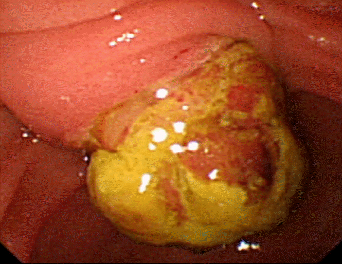
Duodenoscopic finding shows the yellow exudate-covered ampullary mass with a lobulating contour.

Pathologic features of renal and ampulla of Vater tumors. The nephrectomy specimen shows clear cell renal cell carcinoma comprising ovoid to polygonal clear cells with a regular network of small thin-walled vessels (A, hematoxylin and eosin stain, × 200). The ampulla of Vater tumor shows clear to eosinophilic ovoid cells with marked degenerative changes and an abundant vascular network (B, hematoxylin and eosin stain, × 200). The tumor cells are immunopositive for pancytokeratin (C, cytokeratin immunohistochemical staining, × 200).
스텐트 제거 이후로 17개월 동안 증상 없이 잘 지내던 중 내원 3일 전부터 복통이 발생하여 응급실 통해 입원하였다.
과거력: 특이사항 없었다.
가족력: 특이사항 없었다.
진찰 소견: 내원 당시 혈압 115/70 mmHg, 맥박 92회/분, 호흡수 18회/분, 체온 36.5℃로 측정되었다. 의식은 명료하였고 급성 병색을 보였다. 결막은 창백하지 않았고, 공막에 황달소견은 없었다. 호흡음은 정상으로 수포음 등의 이상 호흡음은 들리지 않았고, 심음은 규칙적이며 심잡음은 들리지 않았다. 상복부에 지속적인 통증 및 압통을 호소하였고, 통증은 등으로 방사되는 양상을 보였다. 복부 팽만과 장음 감소의 소견을 보였으며 반동 압통은 없었다. 피부에 특이소견은 없었고, 신체 검사 소견상 다른 특이소견은 없었다.
검사 소견: 혈액 검사에서 백혈구 4,600/mm3, 혈색소 12.4 g/dL, 헤마토크리트 38.7%, 혈소판 201,000/mm3였다. 혈청 췌장 효소 수치는 아밀라제 892 U/L, 리파제 755 U/L로 정상보다 3배 이상 상승되었다. 총 단백 6.6 g/dL, 알부민 4.1 g/dL, AST 42 IU/L, ALT 25 IU/L, 알칼리인산분해효소(ALP) 49 IU/L, 총 빌리루빈 0.5 mg/dL, γ-GT 18 IU/L, 크레아티닌 1.51 mg/dL, CRP 0.1 mg/dL였다. 전해질 검사에서 Na+ 136 mmol/L, K+ 4.3 mmol/L, Cl- 104 mmol/L, 칼슘 9.1 mg/dL, 인 2.5 mg/dL으로 측정되었다.
췌담도 자기공명영상 검사에서 바터 팽대부 종괴가 다시 커졌으며, 이로 인해 주췌관 및 총담관의 미만성 확장 소견이 관찰되었다(Fig. 4). 기존의 폐 전이는 이전과 비슷하였으며, 다른 부위로의 재발은 없었다.

Magnetic resonance cholangiopancreatography shows the ampullary mass (arrows) and upstream pancreatic and biliary duct dilatation.
치료 및 경과: 내시경 역행성 췌관 조영술에서 췌장 두부쪽의 주췌관 협착으로 상류쪽 췌관이 확장되었고, 이에 대하여 완전 피막형 자가 팽창성 금속 스텐트(직경 6 mm, 길이 8 cm, 태웅메디컬, 한국)를 주췌관 내로 삽입하고 하였다(Fig. 5). 확장된 총담관에 대해서는 췌관내 금속 스텐트 삽입 전에 내시경 조임근 절개술을 시행하였다. 스텐트 삽입 이후 혈청 췌장 효소 수치 정상화되었고, 복통 호전되었다. 식이 진행 후 별다른 증상 없어 항암제(sunitinib) 복용 지속하며 외래 경과관찰 중이다.

Duodenoscopic and endoscopic retrograde pancreatographic findings. A fully-covered self-expandable metallic stent has been inserted into the main pancreatic duct via endoscopic retrograde cholangiopancreatography (A and B).
고 찰
신세포암의 자연 경과는 매우 다양해서 예견하기 힘들다. 신장 절제술로 질병의 퇴치와 완전 치유가 가능한 경우도 있고, 오랜 기간 동안의 잠복기와 예상하지 못한 해부학적 위치에 전이가 발생하는 경우도 있다[3]. 신세포암은 림프성 또는 혈행성으로 전이되며, 복막 전이나 인접 구조물로의 직접 침범에 의해서도 전이될 수 있다[4]. 흔한 전이 장기는 폐(75%), 림프절(36%), 뼈(20%), 간(18%) 등으로 바터 팽대부 전이는 아주 드문 경우로 아직까지 국내에 보고된 바는 없다.
바터 팽대부의 전이성 암은 흔치 않으나, 흑색종과 림프종에 의한 경우가 상대적으로 많다[5]. 바터 팽대부에 전이되는 경우 담관의 폐색으로 황달이, 췌관의 폐색으로 복통을 동반한 급성 췌장염이 발생할 수 있으며, 종양의 궤양화로 인해 위장관 출혈이 발생할 수도 있다[6].
본 증례의 환자는 신세포암으로 인한 신장 절제술 시행 후 10년이 지나서 복통을 주소로 내원하였다. 급성 췌장염 소견으로 발생 원인을 찾고자 내시경 역행성 담췌관 조영술을 시행하였고, 바터 팽대부에 종괴가 관찰되어 조직검사를 시행하였다. 10년 전 수술 결과를 다시 검토하면 절제된 검체에서 우측 신장의 하극에 경계가 좋은 피막으로 둘러 쌓인 난형의 고형 종양이 보였고, 현미경적 검사에서 종양은 투명 세포 신장 세포 암이었다(Fig. 3A). 10년이 지난 후 내원하여 확인된 바터 팽대부 종괴는 투명한 호산구성 비전형 난형세포로 구성되어 있으며, 퇴행성 변화와 작은 혈관들이 풍부하게 관찰되었다(Fig. 3B). 세포들은 pan-cytokeratin 면역 양성을 보였으며, 이는 종양이 전이성 신세포암임을 지지하는 소견이다(Fig. 3C).
신세포암의 바터 팽대부 전이에 대한 치료 선택은 병변의 정도 등에 따라 각각 달라질 수 있다. 수술로는 췌십이지장 절제술이 시행될 수 있다. 의학적으로나 기술적으로 가능하다면 완전히 전이성 병변을 제거하는 것이 치료의 목표가 되어야 한다. 종양이 진행되어 수술이 어려운 경우 방사선 치료, 항암 치료, 면역 자극 치료(인터루킨-2) 등이 시행될 수 있다[7]. 본 환자는 바터 팽대부 전이 외에도 양전자 방출 단층 촬영술 및 흉부 전산화 단층 촬영 결과에서 폐전이가 추가로 의심되어 수술적인 치료를 하지 않고 sunitinib을 이용한 고식적 항암 치료를 시행하고 있다.
알코올의 남용이나 담낭 결석 등도 반복적인 급성 췌장염을 일으킬 수 있지만, 해부학적으로 췌액의 흐름을 방해하는 인자도 췌장염을 유발할 수 있다. 췌관의 폐색을 야기할 수 있는 원인으로는 크게 (1) 선천적인 해부학적 변이(분리 췌장이 가장 흔함), (2) 유두, 주췌관, 유두 근처의 십이지장을 침범하는 악성 종양, (3) 오디 조임근 기능 이상 등으로 나누어 생각해 볼 수 있다. 이런 기계적인 요인들이 이자액이 십이지장으로 흘러가는데 일시적 혹은 지속적으로 폐색을 유도하여 췌관 내 압력을 증가시키고 그 결과 반복적인 췌장염을 야기할 수 있다[8]. 팽대부 종양에 의해 십이지장 주유두 입구가 막히게 되면 위와 같은 기전으로 급성 췌장염을 야기할 수 있는데, 본 증례의 환자도 이러한 경우에 해당된다.
내시경적 스텐트 삽입술은 악성 종양에 의한 췌담관 협착의 치료에 있어서 효과적인 수단으로 알려져 있으며, 플라스틱 스텐트와 자가 팽창성 금속 스텐트(self-expandable metallic stent) 등이 사용되고 있다. 주췌관에는 보통 플라스틱 스텐트를 사용하지만, 수술의 적응증이 되지 않을 경우 플라스틱 스텐트에 비해 자주 교환할 필요가 없는 금속 스텐트를 사용할 수 있다. 이것은 자가 팽창성 금속 스텐트는 플라스틱 스텐트에 비해서 스텐트 내경이 크고, 우월한 개방성을 보이기 때문이다. 그러나 악성 종양이 금속망 내로 증식하여 스텐트 폐색이 발생할 수 있고 스텐트 제거 시 어려움이 있을 수 있는데, 이런 점을 극복하기 위해 완전 피막형 자가 팽창성 금속 스텐트(fully-covered self-expandable metallic stent)가 개발되어 사용되고 있다[9,10]. 향후 치료 효과와 안전성, 제거의 용이함, 비용 대비 효과 등에 대한 대규모의 전향적 연구가 필요한 상태이다.
본 환자의 경우 내원시 총담관 확장 소견이 동반되어 있어 췌관 스텐트 삽입 이전에 담관 내시경 조임근 절개술을 시행한 환자로 췌관 스텐트 삽입 후에 담도 폐쇄의 소견은 없었다. 그러나 일반적으로 직경이 큰 금속 스텐트를 췌관에 삽입한 경우 담도 폐쇄의 증상이 나타날 수 있는데, 이러한 경우 담관 내시경 조임근 절개술 및 필요에 따른 담도 스텐트 삽입을 통하여 회복할 수 있다[10].