담낭에서 유례한 과립막 세포종 1예
Granular Cell Tumor Originating from Gallbladder
Article information
Trans Abstract
Granular cell tumors are rare benign tumors that arise from Schwann cells. They are especially rare in the gallbladder; indeed, only four cases have been reported in the English-language literature. This paper reports a granular cell tumor in the gallbladder and is the first such report in a Korean woman. She was admitted to the hospital with jaundice and fever and was diagnosed with acute hepatitis A. While hospitalized, a well-demarcated round mass was incidentally found in her gallbladder. The acute hepatitis A improved with conservative care, but the mass did not change in size after 4 months. The tumor was resected with the gallbladder during laparoscopic surgery, and was found to be a granular cell tumor. The tumor was composed of sheets or irregular fascicles of large polygonal or spindle cells with plump eosinophilic granular cytoplasm that was immunohistochemically positive for S-100 and neuron-specific enolase and negative for neurofilament. (Korean J Med 2012;83:624-628)
INTRODUCTION
Granular cell tumors (GCTs) are uncommon, relatively benign neoplasms that are thought to originate from Schwannlike mesenchymal cells [1]. GCTs are common in the fourth to sixth decades of life and are rare in children [2]. They can develop in any part of the body, but commonly occur in the skin, subcutis, tongue, and oral cavity [3]. GCTs of the biliary tract are quite rare and typically occur in young African- American women presenting with abdominal pain and jaundice [4,5]. Anatomically, the common bile duct (CBD) is the most frequent site of involvement, followed by the cystic duct and common hepatic duct. GCTs of the gallbladder comprise 1-2% of biliary GCTs [4,5]. To our knowledge, only four granular cell tumors in the gallbladder have been described in the English-language literature [6-9].
We herein present a case of granular cell tumor in the gallbladder of a young female Korean. She was initially diagnosed with acute hepatitis A, which improved with conservative management. A mass-like lesion near the gallbladder was found incidentally and was identified as a granular cell tumor after surgical removal and pathological examination.
CASE REPORT
A 36-year-old woman preseted with a 3-day history of fever and chills. Her medical history was unremarkable; she was taking pheniramine for chronic urticaria. Her conjunctivae were icteric, but she had no abdominal pain. Initial laboratory findings were hemoglobin (Hgb) of 14.4 g/dL (normal, 12-16 g/dL), white blood cell (WBC) count of 6.08 × 10³/μL(normal, 4-10 103/μL), platelets of 164 × 103/μL (normal, 150-450 103/μL), aspartate transaminase of 277 IU/L (normal, 5-40 IU/L), alanine transaminase of 983 IU/L (normal, 5-40 IU/L), total bilirubin of 6.9 mg/dL (normal, 0.2-1.2 mg/dL), alkaline phosphatase of 451 IU/L (normal, 35-123 IU/L), IgM anti-hepatitis A virus (HAV) antibody-positive, IgG anti-HAV antibody-negative, HBsAg-negative, and antihepatitis C virus (HCV)-negative. Abdominal ultrasonography showed a 3-cm round, well-demarcated, isoechoic mass with peripheral hypervascularity in the neck of the gallbladder(Fig. 1A). There were no abnormal findings in the liver, CBD, or pancreas. Abdominal computed tomography (CT) showed a well-demarcated, slightly heterogeneously enhancing lesion in the neck of the gallbladder (Fig. 1C, 1D). Whether the mass was located inside or outside the gallbladder on imaging was uncertain.

(A) Abdominal ultrasound shows a well-demarcated, isoechoic mass with peripheral hypervascularity in the gallbladder neck. (B, C) Abdominal CT shows a well-demarcated, slightly heterogeneously enhancing lesion in the gallbladder.
Initially, the patient was treated conservatively for acute hepatitis A and improved with no complications. Follow-up abdominal CT performed 4 months later showed no change in the previously noted lesion. To confirm the diagnosis, laparoscopic surgery was performed. At surgery, there was an approximately 3-cm ovoid soft tissue mass on the infundibulum of the gallbladder.
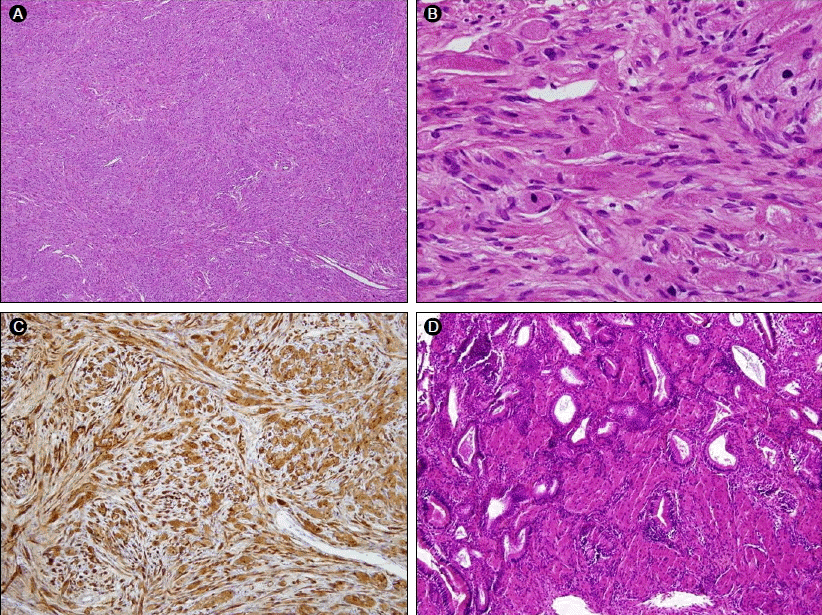
Pathologically, the specimen contained sheets or irregular fascicles of large polygonal or spindle cells with distinct cell borders, small nuclei, and plump eosinophilic granular cytoplasm. Grossly, the mass was thought to be located in the soft tissue outside the gallbladder, but histologically, the tumor cells originated from the mucosal and muscular layers of the gallbladder. The tumor cells were positive for S-100 protein and neuron-specific enolase (NSE), but negative for neurofilament (Fig. 2). Ultimately, the resected tumor was diagnosed as a granular cell tumor in the gallbladder, and the patient has been followed with no complications or tumor recurrence.

(A) The tumor consists of sheets or irregular fascicles of large polygonal or spindled cells (H&E, × 100). (B) Tumor cells have plump, eosinophilic, granular cytoplasm (H&E, × 400). (C) Tumor cells are positive for S-100 protein (S-100 protein immunostaining, × 200). (D) Tumor cells are present in the mucosal and muscular layers of the gallbladder (H&E, × 100).
DISCUSSION
Abrikossoff first described granular cell tumors in 1926 [10]. He called these tumors myoblastomas because he thought that they originated in striated muscle. However, based on immunohistochemical and electron microscopic findings, it is now widely accepted that these tumors arise from Schwann cells [11,12].
GCTs of the biliary tract are very rare, and Coggins reported the first case of a GCT in the biliary tree in 1952 [13]. Approximately 80 such tumors have been reported, accounting for fewer than 1% of all GCTs [4,5]. The differential diagnosis of GCTs in the biliary tract includes cholangiocarcinoma, primary sclerosing cholangitis, biliary stricture, adenoma, papilloma, and choledochal cyst [4,14]. GCTs in the gallbladder are very rare, accounting for 1-2% of biliary GCTs [4]. The first case of a GCT in the gallbladder was reported in 1977, and only four cases of GCTs in the gallbladder have been reported to-date (Table 1). Three cases occurred only in the gallbladder, and one case involved multiple tumors in the gallbladder, cystic duct, and CBD [6-9].
Biliary GCTs usually occur in young African-American women, and the most common symptoms are abdominal pain and jaundice [4]. Little is known about the clinical characteristics of gallbladder GCTs because of their rarity. In the previous three cases of gallbladder GCT, the patients had symptoms such as epigastric or right upper quadrant pain, which was thought to have been caused by gallbladder stones, CBD stones, or a CBD granular cell tumor. In addition, the GCTs seemed to have been found incidentally [6-8]. There was no information on one case with the exception that it occurred in the gallbladder [9]. Cholangiograms were performed in three cases but failed to visualize the gallbladder. Sonograms were performed in two cases and showed a collapsed gallbladder [6-8].
Our case was also found incidentally, and the patient had no specific symptoms. A round, mass-like lesion near the gallbladder should be differentiated from an enlarged lymph node and exophytic liver or gallbladder tumors, including GCTs.
Grossly, GCTs appear as yellow or yellow-whitish solitary nodules and are usually smaller than 3 cm [4]. In our case, the GCT was removed laparoscopically with the gallbladder, so we could not determine its exact gross appearance. Histologically, GCTs consist of large polygonal cells with eosinophilic gran ular cytoplasm and centrally located, small, dark, uniform nuclei. These eosinophilic granules are strongly positive for PAS, S-100, and NSE. Individual cells or small clusters are separated by thin layers of fibrous connective tissue and can infiltrate the surrounding fat or fibrous tissue. Malignant GCTs are very rare, comprising less than 2% of all GCTs, but no malignant GCT has been reported in the biliary tract [4]. The malignant potential of GCTs increases with a size greater than 4 cm, necrosis, pleomorphism, increased mitotic activity, and spindle-shaped cells. It is often difficult to distinguish benign from malignant GCTs. Apparent infiltration of the tumor into the periphery is a normal finding in benign GCTs [15]. In our case, the tumor cells were also found in the mucosa and muscular layer of the gallbladder with no pathological findings indicating malignancy.
The treatment of GCTs is surgical removal with tumor-free margins because they can recur locally with incomplete resection. Regular follow-up is necessary after the surgical removal of GCTs to check for local recurrence and potential malignancy.
