


мДЬ л°†
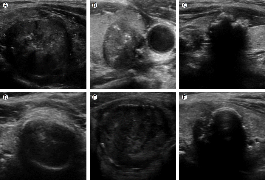
к∞СмГБмД† міИмЭМнММлКФ к∞СмГБмД† к≤∞м†ИмЭШ мІДлЛ® л∞П нПЙк∞АмЧР мЬ†мЪ©нХЬ к≤АмВђл∞©л≤ХмЬЉл°Ь, мЧђлЯђ мЧ∞кµђл•Љ нЖµнХШмЧђ мХЕмД±мЭД мЛЬмВђнХШлКФ мЖМк≤ђмЭі мЮШ мХМ놧솪 мЮИмЦі мєШл£Мк≥ДнЪНмЭД мИШл¶љнХШлКФ лН∞ м§СмЪФнХШлЛ§[1,2]. мХЕмД±мЭД мЛЬмВђнХШлКФ міИмЭМнММ мЖМк≤ђмЬЉл°ЬлКФ лѓЄмДЄмДЭнЪМнЩФ(microcalcification), нШДм†АнХЬ м†АмЧРмљФк≤∞м†И(marked hypoechogenicity), мє®мГБ к≤љк≥Д(spiculated margin), мХЮлТ§л°Ь кЄі л™®мЦС(taller-than-wide shape), к≥†нШХ к≤∞м†И(solid) л∞П лПЩл∞ШлРШлКФ л≥См†Б к≤љлґАл¶ЉнФДм†ИлєДлМА лУ±мЭі мЮИлЛ§[3-5]. лѓЄмДЄмДЭнЪМнЩФлКФ к∞СмГБмД†мХФмЭШ мВђмҐЕм≤і(psammoma body)мЭШ міИмЭМнММнХЩм†Б мЖМк≤ђмЬЉл°Ь мХЕмД±мЭД мЛЬмВђнХШлКФ мЖМк≤ђмЬЉл°Ь мЮШ мХМ놧솪 мЮИмЬЉлВШ, к±∞лМА мДЭнЪМнЩФ(dense calcification)лКФ м£Љл°Ь мДђмЬ†мД± к∞СмГБмД† м°∞мІБмЭШ м¶ЭмЛЭмЬЉл°Ь мґЬнШИмЭілВШ кіімВђк∞А л∞ЬмГЭнХЬ нЫД нШИмХ°мЭі нЭ°мИШлРШл©імДЬ лВілґА лВ≠мД± л≥АнЩФмЩА нХ®кїШ мДЭнЪМнЩФк∞А мІДнЦЙлРШлКФ к≤ГмЬЉл°Ь мХМ놧솪 мЮИмЦі мЦСмД±мЭД мЛЬмВђнХШлКФ к≤ГмЬЉл°Ь мХМ놧솪 мЩФлЛ§[6,7]. нХШмІАлІМ мЭЉлґА мЧ∞кµђмЧРмДЬ к±∞лМАмДЭнЪМнЩФ к≤∞м†ИмЭШ 18.5-70%к∞А мХЕмД±мЭілЭЉк≥† л≥ік≥†нХШк≥† мЮИмЦі, к±∞лМАмДЭнЪМнЩФк∞А мХЕмД±мЭД мЛЬмВђнХШлКФ мЬДнЧШмЖМк≤ђмЭЄмІАмЧР лМАнХімДЬлКФ лЕЉлЮАмЭі мЮИлЛ§[8-10]. мЭімЧР л≥Є мЧ∞кµђмЧРмДЬлКФ к±∞лМАмДЭнЪМнЩФл•Љ лПЩл∞ШнХЬ к∞СмГБмД† к≤∞м†ИмЧРмДЬ мХЕмД±мЭШ лєИлПДмЩА мЮДмГБм†Б л∞П міИмЭМнММ мЖМк≤ђмЭД нЩХмЭЄнХШк≥†мЮР нХШмШАлЛ§.
лМАмГБ л∞П л∞©л≤Х
лМАмГБ
2004лЕД 4мЫФлґАнД∞ 2009лЕД 4мЫФкєМмІА к∞СмГБмД† к≤∞м†Ил°Ь нЩФмИЬм†ДлВ®лМАнХЩкµРл≥СмЫР лВілґДлєДлМАмВђлВік≥Љл•Љ л∞©лђЄнХШмЧђ к∞СмГБмД† міИмЭМнММл•Љ мЛЬнЦЙнХЬ 2,387л™ЕмЭШ нЩШмЮРл•Љ лМАмГБмЬЉл°Ь нХШмШАлЛ§. к∞СмГБмД† м†БмґЬмИ†мЭШ к≥Љк±∞놕мЭі мЮИлКФ 49л™Ек≥Љ 2 mm мЭінХШмЭШ лѓЄмДЄмДЭнЪМнЩФ(microcalcification)л•Љ лПЩл∞ШнХЬ 154л™ЕмЭД м†ЬмЩЄнХЬ 2,184л™ЕмЭШ нЩШмЮРл•Љ лМАмГБмЬЉл°Ь лґДмДЭнХШмШАлЛ§.
л∞©л≤Х
міИмЭМнММ к≤АмВђ
лЛ®мЭЉк≤АмВђмЮРмЧР мЭШнХШмЧђ 10-13 MHz мД†нШХнГРміЙмЮР(linear probe)л•Љ мЭімЪ©нХЬ к≥†нХімГБлПД к∞СмГБмД†міИмЭМнММ(Logiq9, GE)к∞А мЛЬнЦЙлРШмЧИмЬЉл©∞ к∞СмГБмД† к≤∞м†ИмЭШ мДЭнЪМнЩФ мЧђлґАмЧР лФ∞лЭЉ мДЭнЪМнЩФк∞А л™ЕнЩХнХШмІА мХКк±∞лВШ мЧЖлКФ кµ∞к≥Љ к±∞лМАмДЭнЪМнЩФл•Љ лПЩл∞ШнХШлКФ кµ∞мЭД м°∞мВђнХШмШАлЛ§. к±∞лМАмДЭнЪМнЩФлКФ к±∞мєЬмДЭнЪМнЩФ(coarse calcification), л≥АмЧ∞лґАмДЭнЪМнЩФ(peripheral calcification) л∞П лЛђк±АкїНмІИмДЭнЪМнЩФ(egg-shell calcification)л•Љ нПђнХ®нХШмШАлЛ§(Fig. 1).
лѓЄмДЄмє®нЭ°мЭЄмДЄнПђк≤АмВђ
мДЭнЪМнЩФл•Љ лПЩл∞ШнХЬ к≤∞м†ИмЭА нБђкЄ∞мЩА мГБкіАмЧЖмЭі міИмЭМнММ мЬ†лПДнХШ лѓЄмДЄмє®нЭ°мЭЄмИ†мЭД мЛЬнЦЙнХШмШАмЬЉл©∞, мДЭнЪМнЩФл•Љ лПЩл∞ШнХШмІА мХКмЭА к≤∞м†ИмЭі лЛ§мИШ кіАм∞∞лРШлКФ к≤љмЪ∞ к∞АмЮ• нБ∞ к≤∞м†ИмЧРмДЬ к≤АмВђл•Љ мЛЬнЦЙнХШмШАлЛ§. лѓЄмДЄмє®нЭ°мЭЄмДЄнПђк≤АмВђлКФ кµ≠мЖМлІИмЈ® мЧЖмЭі 25-27G л∞ФлКШмЭД мЭімЪ©нХШмЧђ к≤∞м†ИмЭШ мЧђлЯђ лґАмЬДмЧРмДЬ лєДнЭ°мЭЄл≤ХмЬЉл°Ь к≤Ам≤іл•Љ нЪНлУЭнХШмШАмЬЉл©∞ нХШлВШмЭШ к≤∞м†ИмЧРмДЬ м†БмЦілПД 3нЪМмЭШ к≤Ам≤іл•Љ нЪНлУЭнХШмЧђ 3мЮ•мЭШ мКђлЭЉмЭілУЬл•Љ м†ЬмЮСнХШмШАлЛ§. к≤Ам≤ілКФ мКђлЭЉмЭілУЬмЧР м¶ЙмЛЬ лПДлІРнХЬ нЫД 95% мЧРнГДмШђмЧР к≥†м†ХнХШмЧђ Papanicolaou мЧЉмГЙмЭД мЛЬнЦЙнХШмШАлЛ§. лѓЄмДЄмє®нЭ°мЭЄмИ†мЭШ к≤∞к≥ЉлКФ Bethesda system [11]мЧР мЭШк±∞нХШмЧђ мЦСмД± (benign), лєДм†ХнШХ(atypical cells of undetermined significance, AUS), мЧђнПђмҐЕмЦС нШємЭА мЧђнПђмҐЕмЦС мЭШмЛђ(follicular neoplasm/suspicious for a follicular neoplasm), мХЕмД±мЭШмЛђ(suspicious for malignancy), мХЕмД±(malignancy) л∞П лґАм†Бм†ИнХШк±∞лВШ лґИл™ЕнЩХнХЬ мІДлЛ®мЭі мЭіл£®мЦімІД к≤љмЪ∞(unsatisfactory/non-diagnostic)л°Ь кµђлґДнХШмШАмЬЉл©∞ 3нЪМкєМмІА лѓЄмДЄмє®нЭ°мЭЄмИ†мЭД л∞Шл≥µнХЬ к≤љмЪ∞л•Љ нПђнХ®нХШмШАлЛ§. мДЄнПђмІДлЛ®к≤∞к≥Љ мХЕмД±мЖМк≤ђмЭЄ к≤љмЪ∞ к∞СмГБмД† м†БмґЬмИ†мЭД мЛЬнЦЙнХШмЧђ мИШмИ† нЫД л≥Сл¶ђк≤∞к≥ЉмЩА лєДкµРнХШмШАлЛ§.
к≤∞ к≥Љ
к∞СмГБмД† к≤∞м†И 2,184мШИ м§С 163мШИ(7.5%)мЧРмДЬ к±∞лМАмДЭнЪМнЩФ(dense calcification) к≤∞м†ИмЭі мІДлЛ®лРШмЧИлЛ§(к±∞мєЬмДЭнЪМнЩФ 123мШИ, л≥АмЧ∞лґАмДЭнЪМнЩФ 40мШИ). к±∞лМАмДЭнЪМнЩФл•Љ лПЩл∞ШнХЬ кµ∞мЭШ нПЙкЈ† мЧ∞л†ємЭА мДЭнЪМнЩФк∞А мЧЖлКФ к≤∞м†Ик≥Љ лєДкµРнХШмЧђ мЬ†мЭШнХШк≤М лЖТмХШмЬЉл©∞(59.2 ¬± 11.8 vs. 55.2 ¬± 12.3, p< 0.01), лСР кµ∞ к∞ДмЭШ мД±л≥Д, к∞СмГБмД†нШЄл•іл™ђ(Free T4) л∞П к∞СмГБмД†мЮРкЈєнШЄл•іл™ђ(TSH)мЭШ м∞®мЭілКФ мЧЖмЧИлЛ§.
лѓЄмДЄмє®нЭ°мЭЄк≤АмВђмЭШ мДЄнПђмІДлЛ®к≤∞к≥ЉмГБ к±∞лМАмДЭнЪМнЩФкµ∞мЭШ мХЕмД±лєДмЬ®(22.7%)мЭА мДЭнЪМнЩФл•Љ лПЩл∞ШнХШмІА мХКлКФ кµ∞мЭШ мХЕмД± лєДмЬ®(13.4%)к≥Љ лєДкµРнХШмЧђ нЖµк≥ДнХЩм†БмЬЉл°Ь мЬ†мЭШнХШк≤М лЖТмХШмЬЉл©∞(p< 0.01), лєДм†ХнШХ л∞П мХЕмД±мЭШмЛђ(suspicious malignancy)мЭШ лєДмЬ®лПД к±∞лМАмДЭнЪМнЩФкµ∞мЧРмДЬ мЬ†мЭШнХШк≤М лЖТмХШлЛ§(11.0% vs. 2.8%, p< 0.01).
лѓЄмДЄмє®нЭ°мЭЄк≤АмВђмЧРмДЬ лґАм†Бм†ИнХЬ к≤Ам≤ік∞А нЪНлУЭлРШмЧИк±∞лВШ лєДм†ХнШХмЭШ мДЄнПђ к≤∞к≥Љл•Љ л≥імЭілКФ к≤љмЪ∞ 3нЪМкєМмІА лѓЄмДЄмє®нЭ°мЭЄк≤АмВђл•Љ мЛЬнЦЙнХШмШАмЬЉл©∞ нПЙкЈ† мЛЬнЦЙнХЬ нЪЯмИШлКФ к±∞лМАмДЭнЪМнЩФкµ∞мЧРмДЬ мЬ†мЭШнХШк≤М лЖТмХШлЛ§(1.6 ¬± 0.8нЪМ vs. 1.3 ¬± 0.6нЪМ, p< 0.01). к±∞лМАмДЭнЪМнЩФ к≤∞м†Икµ∞мЧРмДЬ м≤Ђ л≤ИмІЄ к≤АмВђ мЛЬ лґАм†Бм†ИнХЬ к≤Ам≤іл°Ь нЩХмЭЄлРЬ к≤∞м†ИмЭі 13мШИ(8.0%)мШАмЬЉл©∞, мДЄ л≤ИмЭШ к≤АмВђл°ЬлПД лґАм†Бм†ИнХЬ к≤Ам≤іл°Ь мІДлЛ®лРЬ к≤љмЪ∞к∞А 2мШИ(1.2%)мШАлЛ§. мДЭнЪМнЩФл•Љ лПЩл∞ШнХШмІА мХКмЭА к≤∞м†ИмЧРмДЬлКФ м≤ШмЭМ к≤АмВђ мЛЬ лґАм†Бм†ИнХЬ к≤Ам≤ік∞А 189мШИ(9.4%)мШАмЬЉл©∞ мДЄл≤ИмЭШ к≤АмВђл°ЬлПД лґАм†Бм†ИнХЬ к≤Ам≤іл°Ь мІДлЛ®лРЬ к≤љмЪ∞к∞А 5мШИ(0.2%)мШАлЛ§(Table 1).
к±∞лМАмДЭнЪМнЩФкµ∞мЧРмДЬ мДЄнПђк≤АмВђк≤∞к≥ЉмЧРмДЬ мХЕмД±мЬЉл°Ь мІДлЛ®лРЬ 37мШИмЩА мЦСмД±мҐЕмЦС 103мШИ к∞ДмЭШ мЧ∞л†є, мД±л≥Д, Free T4, TSH л∞П к≤∞м†И нБђкЄ∞мЭШ м∞®мЭілКФ кіАм∞∞лРШмІА мХКмХШлЛ§. к±∞мєЬмДЭнЪМнЩФкµ∞мЧРмДЬ л≥АмЧ∞лґА мДЭнЪМнЩФкµ∞мЧР лєДнХі мХЕмД±мЭШ лєИлПДлКФ лЖТмХШмЬЉлВШ нЖµк≥ДнХЩм†БмЬЉл°Ь м∞®мЭілКФ мЧЖмЧИлЛ§(30.1% vs. 16.2%, p= 0.101) (Table 2).
мДЄнПђк≤АмВђмГБ мХЕмД±мЬЉл°Ь мІДлЛ®лРЬ к±∞лМАмДЭнЪМнЩФ к≤∞м†И м§С 33мШИмЧРмДЬ мИШмИ†мЭД мЛЬнЦЙнХШмШАмЬЉл©∞, кЈЄм§С 32мШИ(97.0%)мЧРмДЬ м°∞мІБнХЩм†Б мХЕмД±(мЬ†лСРмХФ 31мШИ, м†ДмЭімХФ1мШИ)мЬЉл°Ь мІДлЛ®лРШмЧИлЛ§. мДЭнЪМнЩФл•Љ лПЩл∞ШнХШмІА мХКмЭА кµ∞мЧРмДЬ мДЄнПђк≤АмВђмЧРмДЬ мХЕмД±мЬЉл°Ь мІДлЛ®лРЬ к≤∞м†И 193мШИмЧРмДЬ мИШмИ†мЭД мЛЬнЦЙнХШмШАмЬЉл©∞ 191мШИ(99.0%)мЧРмДЬ мХЕмД±(мЬ†лСРмХФ 187мШИ, м†ДмЭімХФ 2мШИ, мИШмІИмХФ 2мШИ)мЬЉл°Ь мІДлЛ®лРШмЧИлЛ§.
к≥† м∞∞
к∞СмГБмД†к≤∞м†ИмЭШ 7.5% (163/2,184)мЧРмДЬ к±∞лМАмДЭнЪМнЩФ к≤∞м†ИмЭі кіАм∞∞лРШмЧИмЬЉл©∞, к±∞лМАмДЭнЪМнЩФ к≤∞м†ИмЭШ 22.7% (37/163)к∞А мДЄнПђнХЩм†БмЬЉл°Ь мХЕмД±мЬЉл°Ь мІДлЛ®лРШмЧИлЛ§. к±∞мєЬмДЭнЪМнЩФкµ∞мЭі л≥АмЧ∞лґАмДЭнЪМнЩФ(лЛђк±АкїНмІИмДЭнЪМнЩФ)кµ∞мЧР лєДнХі мХЕмД±мЭШ лєИлПДк∞А лЖТмЭА к≤љнЦ•мЭД л≥імШАмЬЉлВШ нЖµк≥Дм†БмЬЉл°Ь м∞®мЭілКФ кіАм∞∞лРШмІА мХКмХШлЛ§.
к≥†нХімГБлПД к∞СмГБмД† міИмЭМнММмЭШ мЭімЪ©мЭШ м¶Эк∞Ал°Ь к∞СмГБмД† к≤∞м†ИмЭШ мІДлЛ®мЬ®мЭі лЖТмХДм°МмЬЉл©∞ мХЕмД±мЭД мЛЬмВђнХШлКФ міИмЭМнММ мЖМк≤ђмЧР лМАнХЬ лІОмЭА мЧ∞кµђк∞А мЭіл£®мЦім°МлЛ§[12-14]. лШРнХЬ, міИмЭМнММ мЬ†лПДнХШ мДЄнПђ к≤АмВђл•Љ мЛЬнЦЙнХ®мЬЉл°ЬмН® міЙмІДлРШмІА мХКлКФ к≤∞м†ИмЧР м†ХнЩХнХЬ м†СкЈЉмЭі к∞АлК•нХШмЧђ к∞СмГБмД† к≤∞м†ИмЭШ мІДлЛ®мЧР міИмЭМнММлКФ м§СмЪФнХЬ мШБмГБнХЩм†Б к≤АмВђл°Ь мЭімЪ©лРШк≥† мЮИлЛ§[15,16]. к≤∞м†И лВілґАмЭШ мДЭнЪМнЩФ мЖМк≤ђмЭА мХЕмД± к≤∞м†ИлњР мХДлЛИлЭЉ мЦСмД± к≤∞м†ИмЧРмДЬлПД нЭФнХШк≤М кіАм∞∞лРШлКФ мЖМк≤ђмЭілЛ§. лѓЄмДЄмДЭнЪМнЩФлКФ мХЕмД±мЭШ к∞АлК•мД±мЭД мЛЬмВђнХШлКФ мЖМк≤ђмЬЉл°Ь мЮШ мХМ놧솪 мЮИмІАлІМ к±∞лМАмДЭнЪМнЩФмЭШ мХЕмД± мЬДнЧШлПДмЧР лМАнХімДЬлКФ лЕЉлЮАмЭі мЮИлЛ§[5,6,17-20]. л≥Є мЧ∞кµђмЭШ к≤∞к≥Љ к±∞лМАмДЭнЪМнЩФ к≤∞м†ИмЧРмДЬ мДЄнПђнХЩм†Б к≤АмВђмГБ мХЕмД±мЭШ лєИлПДк∞А 22.7%л°Ь мДЭнЪМнЩФл•Љ лПЩл∞ШнХШмІА мХКмЭА к≤∞м†ИмЧР лєДнХі мЬ†мЭШнХШк≤М лЖТмЭА лєИлПДл•Љ л≥імЭік≥† мЮИмЦі лѓЄмДЄмДЭнЪМнЩФлњР мХДлЛИлЭЉ к±∞лМАмДЭнЪМнЩФк≤∞м†И лШРнХЬ мХЕмД±мЭШ мЬДнЧШмЖМк≤ђмЬЉл°Ь к≥†л†§лРШмЦімХЉк≤†лЛ§. нХЬнОЄ, Lu лУ±[21]мЭШ мЧ∞кµђмЧРмДЬ к±∞мєЬ мДЭнЪМнЩФ к≤∞м†ИмЧРмДЬ мХЕмД±мЭШ лєИлПДк∞А 23.9%л°Ь л≥АмЧ∞лґАмДЭнЪМнЩФ к≤∞м†ИмЭШ 5.3%мЧР лєДнХі лЖТмЭА мХЕмД± лєИлПДл•Љ л≥імШАлЛ§. л≥Є мЧ∞кµђмЧРмДЬлКФ к±∞мєЬмДЭнЪМнЩФк≤∞м†ИмЭШ 30.1%, л≥АмЧ∞лґАмДЭнЪМнЩФ(лЛђк±АкїНмІИмДЭнЪМнЩФ)мЭШ 16.2%мЧРмДЬ мХЕмД±мЭШ лєИлПДл•Љ л≥імШАмЬЉлВШ нЖµк≥ДнХЩм†БмЬЉл°Ь м∞®мЭілКФ кіАм∞∞лРШмІА мХКмХД к±∞лМАмДЭнЪМнЩФ мҐЕл•ШмЧР лФ∞л•Є мХЕмД±лєИлПДмЧР лМАнХімДЬлКФ мґФк∞Ам†БмЭЄ мЧ∞кµђк∞А нХДмЪФнХ† к≤ГмЬЉл°Ь л≥імЭЄлЛ§.
лѓЄмДЄмє®нЭ°мЭЄмИ†мЭА мХИм†ДнХШк≥† лє†л•ік≤М к≤∞к≥Љл•Љ нЩХмЭЄнХ† мИШ мЮИмЬЉл©∞ к≤љм†Ьм†БмЭілЭЉлКФ мЮ•м†РмЭі мЮИмЬЉл©∞, м†Бм†ИнХЬ к≤Ам≤ік∞А мЦїмЦімІИ к≤љмЪ∞ лѓЉк∞РлПДмЩА нКємЭілПДлКФ к∞Бк∞Б 93%, 96%л°Ь мХМ놧솪 мЮИмЦі нШДмЮђ к∞СмГБмД† к≤∞м†ИмЭШ к∞Рл≥Д мІДлЛ®мЧР м§СмЪФнХЬ к≤АмВђл°Ь мЭімЪ©лРШк≥† мЮИлЛ§[22]. нХШмІАлІМ 1-11%мЧРмДЬ мЬДмЭМмД± к≤∞к≥Љк∞А л≥ік≥†лРШл©∞, 5-30%мЧРмДЬ лєДмІДлЛ®м†БмЭЄ к≤∞к≥Љл•Љ л≥імЭілКФ лЛ®м†РмЭі мЮИмЬЉл©∞[22,23], нКєнЮИ мДЭнЪМнЩФл•Љ лПЩл∞ШнХШлКФ к≤∞м†ИмЭШ к≤љмЪ∞ лєДмІДлЛ®м†БмЭЄ к≤Ам≤ік∞А нЪНлУЭлРШлКФ к≤љмЪ∞к∞А лЖТмХД мЮђк≤АмВђ л∞П м†Бм†ИнХЬ к≤Ам≤і нЪНлУЭмЭД мЬДнХЬ лŪ놕мЭі нХДмЪФнХШлЛ§. л≥Є мЧ∞кµђмЧРмДЬлПД к±∞лМАмДЭнЪМнЩФ к≤∞м†ИмЭШ к≤љмЪ∞ м≤Ђ лѓЄмДЄмє®нЭ°мЭЄмИ† мЛЬ м†Бм†ИнХЬ к≤Ам≤ік∞А мЦїмЦімІАмІА мХКмЭА к≤љмЪ∞к∞А 8.0%мШАмІАлІМ, лСР м∞®л°АмЭШ мґФк∞А к≤АмВђл°Ь 1.2%кєМмІА м§ДмЭЉ мИШ мЮИмЧИлЛ§.
л≥Є мЧ∞кµђмЭШ м†ЬнХЬм†РмЬЉл°Ь нЫДнЦ•м†Б мЧ∞кµђл°ЬмДЬ л™®лУ† к±∞лМАмДЭнЪМнЩФ к≤∞м†ИмЧРмДЬ м°∞мІБнХЩм†Б нЩХмЭЄмЭі мЭіл£®мЦімІАмІА мХКмХД м†ХнЩХнХЬ мХЕмД±мЭШ лєИлПДл•Љ нЩХмЭЄнХШмІА л™їнХШмШАмЬЉл©∞, мХЕмД±к≥Љ мЦСмД± к≤∞м†ИмЭШ міИмЭМнММм†Б нКємД±мЭШ лєДкµРлКФ мДЄнПђнХЩм†Б к≤АмВђ к≤∞к≥Љл•Љ кЄ∞м§АмЬЉл°Ь кµђлґДнХШмШАлЛ§лКФ м†РмЭі мЮИлЛ§. мЛ§м†Ьл°Ь мДЄнПђнХЩм†Б к≤АмВђмГБ мХЕмД±мЬЉл°Ь мІДлЛ®лРЬ нЩШмЮР м§С 2.7% (1/37)к∞А мИШмИ† нЫД мЦСмД±мЬЉл°Ь мІДлЛ®лРШмЧИк≥†, мИШмИ†мЭД мЛЬнЦЙнХШмІА мХКмЭА мЦСмД± мДЄнПђнХЩм†Б мЖМк≤ђмЭД к∞ЦлКФ к≤љмЪ∞мЧР мЮИмЦімДЬлПД мХЕмД±мЭі нПђнХ®лРШмЦі мЮИмЭД к≤ГмЬЉл°Ь мГЭк∞БлРЬлЛ§. лШРнХЬ л≥Є мЧ∞кµђмЧРмДЬ лєДм†ХнШХ(AUS)мЭШ лєИлПДк∞А мДЭнЪМнЩФл•Љ лПЩл∞ШнХШмІА мХКлКФ кµ∞мЧРмДЬ 1.8%, к±∞лМАмДЭнЪМнЩФкµ∞мЧРмДЬ 6.1%л°Ь, нГА мЧ∞кµђмЧРмДЬ л≥ік≥†лРШлКФ 5-42%мЧР лєДнХШмЧђ лєДкµРм†Б лВЃмЭА лєИлПДл•Љ л≥імЭік≥† мЮИлЛ§[24]. м≤ШмЭМ мДЄнПђнХЩм†Б к≤АмВђмЧРмДЬ лєДм†ХнШХкµ∞мЬЉл°Ь мІДлЛ®лРЬ к≤љмЪ∞ мґФк∞А лѓЄмДЄмє®нЭ°мЭЄмИ†мЭД мЛЬнЦЙнХШмШАкЄ∞ лХМлђЄмЭіл©∞, к±∞лМАмДЭнЪМнЩФкµ∞мЧРмДЬ мґФк∞Ам†БмЭЄ мДЄнПђк≤АмВђмЭШ лєИлПДк∞А лЖТмХД мДЭнЪМнЩФкµ∞мЭШ мХЕмД±мЭШ лєИлПДл•Љ лЖТмШАмЭД к∞АлК•мД±мЭімЮИк≤†лЛ§.
к≤∞л°†мЬЉл°Ь к±∞лМАмДЭнЪМнЩФк≤∞м†ИмЭА мХЕмД±мЭШ мЬДнЧШлПДк∞А лЖТмЬЉл©∞, мДЄнПђк≤АмВђ мЛЬ лґАм†Бм†ИнХЬ к≤Ам≤імЭШ нЪНлУЭмЭі лНФ нЭФнХШлѓАл°Ь к±∞лМАмДЭнЪМнЩФ к≤∞м†ИмЭі кіАм∞∞лРШлКФ к≤љмЪ∞ мХЕмД±мЧђлґАл•Љ мІДлЛ®нХШкЄ∞ мЬДнХЬ л≥ілЛ§ м£ЉмЭШ кєКмЭА к≤АмВђ л∞П мґФм†Б кіАм∞∞мЭі мЪФкµђлРШк≤†лЛ§. нЦ•нЫД к±∞лМАмДЭнЪМнЩФмЧРмДЬ мХЕмД±мЭД мЛЬмВђнХ† мИШ мЮИлКФ мДЄлґА мЖМк≤ђмЧР лМАнХЬ мґФк∞Ам†БмЭЄ мЧ∞кµђк∞А нХДмЪФнХШк≤†лЛ§.
 PDF Links
 PubReader
 ePub Link
 Full text via DOI
 Download Citation
 Print
Print


