


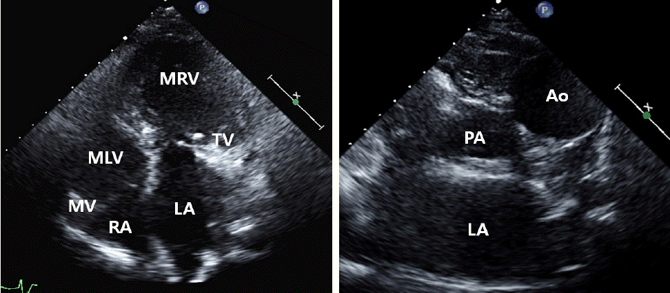
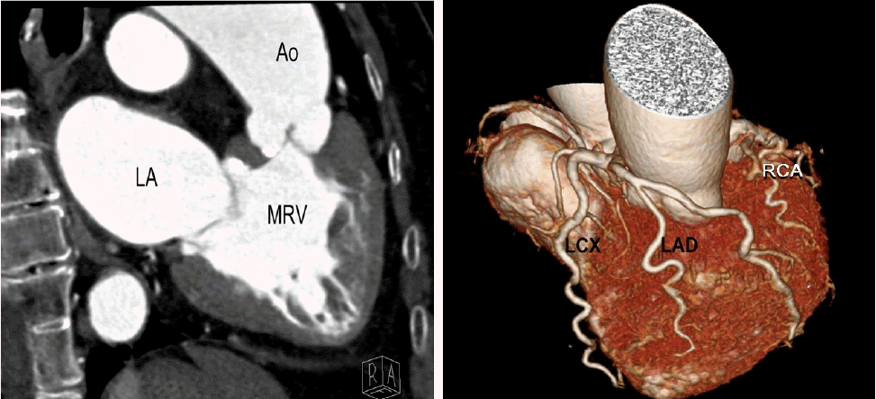
91ýä© ýù¼ý×ÉÛ░Ç Ýÿ©ÝØíÛ│ñÙ×Çý£╝Ùí£ Ùé┤ýøÉÝòÿýÿÇÙïñ. ýêÿýï¡ Ùàä ýáäÙÂÇÝä░ ýºÇýåìÙÉ£ Ýÿ©ÝØíÛ│ñÙ×Çý£╝Ùí£ Û░£ýØ©ýØÿýøÉýùÉýä£ ýï¼ÙÂÇýáäýªØý£╝Ùí£ ý╣ÿÙúîÙ░øÛ│á ý×êýùêý£╝Ù®░ ýÁ£ÛÀ╝ Ýÿ©ÝØíÛ│ñÙ×ÇýØ┤ ýï¼Ýò┤ýíîÙïñ. Ýÿêýù¡ÝòÖýáü ýåîÛ▓¼ýØÇ ýáòýâüýØ┤ýùêý£╝Ù®░, ýØ┤ÝòÖýáü Û▓Çýé¼ýâü ÝØëÛ│¿ýóîýù░ýùÉýä£ II/IV ýØ┤ýÖäÛ©░ ýï¼ý×íýØîýØ┤ ý▓¡ýºäÙÉÿýùêÙïñ. Û▓¢ÝØëÙÂÇ ýï¼ý┤êýØîÝîî Û▓Çýé¼ýâü ýóîý©íýùÉ ý£äý╣ÿÝò£ ýï¼ýïñýØ┤ ÝÖòý×ÑÙÉÿýùêÛ│á(ýØ┤ýÖäÛ©░ÙºÉ ýºüÛ▓¢: 61 mm) ÛÁ¼ÝÿêÙÑáýØ┤ 48%Ùí£ Û░ÉýåîÙÉÿýû┤ ý×êýùêý£╝Ù®░, ýñæÙô▒ÙÅäýØÿ ÙîÇÙÅÖÙºÑÝîÉ ýù¡ÙÑÿÛ░Ç Û┤Çý░░ÙÉÿýùêÙïñ. ÙÿÉÝò£ ýóîý©í Ù░®ýïñÝîÉÙºëýØ┤ ýÜ░ý©í Ù░®ýïñÝîÉÙºëýùÉ Ù╣äÝòÿýù¼ ýï¼ý▓¿ÙÂÇÙí£ ÝÄ©ý£äÙÉÿýû┤ ý×êýùêý£╝Ù®░, ýï¼ýïñ Ùé┤ bandýÖÇ Ù╣äÝøäÙÉ£ ý×öÛ©░ÙæÑÝÿòýä▒(trabeculation)ýØä Û░Çýºä ÝÿòÝâ£ÝòÖýáü ýÜ░ýï¼ýïñýØ┤ ýóîý©íýï¼ý×ÑýùÉýä£ Û┤Çý░░ÙÉÿýùêÙïñ(Fig. 1). 3ý░¿ýøÉ ýï¼ý×Ñ CT ýí░ýÿüýêáýâü ÝÅÉÙÅÖÙºÑÙ│┤Ùïñ ýò×ý¬¢ýùÉ ÙîÇÙÅÖÙºÑýØ┤ ý£äý╣ÿÝòÿÛ│á ý×êýùêý£╝Ù®░ ýáòýâüýáüýØ© ýÜ░ýï¼ýïñ Ù│┤Ùïñ Ù╣äÝøäÙÉ£ ÝÿòÝâ£ÙÑ╝ Ù│┤ýØ┤Ù®░, ý×öÛ©░ÙæÑÝÿòýä▒(trabeculation)ýØ┤ ýªØÛ░ÇÙÉ£ ÝÿòÝâ£ÝòÖýáü ýÜ░ýï¼ýïñýØ┤ ýé╝ý▓¿ÝîÉýØä ÝåÁÝò┤ ýóîýï¼Ù░®Û│╝ ýù░Û▓░ÙÉÿýû┤ ý×êýùêÙïñ. ÙîÇÙÅÖÙºÑÝîÉÙºë ýñæýùÉýä£ Ù╣äÛ┤ÇÙÅÖÙºÑ ý▓¿ÝîÉýØ┤ ýò×ý¬¢ý£╝Ùí£ ý£äý╣ÿÝòÿÛ│á ý×êýùêý£╝Ù®░, Ù░£ýé┤Ù░öÙÅÖýØÿ ýÜ░ý©íýùÉ ý£äý╣ÿÝò£ ýóîÛ┤ÇÙÅÖÙºÑ ý▓¿ÝîÉ ýâüÙÂÇýùÉýä£ ÝÿòÝâ£ÝòÖýáü ýóîÛ┤ÇÙÅÖÙºÑýØ┤ ÙéÿýÖÇýä£ ýóîýáäÝòÿÝûëýºÇýÖÇ ýóîÝÜíýºÇ Û┤ÇÙÅÖÙºÑý£╝Ùí£ ÙÂäýºÇ Ýòÿýù¼ ýÜ░ý©íýùÉ ý£äý╣ÿÝò£ ÝÿòÝâ£ÝòÖýáü ýóîýï¼ýïñýØÿ ÝÿêýòíÛ│ÁÛ©ëýØä Ùï┤Ùï╣ÝòÿýÿÇÙïñ. ÝÿòÝâ£ÝòÖýáü ýÜ░Û┤ÇÙÅÖÙºÑýØÇ ýóîý©íýùÉ ý£äý╣ÿÝò£ Ù░£ýé┤Ù░öÙÅÖ ýÜ░Û┤ÇÙÅÖÙºÑ ý▓¿ÝîÉ ÙÂÇý£äýùÉýä£ Û©░ýï£Ýòÿýù¼ ýóîý©íýùÉ ý£äý╣ÿÝò£ ÝÿòÝâ£ÝòÖýáü ýÜ░ýï¼ýïñýØÿ ÝÿêýòíÛ│ÁÛ©ëýØä Ùï┤Ùï╣ÝòÿýÿÇÙïñ(Fig. 2). ÝÖÿý×ÉÙèö ýäáý▓£ýä▒ ýêÿýáò ÙîÇÝÿêÛ┤Ç ýáäý£ä(congenitally corrected transposition of the great arteries)Ùí£ ýºäÙï¿ÙÉÿýùêý£╝Ù®░ ýØ┤Ùç¿ýᣠýé¼ýÜ® Ùô▒ýØÿ ýï¼ÙÂÇýáäýùÉ ÙîÇÝò£ Ù│┤ýí┤ýáü ý╣ÿÙúî Ýøä ýªØýâüýØ┤ Ýÿ©ýáäÙÉÿýû┤ Ýç┤ýøÉÝòÿýÿÇÙïñ.
ýäáý▓£ýä▒ ýêÿýáò ÙîÇÝÿêÛ┤Ç ýáäý£äÙèö ýäáý▓£ýä▒ ýï¼ý×ÑÙ│æýØÿ ýò¢ 1%ÙÑ╝ ý░¿ýºÇÝòÿÙèö ÙºñýÜ░ ÝؼÛÀÇÝò£ ýºêÝÖÿý£╝Ùí£, ýÜ░ýï¼Ù░®ý£╝Ùí£ Ùôñýû┤ýÿ¿ ýáäýïáýê£ÝÖÿ ýáòÙºÑÝÿêýØ┤ ýØ┤ý▓¿ÝîÉýØä ÝåÁÝò┤ ýÜ░ý©íýùÉ ý£äý╣ÿÝò£ ÝÿòÝâ£ÝòÖýáü ýóîýï¼ýïñÙí£ Ùôñýû┤ýÖÇýä£ ÝøäÙ░®ýùÉ ý£äý╣ÿÝò£ ÝÅÉÙÅÖÙºÑý£╝Ùí£ ÙéÿÛ░ÇÙ®░, ÝÅÉýê£ÝÖÿýØä ÝåÁÛ│╝Ýò£ ÝÅÉýáòÙºÑÝÿêýØ┤ ýóîýï¼Ù░®, ýé╝ý▓¿ÝîÉýØä Û▒░ý│É ýóîý©íýùÉ ý£äý╣ÿÝò£ ÝÿòÝâ£ÝòÖýáü ýÜ░ýï¼ýïñÙí£ Ùôñýû┤ýÖÇýä£ ýóîý©í ýáäÙ░®ýùÉ ý£äý╣ÿÝò£ ÙîÇÙÅÖÙºÑý£╝Ùí£ ÙéÿÛ░ÇÛ▓î ÙÉ£Ùïñ. ÛÀ©Ùƒ¼Ù»ÇÙí£ ÝÿêýòíýØÿ ýê£ÝÖÿýØÇ ÔÇ£ýâØÙª¼ÝòÖýáüÔÇØý£╝Ùí£ ÔÇ£ÛÁÉýáòÔÇØÙÉÿýùêýºÇÙºî ÝÿòÝâ£ÝòÖýáüýØ© ýÜ░ýï¼ýïñýØ┤ ýáäýïáýê£ÝÖÿýØä Ùï┤Ùï╣ÝòÿÛ▓î ÙÉ£Ùïñ. ýØ┤ ÝÖÿý×ÉýùÉýä£ýÖÇ Û░ÖýØ┤ ÙÅÖÙ░ÿÙÉ£ Û©░ÝÿòýØ┤ ýùåÛ│á Ù╣äÛÁÉýáü ýï¼ýïñ ýêÿýÂòÛ©░ÙèÑýØ┤ ý£áýºÇÙÉÿýû┤ 60-70ÙîÇýùÉ Û▓Çýé¼ýâü ýÜ░ýù░Ý×ê Ù░£Û▓¼ÙÉÿÙèö Û▓¢ýÜ░ÙÅä Ùô£Ù¼╝Û▓î ý×êý£╝Ùéÿ, ýâüÙï╣ýêÿýùÉýä£ ýï¼ýïñýñæÛ▓® Û▓░ýåÉ, ÝÅÉÙÅÖÙºÑ Ýÿæý░®, ýé╝ý▓¿ÝîÉýØÿ EbsteinÝÿò Û©░Ýÿò, ýï¼Ù░®ýñæÛ▓® Û▓░ýåÉ, ÙîÇÙÅÖÙºÑ ýÂòý░® Ùô▒ýØÿ ÙÅÖÙ░ÿÙÉ£ ýï¼ý×Ñ Û©░ÝÿòýØ┤ ÝØöÝ×ê Ù│æÙ░£ÝòÿÙ®░ ý▓┤ýê£ÝÖÿýØäÙï┤Ùï╣ÝòÿÙèö ÝÿòÝâ£ÝòÖýáü ýÜ░ýï¼ýïñýØÿ Û©░ÙèÑÙÂÇýáä, ýé╝ý▓¿ÝîÉ ýù¡ÙÑÿ, ýÖäýáäÙ░®ýïñý░¿Ùï¿, WPW ýªØÝøäÛÁ░, ýï¼Ù░®ýä©ÙÅÖ Ù░Å ýí░ÙÅÖ Ùô▒ýØ┤ ÝØöÝ×ê Ù░£ýâØÝò£Ùïñ[1]. Û▓¢ÝØëÙÂÇ ýï¼ý┤êýØîÝîîýâü 1) ýï¼ý▓¿4Ù░®ÙÅäýùÉýä£ ýï¼ý▓¿ÙÂÇýùÉ ÛÀ╝ýáæÝò┤ ý×êÙèö ýé╝ý▓¿ÝîÉýØ┤ ýÜ░ý©íýØ┤ ýòäÙïî ýóîý©íýùÉýä£ Û┤Çý░░ÙÉÿÙèö Û▓¢ýÜ░ ýºäÙï¿ýØÿ Ùï¿ýä£Û░Ç ÙÉÿÙ®░ 2) Ù╣äýáòýâüýáüý£╝Ùí£ ÙÆñý¬¢ýùÉ ý£äý╣ÿÝò£ ÝÅÉÙÅÖÙºÑÝîÉÛ│╝ ýò×ý¬¢ýùÉ ý£äý╣ÿÝò£ ÙîÇÙÅÖÙºÑÝîÉýØ┤ ÝØëÛ│¿ýù░Ùï¿ýÂòÙï¿Ù®┤ÙÅäýØÿ ÙîÇÙÅÖÙºÑÝîÉ Ùï¿Ù®┤ýùÉýä£ ÙÅÖýï£ýùÉ Û┤Çý░░ÙÉÿÛ│á, 3) ÝØëÛ│¿ýù░ý×ÑýÂòÙï¿Ù®┤ýùÉýä£ ÙæÉÛ░£ýØÿ ÙîÇÝÿêÛ┤ÇýØ┤ ÙÅÖýï£ýùÉ ÝÅëÝûëÝòÿÛ▓î Û┤Çý░░ÙÉÿÙèö Û▓âýØ┤ Ýè╣ýºòýáüýØ┤Ùïñ[2]. ýÁ£ÛÀ╝ýùÉÙèö ýé╝ý░¿ýøÉ ýï¼ý×Ñ CT ÝÿêÛ┤Çý┤¼ýÿüýØ┤ Ù░£ýáäÝò¿ýùÉ Ùö░ÙØ╝ ýØ┤ Ù│æýØä ýºäÙï¿ÝòÿÛ│á ýØ┤Ýò┤ÝòÿÙèö Ùì░ ÙºÄýØÇ ÙÅäýøÇýØä ýñÇÙïñ[3].
 PDF Links
 PubReader
 ePub Link
 Full text via DOI
 Download Citation
 Print
Print


