Korean J Med > Volume 80(5); 2011 > Article
과민성장증후군의 병태생리와 진단

Abstract

Irritable bowel syndrome (IBS) is a very common functional gastrointestinal disorder characterized by abdominal pain or discomfort with bowel habit changes. However, its pathogenesis and pathyphysiology have been poorly understood. There were many robust functional and anatomical evidences in Korea for visceral hypersensitivity, low-grade inflammation and immunologic alterations, brain-gut interaction and genetic factors in the development of IBS. The link between pathophysiologic changes and gastrointestinal symptoms would be the strong basis for the relevance of target therapy in this condition. The ROME III criteria represent the current gold standard for the identification of characteristic and supportive symptoms of IBS and for the categorization of IBS patients into symptom-based subgroups with exclusion of organic diseases. There were some limitations in use of Rome III criteria in the clinical practice and research fields. Test of Rome III are needed, especially in Korea. (Korean J Med 2011;80:491-498)

서 론

과민성장증후군(Irritable bowel syndrome, IBS)은 기질적인 이상 없이 배변 습관의 변화를 동반한 복통 및 복부 불편감을 특징으로 하는 기능성 위장관 질환이다[1-3]. 이는 가장 흔한 위장관 질환 중의 하나로 그 유병률은 미국 인구에서 10-22%, 아시아 인구에서 5-10%로 보고되고 있다[2]. IBS 환자들은 장 증상으로 인해 일상생활에서 불편을 많이 겪을 뿐 아니라 질병이 다른 기질적 질환을 제외한 후 진단되고 질병 자체를 입증할 수 있는 생체징후가 없어 IBS라는 진단에 대해 만족하지 않거나 이해가 부족한 경우가 종종 있다. 또한 증상이 악화와 호전을 반복하고 효과적인 약물 치료가 없어 치료에 만족하지 못하고 지속적으로 의료 기관을 이용하여 높은 사회 경제적 비용을 발생시키는데[3], 이는 일부 질병의 원인과 병태생리의 불명확성에 기인한다. 제한적이지만 과민성장증후군의 원인 및 병태생리에 대해서 다양한 가설이 제시되고 연구가 진행되어 왔다. 본 고찰에서는 국내에서 최근까지 보고된 IBS의 발생기전에 관한 연구를 정리하고, 최근 중요한 병태생리의 하나로 알려진 뇌-장관 상호작용 및 유전 관련 자료에 관한 연구결과와 함께 IBS의 진단체계를 고찰하고자 하였다.

병태생리

내장 과민성(visceral hypersensitivity)

이제까지 가장 잘 알려진 병태생리의 하나로 바로스타트를 이용한 풍선확장법 연구에 의하면 IBS 환자의 상당수가 풍선 확장 시 정상인에 비해 적은 부피나 압력에서 불편감을 느낀다고 알려져 있다. 즉, 자극에 대한 직장의 내장 민감도가 증가해 있는 내장 과민성(hypersensitivity)이 특징으로, 좌측대장 혹은 직장의 통증에 대한 감각 역치가 의미있게 낮아져 있다. 국내에서도 활발한 연구가 진행되어 있는데(Table 1), 내장 과민성은 과민성장증후군의 아형에 따라 달리 나타나는데, 주로 설사우세형에서 나타나는 것으로 알려져 있으나[4-11], 변비형 아형에서도 보고된 바 있다[8]. 내장 과민성은 과민성장증후군의 모든 환자에서 나타나는 것은 아니고 대체적으로 약 반수정도 나타나는데, 남녀 간 성 차이에 따라 달라서 여성에서 직장 확장 시 불편감 역치가 유의하게 낮았다는 연구가 있으나 아직까지 국내 자료는 미흡한 실정이다. 내장 과민성의 유발인자로서 심리적 스트레스, 장관의 염증 등이 알려진 인자이다. 과민성장증후군에서 위 장관을 조절하는 자율신경계는 감정상태, 우울증이나 불안증에 의해 영향을 받는다고 알려져 있다. 직장 풍선을 확장하는 동안 과민성장증후군 환자에서 β-adrenergic receptor 민감도를 측정한 결과, 대조군과 유의한 차이가 있었고 우울 증상 정도와 유의한 상관관계가 있었다[5].

뇌-장관 상호작용(brain-gut interaction)

뇌-장관 상호작용은 건강한 사람들과 환자 모두에서 중요한 작용을 한다. 이는 건강한 사람들에서 장내 면역 체계를 조절하며 신체적 혹은 심리적인 상태에 따라 위장관 운동을 조화롭게 조절하는 역할을 한다[12]. 위 장관은 구심성 신경인 구심성 척수신경과 구심성 미주신경의 이중 신경 구조를 통해 중추신경계로 신호을 전달하고, 이러한 신경의 전달은 원심성 신호전달 체계를 통해 위 장관의 운동기능이나 장내 면역체계 등에 변화를 일으켜 결과적으로 다양한 기능성 위장관 질환을 유발한다.
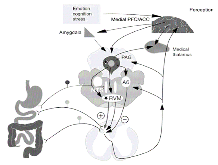
위 장관은 자체적인 신경 체계를 통해 자율적으로 기본적인 위장관 운동기능, 분비기능 및 혈류 등을 조절하는 신경 조절 시스템을 유지한다[12]. 개체 내에서 항상성 유지를 위해 위장관 신경계는 중추 신경계와도 지속적인 상호 신호 전달 체계를 유지하며, 구심성 신경섬유가 내장 감각을 중추신경계로 전달하고 이에 따라 감정, 통증, 면역 반응 등이 나타나게 되면서 항상성을 유지하게 된다[12]. 또한 반대로 감정 상태의 변화나 환경의 변화를 시작으로 뇌 피질-변연계에서 위 장관으로 하행성 신호를 보내어 위 장관 증상을 유발하기도 한다. 과민성장증후군에서는 이러한 신경 체계 조절의 이상, 장 운동성의 변화, 내장 감각의 감각능의 항진이 특징이다[12-14]. 이러한 신경 연결망에 주로 작용하는 대표적인 신경전달 물질이 세로토닌(serotonin, 5-hydroxytryptamine, 5-HT)으로, 위 장관에 주로 작용하는 5-HT 수용체는 5-HT3, 5-HT4 수용체 등이다. 이외에도 CCK antagonist, opioid agonist, NK antagonist 등 여러 신경전달 물질이 관여하며 이를 표적으로 하는 약제들이 치료에 점차 사용되고 있다(Fig. 1) [15].
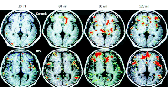
최근 들어 과민성장증후군의 병태생리 중 부적절한 뇌-장관 상호작용과 관련하여 자기공명촬영술이나 기능성 뇌 자기공명 영상(functional brain MRI)에 대한 연구들이 많이 시행되고 있다. 그중 기능성 뇌 자기공명 영상은 국소적인 뇌 혈류의 변화를 측정하는 데 유용한 도구로, 국소적인 대사 작용의 변화를 반영할 수 있어 내장 자극에 의한 국소적인 뇌 혈류의 변화를 측정하는데에 여러 연구에서 사용되었다[13,14]. 이들 연구에서는 내장 감각의 항진을 과민성 장증후군의 주된 병태생리로 생각하여 장에 가해지는 자극에 따라 발생하는 뇌 신호가 과민성장증후군 환자와 정상인에서 차이를 보일 것이라고 생각하였다. 이들은 직장에 풍선을 이용하여 순응도(compliance)를 측정하는 방법으로 장관을 자극하고, 이에 따라 뇌에서 발생하는 신호를 감지하여 그 정도를 정상인과 비교하였다[13,14]. 과민성장증후군 환자에서 직장 자극에 대하여 통증을 감지하는 부분인 anterior cingulate, prefrontal insular cortex, thalamus 모두에서 대조군에 비해 신호가 증가되는 것을 관찰할 수 있었고, 특히 anterior cingulated cortex에서 대조군에 비해 높은 신호증강이 발생하였다[13]. Yuan 등[14]의 연구에 의하면 120 mL의 풍선 확장 자극에 의해 대조군에 비해 과민성장증후군 환자의 anterior cingulate, prefrontal insular cortex, thalamus 모두에서 뚜렷한 신호 증가를 보였다. 이들은 모두 통증성 자극과 비 통증성 자극에 대하여 정상인에 비하여 뇌의 통증을 감지하는 부분에서의 신호가 항진되어 있는 것을 확인하였다(Fig. 2) [14].

감염 후 과민성장증후군(post-infectious irritable bowel syndrome)

내장과민성은 과민성장증후군의 잘 알려진 병태생리로 점막 구심성 신경에 대한 말초성 감작과 외상수용체의 활성화가 중요한 인자이다. 점막 내 저 등급 염증으로 장에 분포하는 구심성 신경말단이 활성화되어 내장 과민성을 유발할 수 있는데, 감염 후 과민성장증후군은 이전에는 과민성장증후군이 없던 환자들에서 위장관 감염이 있은 후에 갑자기 과민성장증후군이 발생하는 것을 의미한다. 현재까지의 보고들에 의하면 급성 위장염을 겪은 환자들 중 3.6%에서 31.6%까지 감염 후 과민성장증후군이 발생하는 것으로 보인다[16]. 또 최근 메타분석에 의하면 급성 위장염을 겪은 환자들에서는 과민성장증후군이 발생할 확률이 7배 정도 높다[17]. 급성 감염 후 염증이 소실된 후에 발생하는 감염 후 과민성장증후군의 위험인자로는 설사의 기간, 여성 및 건강 염려증 등과 같은 정신 신경학적 요인 등이 알려져 있다. 국내에서도 급성 이질, 급성 장염, Clostridium difficile 감염 이후 발병한 감염 후 과민성장증후군이 보고되었고, 여성, 급성 염증기에 세균이 배양되거나 장염의 중증도가 심할수록 감염 후 과민성장증후군이 호발하였다[18-21]. 과민성장증후군 환자의 대장 전 층 조직검사에서 장근 신경총 및 근육층에 비만세포의 침윤이 관찰되었는데, 급성 이질 후 발생한 감염 후 과민성장증후군 환자의 대장내시경 생검 조직에서 5-HT를 포함한 Enterochromaffin (EC) cells, PYY를 포함한 EC cells, 점막 내 상피세포, CD3 림프구, CD8 림프구 및 비만세포 등이 대조군에 비해 유의하게 증가한 것을 보고하였다[22].
최근 위장관내 미생물 무리(microbiota)는 과민성장증후군의 발생기전에 중요한 역할을 한다는 근거가 제시되고 있는데, 그중 하나가 감염 후 과민성장증후군 모델이다[16,23,24]. 위장관 감염 이후 과민성장증후군이 발생하는 기전은 장 상재균의 변화, 소장의 투과성 변화, 또 일부에서는 유전적인 인자가 원인으로 제시되고 있다[16]. 과민성장증후군 환자들에서 실제로 장내 미생물의 변화가 발견되며, 그 증상이 미생물을 목표로 한 치료에 의해 호전되는 것이 과민성장증후군 발생에서 미생물의 역할을 증명하기도 한다[23].

유전적 요인

과민성장증후군의 발생 원인으로 고려되는 것 중, 유전적인 요소에 대한 연구가 최근에 많이 진행되고 있다. 많은 연구들에서 과민성장증후군 환자의 가족 내에서 유병률이 더 높음을 확인하였고, 한 연구에서는 나이, 성별, 사회적 지위 등을 모두 짝지은 대조군에 비해 과민성장증후군 환자에서 뚜렷한 가족력이 있음을 확인하였으며(2% vs. 33%) [25], 또 다른 연구에서는 배우자의 친척들에 비하여 환자와 혈연 관계가 있는 친척에서 유병률이 유의하게 높았다(7% vs. 17%) [26]. 일란성 쌍둥이 연구는 환경과 유전의 영향을 비교하는 고전적인 방법으로서, 1998년과 2001년에 각각 이루어진 연구에서 이란성 쌍둥이에 비교하여 일란성 쌍둥이에서 과민성장증후군의 일치율이 더 높은 것을 확인하였다[27,28]. Morris 등[27]에 의하면 이란성 쌍둥이보다 일란성 쌍둥이에서 같은 위장관 증상을 가질 확률이 높았으며(33.3% vs. 13.3%), Levy 등[28]에 의해 이루어진 연구에서도 일란성 쌍둥이에서 일치율이 유의하게 높았다(17.2% vs. 8.4%). 이 연구에서는 그 어머니가 과민성장증후군을 가진 경우와 그렇지 않은 경우를 비교하였고, 일란성 쌍둥이와 이란성 쌍둥이 모두에서 어머니가 과민성장증후군을 가진 경우가 더 높은 발생률(일란성 쌍둥이, 17.1% vs. 6.7%; 이란성 쌍둥이, 15.2% vs. 6.7%)을 보여, 유전적인 요소 외에 양육 등 환경적인 요소가 발생에 함께 작용한다는 점을 시사했다. 이러한 유전적인 요소들과 관련하여 최근까지 밝혀진 과민성장증후군의 발현과 관련된 유전자에는 serotonin transporter, 5-HT2A receptor, norepinephrine transporter, IL-10 등이 있으며 여러 연구들에 의해 이와 관련된 유전적 다형성이 밝혀져 있다[29].
국내 자료를 살펴보면, Park 등[30]은 뇌-장관 상호작용에 영향을 줄 수 있는 내인성 물질로 cholecystokinin (CCK)의 유전적 다형성이 과민성장증후군과 연관된다고 보고하였다. CCK1 receptor intron 1 779 T > C polymorphic type은 변비 우세형 및 혼합형 과민성장증후군에서 유의하게 흔히 관찰되어 교차비가 2.43 (95% 신뢰구간 1.01-5.86)이었다.
이외 가능성이 있는 연관 유전자로는 serotonin transporter (SERT) 유전적 다형성과 G-protein beta3 C825T 유전적 다형성 등이 보고된 바 있으나 추후 연구가 더 필요하다[30,31].

진 단

과민성장증후군은 유사한 증상을 유발할 수 있는 대장암이나 염증성 장질환과 같은 기질적 질환을 배제한 후 특징적인 증상에 기반하여 진단한다. 직장출혈, 체중감소, 빈혈, 50세 이상의 고령에서 증상이 시작되었을 때, 혹은 대장암이나 염증성 장질환 등의 가족력이 있을 때에는 기질적 질환의 동반 여부를 확인해야 하나 진단의 민감도나 특이도는 낮다. 또한 기질적 질환의 동반 여부를 확인하기 위해 진단 당시에 여러 가지 생화학적 검사나 내시경, 소장내 세균과증식을 확인하기 위한 수소호기검사 등이 이루어지고 있으나 현재까지 선별 검사로 그 유용성이 인정된 것은 일반혈액검사와 C 반응 단백 정도이다[32].
과민성장증후군은 진단을 위한 생물학적 표지자가 없기 때문에 기질적 질환을 배제한 후 특징적인 증상에 근거하여 기준을 만들어 진단과정을 표준화하려는 노력이 이루어져 왔으며 대표적인 진단 기준으로 manning 기준, 로마 I, II, III 기준이 있다(Table 2) [1].
로마 기준은 1988년 다국적 실행위원회(multinational working team committee)가 로마에서 열린 국제소화기병학회에서 기능성 위장관 질환의 진단기준을 만들었기 때문에 붙여진 이름이다. 지난 22년간 총 3차례의 개정이 있었으며, 로마 III 기준이 가장 최근인 2006년에 발표되었다. 개정된 로마기준은 주로는 증상의 회상 기간(recall time)이 지난 12개월에서 최근 3개월간의 증상을 질문함으로써 그 신뢰도를 높이고자 하였으며, 새로 개정된 로마기준 III를 이용한 연구가 보고되고 있다. 국내에서 로마기준 II와 III를 동시에 실시한 인구기반 연구를 살펴보면, 같은 연구자가 동일한 방법으로 15세 이상 성인 남녀 1,009명을 대상으로 로마기준 II와 III에 해당하는 전화설문을 실시한 결과, 로마기준 II를 적용한 IBS 유병률은 남자 6.4%, 여자 9.6%였으나 성별에 따른 통계적인 차이는 없었고, 로마기준 III에 의해서는 여성이 11.0%, 남성이 7.0%로, 로마기준 III를 적용하면 로마기준 II에 비해 유병률이 높고 유의하게 여성에서 호발했다(p= 0.025) [33,34].
저자들은 이전의 연구에 비해 로마기준 III를 적용한 최근 연구에서 IBS가 여성에서 뚜렷하게 호발하는 것이 여성에서 직업의 변화와 연관될 수 있다고 제시하였는데, 실제로 가정주부의 비율이 줄고(64.5% vs. 57.4%) 대신 직장생활을 하는 여성 인구가 7.1%로 증가하였다. 그러나 이는 진단기준의 변화로 인한 것인지 사회인구학적 특징에 변화에 의한 것인지 확실하지 않고, 국내외에서 로마기준 III에 대한 타당성(validation)에 대한 검증 자료는 매우 제한적이다.

결 론

과민성장증후군은 흔한 기능성 위장관 질환으로 그 원인과 병태생리가 아직까지 명확하게 밝혀지지 않았다. 현재까지 중요하게 생각되는 병태생리로는 내장 과민성, 뇌-장관 상호작용, 감염 후 과민성장증후군에서 관찰된 저등급 염증 혹은 미생물 환경 변화과 연관된 면역 이상반응, 유전 소인 등이 있으며 국내에서도 이에 대한 활발한 연구가 진행되어 왔다.
과민성장증후군은 환자의 증상 호소에 근거하여 진단하는 기능성 위장관 질환으로서 뚜렷한 생체 징후 없이 진단이 이루어지므로 이를 표준화하기 위한 노력이 지속되어 왔다. 2006년 로마 III가 개정된 이래로 이는 임상에서 실제로 적용되고 있으며, 다양한 연구를 근간으로 로마기준 III의 제한점에 대해서도 논의가 시작되고 있다. 향후 지속적인 연구를 통한 과민성장증후군의 병태생리에 대한 이해는 합리적인 진단과 치료의 근거가 될 것으로 기대한다.

REFERENCES

1. 박정호, 변정식, 신운건 등. 과민성장증후군의 진단: 체계적인 문헌고찰. 대한소화기학회지 2010;55:308–315.
crossref pmid

2. Liu J, Hou X. A review of the irritable bowel syndrome investigation on epidemiology, pathogenesis and pathophysiology in China. J Gastroenterol Hepatol 2011;26:881–893.
crossref pmid

3. Shin F, Motoyori K. Gene, environment, and brain-gut interaction in irritable bowel syndrome. J Gastroenterol Hepatol 2011;26:110–115.
crossref pmid

4. Moon W, Lee OY, Kim JM, et al. Rectal accommodation in irritable bowel syndrome. Korean J Neurogastroenterol Motil 2007;13:132–137.


5. Yu BH, Kang EH, Kim HS, et al. Beta adrenergic receptor sensitivity, mood state and colonic sensation in patients with irritable bowel syndrome. Korean J Neurogastroenterol Motil 2006;12:63–68.


6. Jun DW, Lee OY, Yang SY, et al. Differences of sigmoid accommodation according to the subtype of irritable bowel syndrome. Korean J Neurogastroenterol Motil 2005;11:129–134.


7. Yang JH, Lee JS, Hong SJ, et al. The rectal perception in patients with constipation predominant irritable bowel syndrome and functional constipation. Korean J Neurogastroenterol Motil 2005;11:58–65.


8. Lee OY, Yoon BC, Choi HS. Sigmoid discomfort thresholds in irritable bowel syndrome. Korean J Neurogastroenterol Motil 2005;11:38–43.


9. Kang EH, Phee PL, Lee JH, et al. Beta adrenergic receptor function and anxiety in patients with irritable bowel syndrome. J Korean Neuropsyhiatr Assoc 2004;43:40–45.


10. Kim HS, Phee PL, Lee JH, et al. The role of rectal hypersensitivity in the diagnosis of irritable bowel syndrome. Korean J Neurogastroenterol Motil 2003;9:119–124.


11. Lee KJ, Kim JH, Hahm KB, et al. The factors related to rectal hypersensitivity in patients with diarrhea predominant irritable bowel syndrome. Korean J Gastroenterol 2001;37:173–178.


12. Mayer EA, Tillisch K, Bradesi S, et al. Review article: modulation of the brain-gut axis as a therapeutic approach in gastrointestinal disease. Aliment Pharmacol Ther 2006;24:919–933.
crossref pmid

13. Merts H, Morgan V, Tanner G, et al. Regional cerebral activation in irritable bowel syndrome and control subjects with painful and nonpainful rectal distension. Gastroenterology 2000;118:842–848.
crossref pmid

14. Yuan YZ, Tao RJ, Xu B, et al. Functional brain imaging in irritable bowel syndrome with rectal balloon distention by using fMRI. World J Gastroenterol 2003;5:1356–1360.
pmid

15. Mach T. The brain-gut axis in irritable bowel syndrome-clinical aspects. Med Sci Monit 2004;10:125–131.
pmid

16. Uday CG, Prabhat R. Post-infectious irritable bowel syndrome: The past, the present and the future. Gastroenterology 2011;26:94–101.
crossref pmid

17. Halvorson HA, Schlett CD, Riddle MS. Postinfectious irritable bowel syndrome-a meta-analysis. Am J Gastroenterol 2006;101:1894–1899.
crossref pmid

18. Park HS, Han DS, Hyun YS, et al. Postinfectious irritable bowel syndrome after clostridium difficile infection. Intest Res 2009;7:100–104.


19. La JH, Kim TW, Sung TS, et al. Role of mucosal mast cells in visceral hypersensitivity in a rat model of irritable bowel syndrome. J Vet Sci 2004;5:314–324.
pmid

20. Lee JS, Jung SA, Kim SE, et al. The frequency of post infectious irritable bowel syndrome (PI-IBS) in patients with acute diarrhea and a comparison with colonoscopic findings. Korean J Neurogastroenterol Motil 2004;10:43–48.


21. Park CH, Joo YE, Choi SK, et al. Activated mast cells infiltrate in close proximity to enteric nerves in diarrhea-predominant irritable bowel syndrome. J Korean Med Sci 2003;18:204–210.
pmid pmc

22. Kim HS, Lim HJ, Park H, et al. Increased immunoendocrine cells in intestinal mucosa of postinfectious irritable bowel syndrome patients 3 years after acute shigella infection: an observation in a small case control study. Yonsei Med J 2010;51:45–51.
crossref

23. Anne S, Willem MV, Airi P. Gastrointestinal microbiota in irritable bowel syndrome: present state and perspectives. Microbiology 2010;156:3205–3215.
crossref pmid

24. Bolino CM, Bercik P. Pathogenic factors involved in the development of irritable bowel syndrome: focus on a microbial role. Infect Dis Clin North Am 2010;24:961–975.
crossref pmid

25. Whorwell PJ, McVallum M, Creed FH, et al. Non-colonic features of irritable bowel syndrome. Gut 1986;27:37–40.
crossref pmid pmc

26. Kalantar JS, Locke GR, Zinsmeister AR, et al. Familial aggregation of irritable bowel syndrome: a prospective study. Gut 2003;52:1703–1707.
crossref pmid pmc

27. Morris YA, Talley NJ, Boyce PM, et al. Evidence of a genetic contribution to functional bowel disorder. Am J Gastroenterol 1998;93:1311–1317.
crossref pmid

28. Levy RL, Jones KR, Whitehead WE, et al. Irritable bowel syndrome in twins: heredity and social learning both contribute to etiology. Gastroenterology 2001;121:799–804.
crossref pmid

29. Yuri AS, Gloria M, Petersen G, et al. The genetics of irritable bowel syndrome. Clin Gastroenterol Hepatol 2005;3:1057–1065.
crossref pmid

30. Park SY, Rew JS, Lee SM, et al. Association of CCK2 receptor gene polymorphisms and irritable bowel syndrome in Korean. J Neurogastroenterol Moril 2010;16:71–76.
crossref pmid pmc

31. Lee HJ, Lee SY, Choi JE, et al. G protein beta3 subunit, interleukin-10, and tumor necrosis factor-alpha gene polymorphisms in Koreans with irritable bowel syndrome. Neurogastroenterol Motil 2010;22:758–763.
crossref pmid

32. Cash BD, Schoenfeld P, Chey WD. The utility of diagnostic tests in irritable bowel syndrome patients: a systematic review. Am J Gastroenterol 2002;97:2812–2819.
crossref pmid

33. Han SH, Lee OY, Bae SC, et al. Prevalence of irritable bowel syndrome in Korea: population-based survey using the Rome II criteria. J Gastroenterol Hepatol 2006;21:1687–1692.
crossref pmid

34. Park DW, Lee OY, Shim SG, et al. The differences in prevalence and sociodemographic characteristics of irritable bowel syndrome according to Rome II and Rome III. J Neurogastroenterol Motil 2010;16:186–193.
crossref pmid pmc

35. Jeong JB, Yang YM, Jeon WJ, et al. Postprandial colonic motor activity in patients with irritable bowel syndrome. Korean J Gastrointest Motil 2000;6:20–30.


36. Lee OY. Asian motility studies in irritable bowel syndrome. J Neurogastroenterol Motil 2010;16:120–130.
crossref pmid pmc

37. Park SY, Park MH, Yoon KW, et al. Plasma 5-Hydroxytryptamine concentration and its correlation with psychopathology in patients with irritable bowel syndrome. Gut Liver 2009;3:26–30.
crossref pmid pmc

38. Park HY, Jang JH, Lee YJ, et al. Serotonin transporter gene and G-protein beta 3 C825T gene polymorphism in patient with functional dyspepsia and irritable bowel syndrome. Korean J Neurogastroenterol Motil 2009;15:58–64.


39. Kim HS, Park HJ, Lim JH, et al. The role of pro-and antiinflammatory cytokine gene polymorphisms in the development of post-infectious irritable bowel syndrome. Korean J Neurogastroenterol Motil 2006;12:41–47.


40. Park H, Lee DY, Kim MS, et al. Serotonin transporter gene polymorphism and its relationship to plasma serotonin in irritable bowel syndrome. Korean J Neurogastroenterol Motil 2004;10:125–129.


41. Lee DY, Park H, Kim WH, et al. Serotonin transporter gene polymorphism in healthy adults and patients with irritable bowel syndrome. Korean J Gastroenterol 2004;43:18–22.
pmid

42. Park JH, Park DI, Kim JH, et al. The relationship between small-intestinal bacterial overgrowth and intestinal permeability in patients with irritable bowel syndrome. Gut Liver 2009;3:174–179.
crossref pmid pmc

43. Hong KS, Kang HW, Im JP, et al. Effect of probiotics on symptoms in Korean adults with irritable bowel syndrome. Gut Liver 2009;3:101–107.
crossref pmid pmc

44. Eum J, Rhee KH, Cho YK, et al. Therapeutic effects of Agio® 18g per day in patients with constipation dominant irritable bowel syndrome and chronic constipation. Korean J Neurogastroenterol Motil 2008;14:115–121.


45. Paik CN, Choi MG, Nam KW, et al. The prevalence of small intestinal bacterial overgrowth in Korean patients with irritable bowel syndrome. Korean J Neurogastroenterol Motil 2007;13:38–44.


46. Kim YG, Moon JT, Lee KM, et al. The effect of probiotcs on symptoms of irritable bowel syndrome. Korean J Gastroenterol 2006;47:413–419.
pmid

47. Kim HS, Kim MS, Ji SW, et al. The development of irritable bowel syndrome after Shigella infection: 3 year follow up study. Korean J Gastroenterol 2006;47:300–305.
pmid

48. Choi JH, Nah BK, Leem JM, et al. Mucosal mast cells in irritable bowel syndrome. Korean J Neurogastroenterol Motil 2004;10:57–62.


Brain-gut interaction. A6, locus coruleus; RVM, rostra ventral medulla; NTS, nucleus tractus solitaries [12].
/upload/thumbnails/kjm-80-5-491-1f1.gif
Figure 1.
Functional brain map. In patients with IBS, the average percentage area of interest increases in parallel with rectal distention volumes in the anterior cingulate cortex, prefrontal cortex, insular cortex, and thalamus [14].
/upload/thumbnails/kjm-80-5-491-1f2.gif
Figure 2.
Table 1.
Pathophysiology of irritable bowel syndrome in Korea
Focused pathophysiology Authors Year Main finding Ref.
Visceral hypersensitivity Moon W et al. 2007 Lowered rectal accommodation was significant in diarrhea predominant- IBS patients 4
Jun DW et al. 2005 Lowered rectal accommodation was significant in diarrhea predominant- IBS patients 6
Lee OY et al. 2005 Sigmoid discomfort threshold for the patients with IBS was significantly lower than control 7
Yang JH et al. 2005 Male IBS-C patients showed visceral hypersensitivity to low grade stimuli, female IBS-C patients showed visceral hypersensitivity to high grade stimuli, suggesting a differential pathophysiologic mechanism of visceral hypersensitivity in different gender with IBS-C 8
Kim HS et al. 2003 Rectal hypersensitivity is a reliable biologic marker to differentiate IBS from normal subjects. However, individual symptoms were not associated with rectal hypersensitivity. 10
Lee KJ et al. 2001 Rectal hypersensitivity in IBS-D patients was significantly related to age and parasympathetic function 11
Jeong JB et al. 2000 Postprandial intraluminal pressure differences play a role in the patho- physiology of IBS 35
Psychological disturbance, Brain-gut interaction: Lee OY 2010 Many conflicting results about altered motility were found among many Asian studies 36
Park SY et al. 2009 5-HT might play a critical role in IBS, and psychopathological factors are correlated with IBS 37
Yu BH et al. 2006 Beta adrenergic receptor sensitivity is increased in IBS patients who do not have depressive or anxiety disorders. 5
Kang EH et al. 2004 Beta adrenergic receptor function is increased in patients with IBS, which could affect increased anxiety levels 9
Genetics Park SY et al. 2010 CCK receptor polypmorphisms were associated with IBS 30
Park HY et al. 2009 Serotonin transporter, G-protein beta3 polymorphism does not appear to be associated with IBS and functional dyspepsia in Korea 38
Kim HS et al. 2006 Genetic polymorphisms of TNF-alpha, IL-10, TGF-alpha1 have no significant role in the development of PI-IBS 39
Park H et al. 2004 IBS patients have significantly higher fasting plasma serotonin levels than controls, irrespective of genetic polymorphism at promoter of serotonin transporter protein 40
Lee DY et al. 2004 There was no relationship between serotonin transporter gene polymorphism and IBS 41
Immune, inflammation, infection Kim HS et al. 2010 Immunoendocrine cells were increased in PI-IBS 22
Park HS et al. 2009 PI-IBS frequently follows after Clostridium difficile infection, predominantly diarrhea type 18
Park JH et al. 2009 Intestinal permeability was increased in patients with IBS, but this finding did not correlated with the occurrence of SIBO 42
Hong KS et al. 2009 Composite probiotics containing Bifidobacterium bifidum BGN4, Lactobacillus acidophilus AD031, and other species are safe and effective 43
Eum J et al. 2008 Agio® 18 g per day for 21days seems to be effective and safe in patients with constipation-predominant IBS 44
Paik CN et al. 2007 The prevalence of SIBO as diagnosed with using LBT was significantly higher in Korean IBS patients 45
Kim YG et al. 2006 Medilac DS® is a safe and useful probiotic agent for the treatment of abdominal pain in patients with IBS 46
Kim HS et al. 2006 About a quarter of PI-IBS patients are recovered over 3 years, previous functional bowel disorder is a risk factor after 3 years 47
La JH et al. 2005 Higher degranulation rate of mucosal mast cell is responsible for visceral hypersensitivity in this model IBS 19
Choi JH et al. 2004 IBS patients have higher mast cell count, especially in IBS-C patients 48
Lee JS et al. 2004 PI-IBS is frequent in positive bacterial culture, presence of endoscopic colitis, wider extent and more severe colitis 20
Park CH et al. 2003 Activated mast cell in close proximity to enteric nerves were significantly increased in both cecum and rectum of the IBS group 21
Table 2.
The diagnostic criteria of irritable bowel syndrome
Diagnostic criteria Symptom, sign, laboratory investigations included in criteria
Manning (1978) IBS is defined as the symptoms given below with no duration of symptoms described. The number of symptoms that need to be present to diagnose IBS is not reported in the paper, but a threshold of three positive is the most commonly used:
 1. Abdominal pain relieved by defecation
 2. More frequent stools with onset of pain
 3. Looser stools with onset of pain
 4. Mucus per rectum
 5. Feeling of incomplete emptying
 6. Patient-reported visible abdominal distension
Rome I (1990) Abdominal pain or discomfort relieved with defecation, or associated with a change in stool frequency or con- sistency, PLUS two or more of the following on at least 25% of occasions or days for three months:
 1. Altered stool frequency
 2. Altered stool form
 3. Altered stool passage
 4. Passage of mucus
 5. Bloating or distension
Rome II (1999) Abdominal discomfort or pain that has two of three features for 12 weeks (need not be consecutive) in the last one year:
 1. Relieved with defecation
 2. Onset associated with a change in frequency of stool
 3. Onset associated with a change in form of stool
Rome III (2006) Recurrent abdominal pain or discomfort three days per month in the last three months associated with two or more of:
 1. Improvement with defecation
 2. Onset associated with a change in frequency of stool
 3. Onset associated with a change in form of stool
TOOLS
METRICS Graph View
  • 6,876 View
  • 153 Download

Editorial Office
101-2501, Lotte Castle President, 109 Mapo-daero, Mapo-gu, Seoul 04146, Korea
Tel: +82-2-2271-6791    Fax: +82-2-790-0993    E-mail: kaim@kams.or.kr                

Copyright © 2024 by The Korean Association of Internal Medicine.

Developed in M2PI

Close layer
prev next