단독 고립성 결절로 진단된 좌하엽으로의 비정상적인 체순환 1예
A Case of Anomalous Systemic Arterial Supply to the Left Lower Lobe without Sequestration
Article information
Korean J Med. 2011;80(4):402-404
Publication date (electronic) : 2011 April 1
1Department of Internal Medicine, Naval Pohang Hospital, Pohang, Korea
2Department of Internal Medicine, Konkuk University Hospital, Konkuk University College of Medicine, Seoul, Korea
1해군포항병원 내과
2건국대학교 의과대학 건국대학교병원 내과
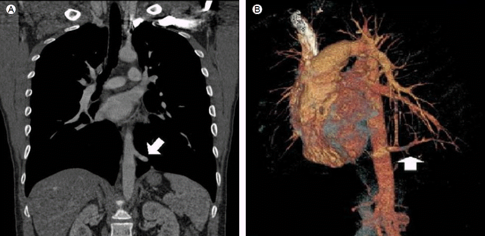
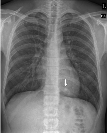
21세 남자 환자가 신체검사에서 심장 음영 후부에 단독 고립성 결절이 발견되어 내원하였다. 환자는 비흡연자로 현재 호흡기 증상은 없었으며 과거력 및 가족력도 특이 소견 없었다. 활력징후는 혈압 124/66 mmHg, 맥박 60회, 호흡수 16회, 체온 36.7℃였고, 흉부진찰에서 특이 소견 보이지 않았다. 단순 방사선 검사에서는 심장 후부에 약 1.2 × 0.9 cm 크기의 원형의 결절이 관찰되었다. 조영증강 흉부 전산화 단층 촬영에서는 좌하엽의 후저분절에 하행대동맥에서 기시하는 굵은 동맥 구조가 관찰되며 엽간동맥 이하로 정상적인 폐동맥은 관찰되지 않았다(-). 환자는 좌하엽으로의 비정상적인 체순환으로 진단뒤 수술적 문제상의 위해 타병원 흉부외과로 전원되었다.
Chest PA film shows nodular lesion (arrow) in the retrocardiac area of left lower lobe.
Chest contrast enhanced CT show anomalous systemic artery originating from the descending aorta suppling the posterior basal segment of left lower lobe (arrow) (A: mediastinal window, B: lung window).
Coronal multiplanar reconstruction image (A) and 3-D reconstriction of multidetector CT scan (B) show aberrant artery originated from thoracic aorta (arrow).
폐분획증이 없는 좌하엽으로의 비정상적인 체순환은 매우 드문 질환으로 1940년 Harris와 Lewis에 의해 최초로 보고된 이래 매우 적은 예가 보고되었다. 1946년 Pryce에 의해 폐로 가는 비정상적인 폐혈관에 대한 분류가 이루어졌으며, 좌하엽으로의 비정상적인 체순환은 비록 폐분획증은 없으나 type I으로 분류될 수 있다. 발생 원인에 대해서는 논란이 많으나 배아기에 대동맥의 후새궁(postbrachial arch)이 주폐동맥이 발달하기 전에 비정상적으로 잔존한 결과로 보인다[1,2].
가장 정확한 검사법은 혈관조영술로 병변이 있는 폐엽의 정상 폐동맥의 분지를 확인하고, 이상 기시 체혈관의 폐엽 내 모세 혈관 상에서 폐정맥으로의 연결 관계를 설명할 수 있을 때 진단이 가능하다. 하지만 최근 전산화 단층 촬영 혈관조영술(CT angiography) 기술의 발달로 이를 통하여 비교적 정확한 혈관 관계를 알 수 있다[2].
치료는 폐동맥 고혈압으로 인한 객혈과 좌심실 과부하로 인한 심부전의 잠재적 위험성 때문에 이 질환을 가진 모든 환자는 수술의 적응증이 되며, 대부분의 보고에서는 폐엽 절제술이 선호되고 있다[3].
References
1. Iizasa T, Haga Y, Hiroshima K, Fujisawa T. Systemic arterial supply to the left basal segment without the pulmonary artery: four consecutive cases. Eur J Cardiothorac Surg 2003;23:847–849.
2. Hong SB, Na KJ, Park JM, Ahn BH, Kim SH. Anomalous systemic arterial supply to normal basal segments of left lower lobe without sequestration. Korean J Thorac Cardiovasc Surg 2005;38:510–513.
3. Kim H, Chung WS, Jang HJ, Kang JH, Kim YH, Kim JH. Anomalous systemic arterial supply to the left basal segments without sequestration from descending thoracic aorta: a case report. Korean J Thorac Cardiovasc Surg 2008;41:512–515.
Article information Continued
Copyright ⓒ 2011 The Korean Association of Internal Medicine
Figure 1.
Chest PA film shows nodular lesion (arrow) in the retrocardiac area of left lower lobe.
Figure 2.
Chest contrast enhanced CT show anomalous systemic artery originating from the descending aorta suppling the posterior basal segment of left lower lobe (arrow) (A: mediastinal window, B: lung window).
Figure 3.
Coronal multiplanar reconstruction image (A) and 3-D reconstriction of multidetector CT scan (B) show aberrant artery originated from thoracic aorta (arrow).


