복막투석의 합병증으로 발생한 복막-후복막 누출 1예
Peritoneal-Retroperitoneal-Scrotal Leakage as a Complication of Peritoneal Dialysis
Article information
Abstract
복막투석액의 누출은 복막투석 환자의 10% 미만에서 발생하는 합병증으로, 투석액은 도관출구, 흉강, 복부 탈장, 복벽으로 누출될 수 있으며, 개방성 초상돌기를 통한 외부생식기로도 누출될 수 있다. 저자들은 흔히 발생하는 누출경로가 아닌 후복막으로 투석액이 누출되는 드문 예를 경험하였다. 배액감소로 내원한 환자에서 CT peritoneography를 시행함으로써, 투석액이 후복막으로 누출된 후 누출된 투석액이 우측 고환까지 이동함을 확인하였기에 이를 보고하는 바이다.
Trans Abstract
We report a case of retroperitoneal and scrotal dialysate leakage resulting from peritoneal-retroperitoneal communication in a patient on peritoneal dialysis (PD). The ultrafiltration volume was reduced and the scrotum became enlarged in a patient who had been undergoing PD for 4 years. Retroperitoneal and scrotal leakage of dialysate was confirmed by computed tomography (CT) performed 1 hour after the intraperitoneal infusion of contrast-containing dialysate. The PD was halted and the patient was transferred to hemodialysis (HD). One month after the transfer to HD, the PD was resumed and there were no signs of extraperitoneal leakage. (Korean J Med 2011;80:108-112)
서 론
복막투석액의 누출은 복막투석 환자의 10% 미만에서 발생할 수 있는 합병증이며, 복벽 및 생식기의 부종 또는 호흡곤란 등의 증상으로 나타날 수 있다[1].
복막투석액은 도관출구, 흉강, 개방성 초상돌기(patent processus vaginalis), 복부 탈장 또는 복벽 등으로 누출될 수 있으며[2,3], 종격동으로의 누출도 보고된 바 있다[4]. 후복막으로의 누출은 국외에서 보고된 예는 있으나[5,6] 국내에서는 아직 보고된 바가 없다.
저자들은 복막투석 환자에서 복막-후복막결손을 통해 투석액이 후복막으로 누출된 후, 다시 우측 고환까지 이동한 예를 경험하였기에 이를 보고하는 바이다.
증 례
환 자: 남자, 33세
주 소: 3일간의 복부팽만감
현병력: IgA 신증에 의한 말기신부전으로 내원 4년 전 지속성 외래 복막투석을 시작하였으며, 내원 2년 전부터 지속성 교환기 복막투석(continuous cycling peritoneal dialysis, CCPD)을 시행해 온 환자이다. 환자는 몸무게 77.4 kg, 키 179.5 cm, 체표면적 1.965 m2이며, 평소 한외여과량은 1,000~1,500 mL로 유지되었다.
내원 4일 전부터 배액량이 감소하면서 우측 복부팽만감이 발생하였고, 내원 1일 전부터 우측 고환이 종대되어 내원하였으며, 내원 시 호흡곤란이나 하지 부종은 없었다.
과거력: 내원 10년 전 부갑상선암으로 좌측 부갑상선제거수술 및 좌측 갑상선엽절제수술을 시행하였으며, 내원 5년 전 부갑상선암 재발로 종양절제수술 후 방사선치료를 시행하였다.
가족력: 특별한 이상은 없었다.
이학적 소견: 입원 당시 활력징후는 혈압 130/80 mmHg, 맥박 88회/분, 체온 36.2℃, 호흡수 16회/분이었고, 만성 병색을 보였다. 흉부 청진 시 특이소견은 없었으며, 복부는 전반적으로 팽만되어 있었으나 촉지되는 종괴는 없었고, 압통 및 반발통은 없었다. 도관 출구 부위의 염증이나 누출 소견은 없었으며 사지에 함요부종은 없었으며 우측 고환이 종대되어 있었다.
검사실 소견: 말초혈액검사에서 혈색소 8.3 g/dL, 백혈구 3,710/μL, 혈소판 318,000/μL, 적혈구 침강속도 15 mm/hr였으며, 일반 화학검사상 혈청 총단백량 6.2 g/dL, 혈청 알부민 3.8 g/dL, AST 15 IU/L, ALT 18 IU/L, 혈청 요소 질소 65.9 mg/dL, 혈청 크레아티닌 16.67 mg/dL, C-반응성단백 0.1 mg/dL였다. 흉부 X-선 검사상 폐부종의 소견은 없었으며, 단순복부촬영상 도관끝은 골반 내에 정상적으로 위치하고 있었다. 배액한 복막투석액은 맑았으며, 세포수는 0/mm3이었으며, 내원 후 시행한 4.25% 포도당 용액을 이용한 복막평형검사에서 4시간째, 순수 한외여과량은 520 mL였으며 D/Pcreatinine은 0.82로 고투과성 복막 기능을 보였다.
경과 및 치료: 환자는 내원 시 우측 복부팽만감을 호소하였으며 우측 고환이 종대되어 있어서 복막투석액의 복벽 및 고환으로의 누출을 의심할 수 있었으며, 이를 확인하기 위하여 복부 전산화 단층촬영을 시행하였다. 조영제를 사용하지 않은 복부 전산화 단층촬영상 우측 후복막강에 수분으로 의심되는 음영이 관찰되었으며, 이를 확인하기 위하여 투석액 2 L에 조영제 100 mL를 섞어 복강 내에 주입하고 1시간이 경과한 후 복부 전산화 단층 촬영을 다시 시행하였다[7].
촬영을 통하여 조영제를 포함한 복막투석액이 right posterior pararenal space, right anterior pararenal space, 우측 복벽의 extraperitoneal space, right paravesical space로 누출되어 있음을 확인하였으며, 우측 정삭 주위조직을 통해 우측 음낭까지 투석액이 누출되어 있음을 확인하였다(그림 1, 2).
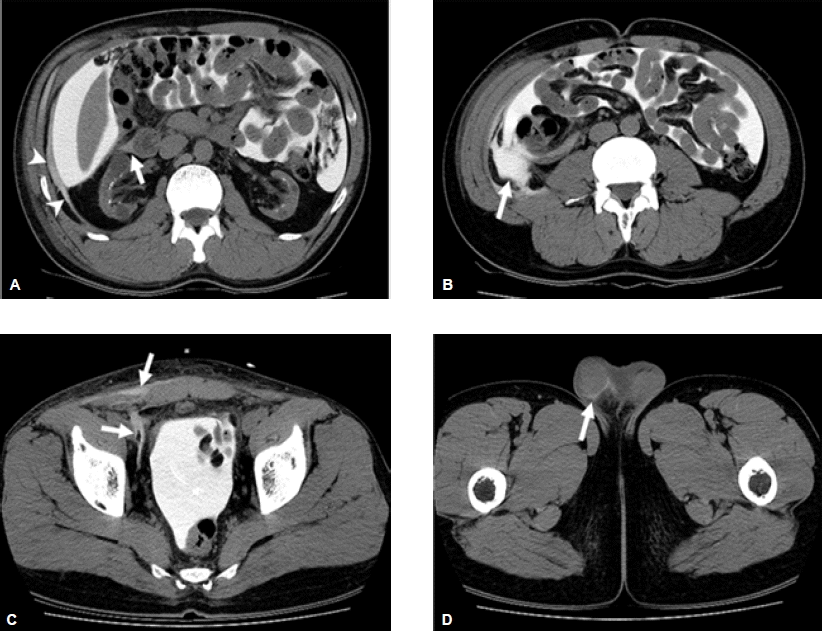
CT scans obtained 1 hour after intraperitoneal infusion of contrast-mixed dialysate. (A) It shows abnormal high density and strand (arrowheads) in the right posterior pararenal space outside of the lateral conal fascia. Scanty abnormal high density (arrow) is also noted in the right anterior pararenal space around the duodenum. (B) Retroperitoneal leakage is marked with an arrow. (C) CT shows leakage into the right anterior extraperitoneal space of abdominal wall, and right paravesical space. (D) Extension of leaked dialysate to the right scrotum going along the spermatic cord was noted.
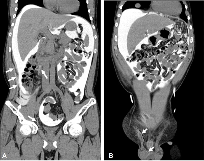
Coronal reformatted image shows fluid collections in the peritoneal space and in the retroperitoneal space, right paravesical space (A). Leaked fluid is extended to the right scrotum via the spermatic cord (B).
이에 복막-후복막 누출로 진단하고 환자는 복막투석을 중지하였으며, 혈액투석용 경정맥 도관 삽관 후 혈액투석으로 전환하였다. 혈액투석 시행 1개월 후 복막투석(CCPD)을 다시 시행하였을때, 정상적인 배액량이 유지되고, 복벽 및 고환 종대 등의 누출소견이 보이지 않아 복막투석을 지속할 수 있었다.
고 찰
복막투석의 합병증 중 탈장 및 투석액누출은 복막투석액에 의한 복강 내 압력의 증가로 발생할 수 있다. 투석액누출은 복벽 또는 생식기의 부종 또는 호흡곤란 등의 증상으로 나타나며, 복막투석 환자의 10% 미만에서 발생한다[1].
투석액 누출은 복막투석 도관 삽관 30일 이내에 발생하는 조기 복강 관류액 누출과 30일 이후에 발생하는 후기 복강 관류액 누출로 나뉘어 지며, 이들은 다른 임상양상으로 나타난다[8].
도관삽관 30일 이내에 일어나는 조기 복강 관류액 누출은 일반적으로 도관설치와 연관되어 발생하며 도관 출구부위 또는 수술 봉합부위로의 체외누출이 대표적이다. 30일 이후에 나타나는 후기 복강 관류액 누출은 대부분 복강 내 압력증가에 의한 복막의 물리적 손상으로 발생하며 탈장 또는 흉강, 복벽, 외부생식기 등으로 체내누출로 나타난다[2]. 투석액의 피하 누출은 조기 및 후기 누출 모두에서 나타날 수 있다[3].
복강으로부터의 복벽, 흉강 등으로 누출되면 한외여과량이 감소하며, 간질공간으로의 누출은 복벽부종과 연관될 수 있으며 생식기의 부종도 동반될 수 있다. 진단은 조영제가 첨가된 투석액의 복강 내 주입을 포함한 전산화 단층 촬영을 이용하여 진단되며, 방사성핵종복막촬영술을 이용하여 진단할 수도 있다[7,9-11].
소량의 누출인 경우 복막투석액량을 감소시키거나 혈액투석으로의 변경을 통해 일시적인 복막투석중단(통상 2~3주) 또는 간헐적 복막투석을 통한 복막을 쉬게 함으로써 저절로 치유될 수도 있으며, 재발할 경우 혈액투석을 더 연장하거나(4~6주) 수술적 재건이 필요할 수도 있다[3,5,7,12,13]. Leblanc 등[2]에 의하면 조기 복강 관류액 누출은 일시적인 혈액투석으로의 전환 또는 수술적 재건에 의해 치료하였으며, 후기 복강 관류액 누출은 일시적인 혈액투석(29%), 수술(27%), 자동 복막투석으로의 전환(16%), 혈액투석으로의 전환(25%)에 의해 치료하였으며, 재발률은 각각 65%, 25%, 14%, 0%였다. 개방성 초상돌기(patent processus vaginalis)를 통한 누출의 경우에는 수술적 치료가 요구되지만, 복벽을 통한 누출은 수술적 치료가 요구되는 경우는 드물다[1].
이 증례는 복막투석 중 갑작스럽게 발생한 배액감소로 내원한 환자에서 전산화 단층 촬영을 통하여 조영제를 포함한 복막투석액이 후복막강으로 누출되고, 우측 고환까지 복막투석액이 이동됨을 확인함으로써 복막-후복막 누출로 진단된 경우이며, 고환으로의 누출의 원인이 될 수 있는 개방성 초상돌기 또는 서혜부탈장 등의 소견은 관찰되지 않았으며, 후복막강에서 연결된 복막외공간을 통하여 고환까지 투석액의 누출이 연장되어 있음을 확인할 수 있었다. 복막투석액 누출의 유발요인으로는 복부수술, 탈장, 과도한 운동, 투석액량의 증가, 비만, 복막염, 스테로이드의 사용 등이 있을 수 있으나[2,3], 이 증례에서는 뚜렷한 유발요인을 찾을 수 없었다. 이처럼 다른 원인으로 설명되지 않는 복막투석액의 배액감소가 있을 경우에 후복막누출도 원인이 될 수 있다. CT peritoneography를 통해 진단할 수 있으며 혈액투석으로의 일시적 전환이 이 문제를 해결할 수 있는 방법이 될 수 있다.