내시경적 점막하 박리술로 치료한 분문부의 유문선 선종 1예
A Case of Pyloric Gland Adenoma in the Cardia of the Stomach, Treated by Endoscopic Submucosal Dissection
Article information
Abstract
분문부에서 발생한 유문선 선종은 아직 국내에서 보고되지 않은 드문 질환으로 점막하의 위선들이 증식하여 발생하게 된다. 유문선형 선종은 노령의 여자에게서 호발하며 주로 위체부에서 많이 발생하며, 이는 자가면역 위축성 위염이 있는 환자에게서 발생하는 경향이 있는 것으로 알려져 있다. 또한 약 30% 정도에서는 잘 분화된 위형 위선암으로 이행될 가능성이 있기 때문에 반드시 제거를 해야 한다. 본 증례에서는 초기에는 조직검사에서 위염 소견이었으나, 내시경 소견에서 이상이 지속되어 단기간 내에 반복적으로 시행한 내시경 조직 검사에서 유문선 선종이 의심되는 소견을 관찰할 수 있었고, MUC6에 염색됨을 확인하였다. 유문선 선종으로 진단하고 내시경적 점막하 박리술을 시행하여 일괄절제하였으며 이후 12개월째 경과관찰 중이다. 국내에서는 십이지장에 생긴 유문선 선종의 포스터 발표 이외에는 증례 보고가 없어 문헌고찰과 함께 보고하는 바이다.
Trans Abstract
Pyloric gland adenoma is a recently described neoplasia that is very rare. It was first classified as a gastric tumor in 1990. Pyloric gland adenomas occur predominantly in old age, more frequently in women than in men, and they are often found in patients suffering from autoimmune gastritis. The diagnosis can be confirmed by immunohistochemistry, which is strongly positive for MUC6 and MUC5AC, expressed in the superficial layer. A pyloric gland adenoma is a type of gastric tumor, but it has also been reported in the gallbladder, pancreatic duct, duodenum, cervix of the uterus, rectum, and Barrett’s esophagus. In 30% of gastric pyloric adenomas, transition to well-differentiated adenocarcinoma has been noted. Therefore, these lesionsshould be removed. In our case, the lesion was removed by endoscopic submucosal dissection. We report a case of pyloric gland adenoma in the cardia of the stomach showing typical endoscopic and microscopic features. This is the first case of pyloric gland adenoma of the stomach reported in Korea. (Korean J Med 2011;80:78-81)
서 론
유문선 선종은 1976년 Elster에 의해 처음 점막선이 선종양상으로 과증식된 것으로 기술되었다. 1990년 Watanabe 등은 이를 유문선 선종(pyloric gland adenoma)이라고 명명하였으며, 현재 WHO 분류에서 위종양의 한 종류로 분류되어 있다[1]. 유문선 선종은 점막선에서 발생하는 종양으로 조직학적으로 위형선종의 한 종류이다. 이는 드물게 발생하는 신생물로서 대부분 위 점막에서 발생하지만 최근 보고에 의하면 비슷한 병변이 위 이외에도 담낭, 췌담관, 십이지장 및 자궁경부에서도 발생하는 것으로 알려져 있다. 위의 유문선 선종은 체부에서 발생하는 경우가 64%로 가장 빈번하고, 분문부는 8% 정도로 비교적 드물다[2]. 저자들은 건강검진으로 시행한 상부 위장관 내시경에서 발견된 유문선 선종에 대하여 내시경적 점막하 박리술로 치료한 1예를 경험하였고, 현재까지 국내에 유문선 선종의 보고가 없었기에 문헌고찰과 함께 보고하는 바이다.
증 례
환 자: 66세, 남자
주 소: 특이 증상 없음.
현병력: 환자는 5년 전 당뇨와 혈압으로 진단 받고, 약물치료 중이었고, 2007년 건강검진 목적으로 시행한 상부 위장관 내시경 검사 및 추적검사에서 분문부에 발생한 유문형 선종으로 진단함.
과거력 및 가족력: 특이사항 없음.
사회력: 과거에 5갑년의 흡연력과 소주 2병/일의 음주력이 있으나 10년 전부터 금주 및 금연함.
이학적 소견: 활력징후는 혈압 141/80 mmHg, 호흡수 18회/분, 맥박 87회/분, 체온 36.2℃였고, 다른 신체검사에서는 특이소견 없었다.
검사 소견: 입원 후 시행한 일반혈액 검사상에서 백혈구 5,600/mm3, 혈색소 14.1 g/dL, 적혈구 용적률은 39.7%, 혈소판 230,000/mm3였다. 생화학 검사는 공복혈당이 127 mg/dL, AST 41 IU/L, ALT 57 IU/L 이외에 이상 소견은 보이지 않았다.
내시경 검사 및 조직 검사: 2007년 5월 내시경에서 그림 1과 같이 분문부에 약 1.5 cm 크기의 돔형으로 융기된 결절성 병변이 관찰되었으며 중앙부에 흰색조의 변색이 동반되어 조직검사를 시행하였고, 전정부에는 반흔기의 위궤양이 관찰되었으며 십이지장에는 다발성 미란들이 관찰되었다. 분문부에서 시행한 조직검사에서는 활동성 만성 위염 이외에는 이상 소견은 관찰되지 않았으며, Helicobacter pylori 균이 관찰되었다. 3제 요법으로 제균치료를 시행하고 이후 1개월간 omeprazole을 복용한 다음 시행한 내시경 검사 및 7개월 후에 시행한 내시경 검사에서 분문부의 병변은 처음과 변화 없는 양상이었으며, 조직검사에서도 계속 위염 소견이 관찰되었다. 병변확인 후 9개월 경과 후에 시행한 검사에서 육안적 소견은 변화가 없었으나 조직 검사에서 그림 2와 그림 3과 같이 유문선 선종이 의심되는 소견이 관찰되어 MUC6 면역조직화학 염색을 시행하였고, 그림 4와 같은 양성 소견을 확인하였다.
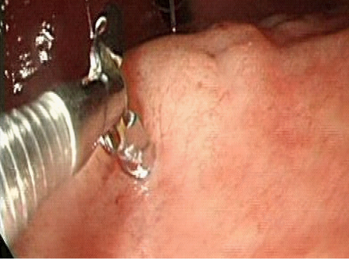
At endoscopy, the pyloric gland adenoma appears as a 1.5 cm nodular/dome-like mucosal elevation with whitish discoloration in the cardia.

Microscopically, the pyloric gland adenoma consists of narrow or cystically dilated tubules, which are fused or branched irregularly in the gastric mucosa (H&E ×20).

Microscopically, the tubules are lined by cuboidal to prismatic columnar epithelial cells that contain basal nuclei and pale eosinophilic cytoplasm (H&E ×100).
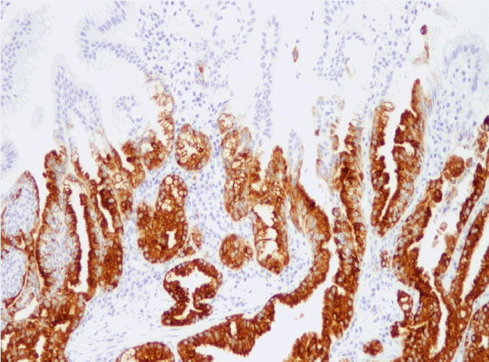
Immunohistochemically, the columnar epithelial cell lining tubules are positive for MUC6 immunohistochemically, indicating pyloric gland adenoma (MUC6 × 100).
치료 및 경과: 병변에 대하여 그림 5와 같이 내시경적 점막하 박리술과 mapping을 시행하였으며 조직검사상 1.5 cm 크기의 유문선 선종 소견이었고, 암성병변 및 이행부위는 관찰되지 않았으며 완전절제가 되었다. 환자는 시술 후 특별한 부작용 없이 퇴원하여 현재 12개월째 경과관찰 중이다.

The lesion was removed successfully by endoscopic submucosal dissection.
고 찰
위선종은 조직학적으로 장 점막형과 위 점막형으로 나뉘며, 위암은 장형에서 더 많이 발생하는 것으로 알려져 있다[3]. 최근까지 위선종이라는 용어는 장형의 평평한 선종을 의미하였고, 위형 선종은 예외적인 것으로 받아들여졌다[4]. 또한 위형 선종은 이론적으로 foveolar형과 유문형으로 나뉘어지나, 실제적으로 foveolar형은 Gastric-foveolar형 선암과 구별하기 어렵고 독일과 일본에서 점막 내 선암으로 진단 내려져왔기 때문에 일반적으로 위형선종은 대부분 유문선 선종이다[5]. 유문선 선종은 점막하 위 점막선에서 발생하는 종양으로 1990년 Watanabe 등[1]에 의해 위종양의 일부로 확인되었다. 진단은 면역조직화학 염색법을 이용하여 장형의 표시자인 MUC2와 CD10에서는 음성 반응을 보이나 위형의 표시자인 Mucin 6와 MUC5AC에 대해 강하게 반응하면서 염색되는 것으로 얻을 수 있다[6]. 1990년부터 2000년까지 위용종 2,778예를 분석한 일본의 한 연구에 의하면 유문선 선종은 90예로 모든 용종 중 2.7%로 흔하지 않은 용종으로 확인되었다[7]. 그러나 이는 유문선 선종의 보고가 매우 드문 것에 비하여는 상당히 높은 비율로 이는 증례보고가 부족했거나 유문선 선종이 제대로 진단되지 못하고 있을 가능성을 시사한다. 그리고 이 보고에 의하면 발생하는 위치는 위체부에서 58예(64%)에서 발생했으며, 분문부에서 발생한 경우는 약 7예(8%)에 불과하였다. 평균연령은 여자가 75세, 남자는 70세 정도였고, 남녀간 발생비율은 55:35 정도였다. 또한 18예(34%)에서 자가면역성 위축성 위염이 존재하였다. 자가 면역성 위축성 위염은 대개 고령과 여자에게서 더 흔하게 발병하는 경향이 있지만 유문선 선종과의 연관성에 대해서는 아직 연구가 더 필요한 상황이다. 또한 이 연구에서 유문선 선종의 약 30%에서 선암으로 이행되는 것으로 보고되었다. 유문선 선종은 위에서 가장 흔하게 발생하지만 십이지장[8], 췌관[9], 담낭 및 담관[10]에서 발생했다는 보고가 있었으며 이후에는 직장[11]과 바렛식도[12]에서 유문선 선종이 발생되었다는 증례 보고가 있었다.
유문선 선종은 일반적으로 내시경을 통해 관찰했을 때 결절성 형태로 돔형으로 융기되어 점막하 종양과 같이 보이며, 조직검사를 시행했을 경우 그림 2와 같이 좁고, 낭종성 모양으로 생긴 길고 작은 관들이 늘어나 있고, 이들이 불규칙한 나뭇가지 모양으로 뻗어 있다. 고배율에서 관찰하면 그림 3과 같이 주로 입방상피세포와 원주상피세포가 이 관들을 둘러싸고 있다.
본 증례에서는 건강 검진 내시경상 육안적 소견이 점막하종양 및 위선종이 의심되었고, 조직검사에서 위염 이외에는 특별한 소견이 관찰되지 않았다. 하지만 실제로 조기 위암과 위샘종의 내시경 생검과 내시경 점막하 박리법 후 병리학적 진단이 변하는 경우가 58.06%로 높다는 보고가 있어 단기간내 내시경 검사를 반복 시행하였고, 처음 내시경을 시행한 후 9개월 정도 지난 후 시행한 조직검사에서 유문선 선종이 확인되었다[13]. 또한 전구암 병변으로 악성으로 이행될 가능성이 있기 때문에 내시경적 점막하 박리술을 시행하였고, 병변은 완전히 제거되었다. 국내에서는 2003년도 십이지장에서 발생한 유문선형 선종 2례를 포스터로 발표하였으나 면역조직화학 염색을 시행하지 않아 확진되지 못한 증례이었고, 이후 문헌으로도 보고되지 않았다[14].