원발성 간세포암 환자에서 부갑상선 간세포암 전이가 병발한 1예
A case of hepatocellular carcinoma combined with solitary parathyroid metastasis
Article information
Abstract
저자들은 현재까지 국내외에 원발성 간세포 암의 전이부위로 부갑상선이 보고된 적이 없고 현미경적으로 tumor-to-tumor 전이(공여 부위: 간세포암, 수여 부위: 부갑상선 선종)가 병발한 1예를 경험하였기에 문헌고찰과 함께 보고하는 바이다.
Trans Abstract
Hepatocellular carcinoma (HCC) is the third leading cause of cancer-related death worldwide1). Extrahepatic metastasis of HCC is now increasing due to prolonged survival. Most extrahepatic HCC occurs in patients with advanced stages. The lung, abdominal lymph nodes, and bone are common sites of extrahepatic metastasis. However, the parathyroid gland has not been reported as a metastatic focus. We report the first case of parathyroid metastasis as the first single metastasis site of HCC and microscopic tumor- to-tumor metastasis to a parathyroid adenoma. (Korean J Med 79:686-690, 2010)
서 론
간세포암종은 전 세계적으로 다섯 번째 많은 암이면서 암 관련 사망 빈도가 세 번째로 높은 암이다. 간세포암의 예후는 최근 영상 진단 및 다양한 치료 기술이 발전되면서 과거에 비하여 많이 향상되어, 간암의 전이가 간암 치료의 중요한 문제로 대두되었다1). 간내 전이의 경우 간내 전이 병소의 재절제, 경동맥화학 색전술(TACE), 경피적 에탄올 주입(PEI) 등을 통한 치료 효과에 대한 보고는 있었다2-4). 그러나 간외 전이는 간내 전이에 비하여 빈도가 낮으며, 아직까지 간내 전이암의 치료에 관심이 집중되어 있는 바, 간외 전이의 치료에 대한 보고는 많지 않는 실정이다5). 간외 전이의 가장 흔한 부위는 폐, 주위 임파절, 근 골격계, 부신, 복막 등으로 알려져 있으며, 그외에도 비장 소장, 대장 및 식도 등 다양한 장기로의 전이가 보고되고 있다6). 그러나 아직까지 간세포암의 부갑상선 전이가 보고된 바는 없다.
이에 저자들은 국내외에 보고된 적이 없는 원발성 간세포암 환자에서 단독 전이 병소로 부갑상선 전이소견과 현미경적으로 tumor-to-tumor metastasis (공여 부위: 간세포암, 수여 부위: 부갑상선 선종)가 병발한 1예를 경험하였기에 문헌고찰과 함께 이를 보고하는 바이다.
증 례
환 자: 심〇〇, 남자, 53세
주 소: 전신 근육통과 열감
현병력: 42세 남자 환자로 내원 5일 전부터 발생한 전신 근육통과 열감을 주소로 내원하였다. 입원 당시 전신 피로감 및 열감과 근육통을 호소하였고, 한달 동안 3 kg의 체중감소가 있었다. 신체검사상 활력징후는 혈압 120/80 mmHg, 맥박수 80회/분, 호흡수 20회/분, 체온 38.4℃였다. 의식은 명료하였고, 경미한 공막황달 소견이 있었으며 복부 진찰에서 심와부와 우상복부에 경도의 압통을 호소하였다.
부와 우상복부에 경도의 압통을 호소하였다. 말초혈액 검사에서 백혈구 6,900/mm3, 혈색소 10.8 g/dL, 적혈구 용적률 31.3%, 혈소판 198,000/mm3이었으며 생화학 검사에서 혈중요소질소 15 mg/dL, 크레아티닌 1.1 mg/dL, 총 단백 6.3 mg/dL, 알부민 2.6 mg/dL, AST 74 IU/L, ALT 138 IU/L, 총 빌리루빈 2.48 mg/dL, LDH 418 U/L, alkaline phosphatase (ALP) 160 IU/L, 프로트롬빈 시간은 15.6초(INR 1.37)였으며 활성화 부분트롬보플라스틴 시간은 33.4초였다.
혈청 검사에서 Anti-HAV 음성, HBsAg 양성, HBsAb 음성, HBeAg 음성 , HBeAb 양성, Anti-HCV 음성이었고, HBV DNA 2,384 copies/mL, 알파태아단백은 40.73 ng/mL, PIVKA-II는 30 mAU/mL이었으며, 항 바이러스제를 투여한 적은 없었다. 동반된 간경변증의 Child-Turcotte-Pugh 분류의 등급은 Child A에 해당하였다.
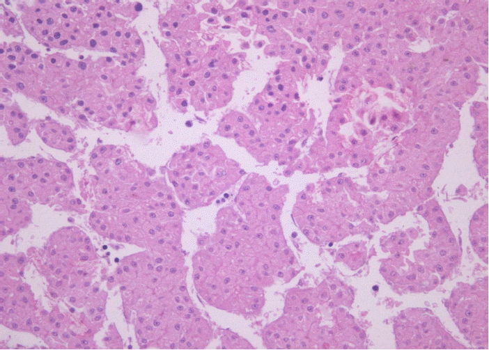
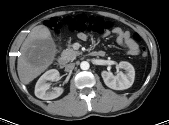
내원 당일 시행한 전산화 단층 촬영에서 간 우엽의 5번에서 6번 분절에 걸쳐 9 cm의 종괴가 관찰되었으며, 5번 분절에서 2.5 cm 크기의 arterial enhancement를 보이는 종괴가 관찰되었다(그림 1). S5의 2.5 cm 병변은 간세포암에 합당하나, S5/6의 9 cm 크기 종괴는 CT상 전형적인 간세포암의 양상을 띠지 않아서, 이 병변에 대한 간조직 생검을 실시하였다. 간 조직생검상 간세포암 진단되었으며(그림 2), PET CT상 원격 전이를 시사하는 소견은 관찰되지 않았고, TNM stage AJCC T3N1M0 IIIC였다.

Liver biopsy: The morphological pattern shows trabecular pattern of growth, corresponds to Grade 2 of the Edmondson-Steiner hepatocellular carcinoma classification (H&E stain, ×200).
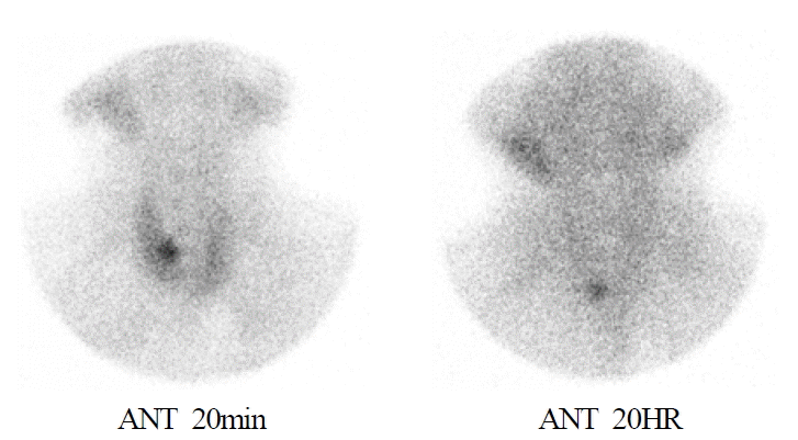
또한 내원 당시 calcium (Ca) 10.5 mg/dL, Ionized Ca 1.72 mmol/L, Phosphorus 1.5 mg/dL, PTH 142.51 pg/mL, TSH 0.4 uIU/mL, freeT4 1.38 ng/dL PTH-RP 1.1 pmol/L 미만의 일차성 부갑상선 항진증 소견을 보였다. 이상 소견의 원인을 확인하기 위해 Tc-99m MIBI 부갑상선 scan을 촬영하였고, 검사상 우측 갑상선 하단부위에 부분적인 hot uptake 소견이 관찰되었다(그림 3). 부갑상선 선종을 의심하고 부갑상선 절제술을 계획하였으나 환자 본인이 무증상으로 수술을 거부하여 우선 간 세포암에 대해 경동맥화학색전술을 먼저 시행하면서 경과관찰하기로 하였다.

Parathyroid scan with Tc-99-m Sestamibi: Initial images (20 min) showed a focal area of increased tracer uptake of the lower pole of the right thyroid lobe. Delayed images (2 HR) showed persistent activity in the same region. Impression was of a parathyroid adenoma in the lower pole of the right thyroid lobe.
내원 4개월 뒤 시행한 검사에서 PTH 159.14 pg/mL, calcium(Ca) 9.9 mg/dL, Phosphorus 1.8 mg/dL로 지속적인 PTH의 상승소견을 보였기에 환자의 수술 동의하에 우측 부갑상선절제술을 시행하였다.
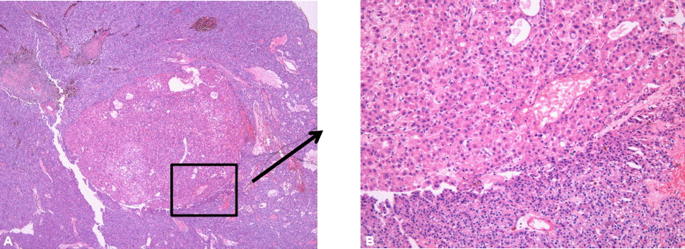
부갑상선 병리 조직 검사상 1.0×1.0 cm 크기의 부갑상선 선종 안에 anti-hepatocyte antibody 면역조직화학염색에 양성인 trabecular pattern의 0.2×0.2 cm 크기의 전이된 간세포암이 존재하는 tumor-to-tumor metastasis (공여 부위로 간세포암, 수여 부위로 부갑상선 선종) 소견이 관찰되었다(그림 4, 5). 따라서 환자는 간세포 암종의 부갑상선 선종과 동반된 부갑상선 전이로 최종 진단되었다.

Right parathyroidectomy : microscopic feature parathyroid gland, representing parathyroid adenoma, is hypercellular and homogenous and measures 1×1 cm. In parathyroid adenoma, there is the 0.2×0.2 cm sized nest of trabecular pattern of hepatocellular carcinoma (A) H&E stain ×40, (B) H&E stain×100.

Immunohistochemical stain for Anti-hepatocyte Antibody: immunostain shows positive to metastatic hepatocellular carcinoma cells (arrow, ×100).
수술 후 3일째 시행한 PTH 5.70 pg/mL calcium (Ca) 8.9 mg/dL, Phosphorus 4.4 mg/dL로 정상소견을 보였다. 후향적으로 확인한 혈청 칼슘 및 부갑상선호르몬 수치가 모두 정상 범위를 보여 현재까지 재발 없이 추적관찰 중이다.
고 찰
간세포암에서 간외 전이는 드물지 않게 동반될 수 있으며, 최근에는 진단기술의 발달과 치료법의 향상에 따른 생존기간의 증가로 간외 전이의 빈도가 더욱 증가하고 있다. 간세포암에서 간외 전이의 빈도는 13.5~64%로 보고되고 있는데6), 간세포암 진단 당시 같이 발견되어 보고되는 경우에는 대략 20~30%로 알려져 있다. 일반적으로 간세포암의 간외 전이 부위는 폐(55%)가 가장 흔하며, 림프절(53%), 골(28%), 부신(11%), 복막(11%) 등으로 비교적 흔하게 전이된다7,8). 현재까지 국내와 외국 문헌으로 보고된 간세포암의 드문 전이부위는 우리나라의 경우 중추신경계(23예), 위장관(22예). 피부(6예), 심장(6예), 안와(5예), 복막(4예), 담낭(3예), 구강(2예), 난소(2예), 유방(1예), 비강(1예), 기관지(1예), 신장(1예), 결막(1예) 순으로 보고되었다9). 또한 대부분의 경우 진단 당시 진행된 간세포암에서 빈번히 원격전이가 발생하는 것으로 보고되었고, 치료 후 간 내 재발 없이 원격전이가 일어난 경우라도 치료 전에 이미 진행된 간세포암이었거나 AFP 수치가 높은 경우(>1000 ng/mL)에 잘 발생하는 것으로 알려져 있다10). 그러나 국내외 문헌에서 본 증례와 같은 간세포암의 부갑상선 전이는 아직까지 보고된 바 없다7-9). 이러한 이유로는 진행성 간암의 짧은 생존률, 부갑상선 전이의 낮은 유병률 및 무증상 등을 들 수 있겠다. 본 환자에서도 동반된 원발성 부갑상선 항진증의 소견이 없었다면, 진단이 어려웠을 것으로 생각된다. 그러므로 진행성 간세포암 환자를 진료할 때 항상 원격 전이 유무를 염두에 둘 필요가 있다.
본 환자는 부갑상 선종에 대해 우측 부갑상선 절제 후 병리 조직상 tumor-to-tumor metastasis가 관찰되었는데, 이러한 “tumor-to-tumor metastasis”는 매우 드문 현상으로 1902년에 Berent에 의해 최초로 보고된 후 현재까지 100여 개의 다양한 증례가 문헌보고되었다11). 1958년 Dobbing12)과 1968년 Campbell13)은 tumor-to-tumor 전이의 진단기준을 제시하였다. ① 1개 이상의 원발성 암이 존재하고 ② 수여(recipient) 종양은 진성 종양(true neoplasm)이어야 하며 ③ 전이(Metastatic) 종양은 host tumor의 전이 임이 증명되어야 한다고 제시하였다. 인접장기전이, 림프종성, 백혈병성 림프절 전이는 진단기준에서 제외하였다.
공여 악성종양 부위는 폐암(52.1%), 전립선암(13%), 갑상선암(8.7%), 두경부 편평상피세포암, 유방암, 위장계암, 빈도 순이며14) 수여(recipient) 악성종양 부위로 신세포암(71.7%)이 가장 흔하며 그 외 육종(6.5%), 전립선암, 췌장암, 유방암, 자궁내막암, 갑상선암, 교아종, 회장 유암종, 신장의 호산성 과립 세포종(renal oncocytoma)이 보고되었다. 이러한 tumor-to-tumor 형태의 전이 발생 원인은 몇 가지 가설만 제시되었을 뿐 뚜렷히 밝혀지지 않았다15). 현재까지 국내외 문헌에서 본 증례와 같은 공여 부위로 간세포암, 수여 부위로 부갑상선 선종은 보고된 바가 없다12-16).
결론적으로 임상의사는 새로 생긴 부갑상선 결절로 내원한 환자를 문진할 때 다른 장기의 악성종양의 병력이 있는지를 확인하고 그러한 병력이 있는 환자에서는 부갑상선 결절이 원발 병소의 전이인지를 반드시 감별해야 한다.
본 증례는 PET-CT 및 전산화 단층 촬영에서 원격전이가 발견되지 않은 간세포 암 환자에서 진단 당시 혈청 calcium 증가, phosporus 감소, PTH 증가, Tc-99m MIBI 부갑상선 스캔상 우측 부갑상선 hot uptake 소견을 보여, 부갑상 선종에 대해 우측 부갑상선 절제술을 시행하였다. 수술 후 부갑상선 조직검사에서 부갑상선 선종 조직 안에 간세포암 소견이 동시에 관찰되어 최종적으로 간 세포암의 부갑상선 전이로 진단된 증례이다.
