 |
 |
| Korean J Med > Volume 92(1); 2017 > Article |
|
мҡ”м•Ҫ
н„ёкі°нҢЎмқҙм—җ мқҳн•ң мІҷ추 кіЁмҲҳм—ј мҰқлЎҖліҙкі к°Җ көӯлӮҙм—җм„ңлҠ” м•„м§Ғ м—Ҷм—Ҳмңјл©°, м „ м„ёкі„м ҒмңјлЎңлҸ„ л§Өмҡ° л“ңл¬јлӢӨ. м Җмһҗл“ӨмқҖ кёүм„ұ л°ұнҳҲлі‘мңјлЎң 3л…„ м „ н•ӯм•” м№ҳлЈҢлҘј л°ӣкі мһ¬л°ңмқҙ м—ҶлҠ” мғҒнғңлЎң м§ҖлӮҙлҚҳ нҷҳмһҗм—җм„ң л°ңмғқн•ң н„ёкі°нҢЎмқҙм—җ мқҳн•ң мІҷ추 кіЁмҲҳм—јмқ„ мЎ°м§Ғнҳ•нғңн•ҷм Ғ л°©лІ• л°Ҹ л©ҙм—ӯмЎ°м§Ғнҷ”н•ҷм—јмғү л°©лІ•мңјлЎң 진лӢЁн•ҳмҳҖкё°м—җ ліҙкі н•ҳлҠ” л°”мқҙлӢӨ
Abstract
Mucormycosis is a rare but fatal disease and usually affects the rhinocerebrum, lungs, traumatic wounds or surgical sites. Vertebral osteomyelitis due to mucormycosis is very rare, with only three cases caused by mucormycosis since 1970 being reported, and none in Korea. Here, we present a case of vertebral osteomyelitis caused by mucormycosis in a 67-year-old woman, having type 2 diabetes mellitus for 10 years, who was in complete remission from acute leukemia after chemotherapy 3 years previously.
н„ёкі°нҢЎмқҙмҰқ(mucormycosis)мқҖ Rhizopus spp, Mucor spp, Cunninghamella spp, Apophysomyces spp, Absidia spp, Saksenaea spp. л“ұм—җ мқҳн•ҙ л°ңмғқн•ҳлҠ” к°җм—јмңјлЎң кіөкІ©м Ғмқҙкі м „кІ©м Ғмқё к°җм—јмқ„ мқјмңјнӮӨлҠ” кІғмңјлЎң м•Ңл Өм ё мһҲлӢӨ. лҢҖл¶Җ분мқҖ л¶Җ비лҸҷ(39%), нҸҗ(24%), н”јл¶Җ(19%)м—җм„ң к°җм—јмқҙ лӮҳнғҖлӮҳл©°, кё°м Җ м§ҲнҷҳмңјлЎңлҠ” м•…м„ұ мў…м–‘, лӢ№лҮЁлі‘, мһҘкё°мқҙмӢқ л“ұмқҙ мһҲлӢӨ[1].
мІҷ추 кіЁмҲҳм—јмқҖ м „мІҙ кіЁмҲҳм—јмқҳ 3-5% м •лҸ„лҘј м°Ём§Җн•ҳлҠ” к°җм—јмңјлЎң мң лҹҪ м§Җм—ӯм—җм„ң л§Өл…„ 10л§Ң лӘ…лӢ№ 0.4-2.4лӘ…мқҙ л°ңмғқн•ңлӢӨ. мӣҗмқёк· мңјлЎң Staphylococcus aureusк°Җ м•Ҫ м Ҳл°ҳ м •лҸ„лҘј м°Ём§Җн•ҳлҠ” к°ҖмһҘ нқ”н•ң к· мқҙл©°, к·ёлһҢ мқҢм„ұ к°„к· (Gram negative rods)мқҙ 7-33%, coagulase-negative staphylococciк°Җ 5-16%лҘј м°Ём§Җн•ңлӢӨ. лҳҗн•ң мң н–ү м§Җм—ӯм—җм„ң Mycobacterium tuberculosis л°Ҹ BrucellaлҸ„ мӣҗмқёк· мңјлЎң мһҗмЈј нҷ•мқёлҗҳкі мһҲлӢӨ[2].
к·ёлҹ¬лӮҳ, н„ёкі°нҢЎмқҙм—җ мқҳн•ң мІҷ추 кіЁмҲҳм—јмқҖ м•„мЈј л“ңл¬ё м§ҲнҷҳмңјлЎң нҳ„мһ¬к№Ңм§Җ л¬ён—ҢкІҖмғүмғҒ 3мҳҲл§Ңмқҙ ліҙкі лҗҳм—ҲлӢӨ. м Җмһҗл“ӨмқҖ 67м„ё м—¬мһҗм—җм„ң н„ёкі°нҢЎмқҙм—җ мқҳн•ң нқү추 л¶Җмң„мқҳ мІҷ추 кіЁмҲҳм—јмқ„ 진лӢЁн•ң мӮ¬лЎҖлҘј кІҪн—ҳн•ҳмҳҖкё°м—җ л¬ён—Ңкі м°°кіј н•Ёк»ҳ ліҙкі н•ҳкі мһҗ н•ңлӢӨ.
нҷҳ мһҗ: 67м„ё м—¬мһҗ
мЈј мҶҢ: 2мЈј м „ л°ңмғқн•ң м–‘мӘҪ н•ҳм§Җ к·јл Ҙ м Җн•ҳ
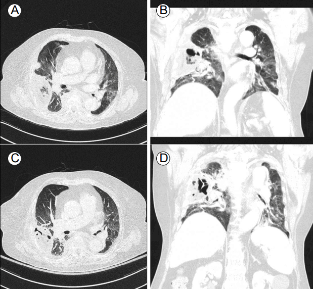
нҳ„лі‘л Ҙ: 3л…„ м „ кёүм„ұ л°ұнҳҲлі‘мңјлЎң мң лҸ„ н•ӯм•”нҷ”н•ҷмҡ”лІ•кіј 1м°ЁлЎҖмқҳ кіөкі н•ӯм•”нҷ”н•ҷмҡ”лІ• нӣ„ мҷ„м „мҷ„нҷ” мғҒнғңлЎң мқҙнӣ„ н•ӯм•” м№ҳлЈҢлҘј мӣҗм№ҳ м•Ҡм•„ мҷёлһҳ 추м Ғ кҙҖм°°л§Ң н•ҳлҚҳ нҷҳмһҗлЎң лӮҙмӣҗ 3мЈј м „ мҡ°мғҒм—Ҫмқҳ кҙҙмӮ¬м„ұ нҸҗл ҙ(Fig. 1)мңјлЎң мһ…мӣҗн•ҳмҳҖлӢӨ. лӢ№мӢң кё°кҙҖм§Җ лӮҙмӢңкІҪмқ„ мӢңн–үн•ҳмҳҖмңјлӮҳ лҸҷм •лҗң к· мқҖ м—Ҷм—Ҳмңјл©°, м„ёк· м„ұ нҸҗл ҙмңјлЎң мғқк°Ғн•ҳкі м—°кі м§Җ мқёк·ј лі‘мӣҗм—җм„ң ceftriaxone, clindamycinмқ„ нҲ¬м•Ҫн•ҳмҳҖлӢӨ. лӮҙмӣҗ 2мЈј м „л¶Җн„° м җм°Ё м–‘мӘҪ н•ҳм§Җ к·јл Ҙ м Җн•ҳк°Җ л°ңмғқн•ҳмҳҖкі , лӮҙмӣҗ 1мЈј м „л¶Җн„° л°°лҮЁ мһҘм• к°Җ лҸҷл°ҳлҗҳм–ҙ лӢӨмӢң м „мӣҗлҗҳм—ҲлӢӨ.
кіјкұ°л Ҙ: 10л…„ м „л¶Җн„° лӢ№лҮЁлі‘мңјлЎң metformin, linagliptinмқ„ ліөмҡ© мӨ‘мқҙкі , 7л…„ м „л¶Җн„° кі нҳҲм••мңјлЎң olmesartanмқ„ ліөмҡ© мӨ‘мқҙм—ҲлӢӨ.
мӢ мІҙ кІҖмӮ¬ мҶҢкІ¬: нҷңл Ҙ 징нӣ„лҠ” нҳҲм•• 149/89 mmHg, л§Ҙл°• мҲҳ 92нҡҢ/분, нҳёнқЎ мҲҳ 20нҡҢ/분, мІҙмҳЁ 37.4в„ғмҳҖлӢӨ. нӮӨлҠ” 152 cm, лӘёл¬ҙкІҢлҠ” 61 kgмқҙм—ҲлӢӨ. л§Ңм„ұ лі‘мғүмқ„ ліҙмҳҖкі мқҳмӢқмқҖ лӘ…лЈҢн•ҳмҳҖлӢӨ. м–‘мӘҪ мғҒм§Җмқҳ к·јл ҘмқҖ grade 5лЎң нҷ•мқёлҗҳм—Ҳкі , м–‘мӘҪ н•ҳм§Җмқҳ к·јл ҘмқҖ кі кҙҖм Ҳ м•„лһҳлЎң лӘЁл‘җ grade 0мқҙм—ҲлӢӨ. к°җк°ҒмқҖ T5-6 level м•„лһҳлЎң л–Ём–ҙм ё мһҲм—Ҳкі , мӢ¬л¶Җкұҙл°ҳмӮ¬лҠ” м •мғҒмңјлЎң лі‘м Ғ л°ҳмӮ¬лҠ” м—Ҷм—ҲлӢӨ.
кІҖмӮ¬мӢӨ мҶҢкІ¬: л§җмҙҲнҳҲм•Ў кІҖмӮ¬м—җм„ң л°ұнҳҲкө¬ 9,000/mm3, нҳҲмғүмҶҢ 10.8 g/dL, нҳҲмҶҢнҢҗ 155,000/mm3мҳҖлӢӨ. Erythrocyte sedimentation rateмқҖ 90 mm/hrмҳҖлӢӨ. нҳҲмІӯ мғқнҷ”н•ҷ кІҖмӮ¬м—җм„ң AST 63 IU/L, ALT 120 UI/L, мҙқ л№ҢлҰ¬лЈЁл№Ҳ 0.4 mg/dL, blood urea nitrogen 14 mg/dL, Cr 0.63 mg/dL, Na 137 mmol/L, K 4.1 mmol/L, Cl 103 mmol/L, C-reactive protein 3.93 mg/dL, нҳҲмІӯ galactomannan assayлҠ” 0.26 (negative)мҳҖлӢӨ.
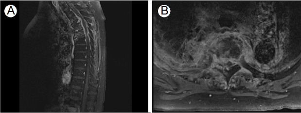
л°©мӮ¬м„ мҶҢкІ¬: нқүл¶Җ лӢЁмҲңмҙ¬мҳҒм—җм„ң мҡ°мёЎ мӨ‘м•ҷ нҸҗм•јм—җ кІҪнҷ”к°Җ мһҲмңјл©° мҡ°мёЎнҺём—җ нқүмҲҳк°Җ мһҲм—ҲлӢӨ. T-spine X-rayм—җм„ң T2-5м—җ кіЁ нҢҢкҙҙмҷҖ л””мҠӨнҒ¬ кіөк°„ нҳ‘м°©мқҙ мһҲм—ҲлӢӨ. нқүл¶Җ м»ҙн“Ён„°лӢЁмёөмҙ¬мҳҒ(computed tomography, CT)м—җм„ң мҡ°мғҒм—Ҫм—җ кІҪнҷ” л°Ҹ кіөлҸҷм„ұ лі‘ліҖмқҙ мһҲм—Ҳкі , мҡ°мёЎ к°ҖмҠҙм—җ нқүмҲҳк°Җ нҷ•мқёлҗҳм—ҲлӢӨ. мқҙлҠ” 3мЈј м „ нқүл¶Җ CTмҷҖ 비көҗн•ҳм—¬ нҒ° ліҖнҷ”лҠ” м—ҶлҠ” мғҒнғңмқҙл©°, reverse halo signмқ„ ліҙмқҙкі мһҲм—ҲлӢӨ. лҳҗн•ң T3, T5мқҳ мІҷ추мІҙк°Җ нҢҢкҙҙлҗҳм—Ҳкі , T2-T5 levelмқҳ мІҷ추мІҙ мЈјліҖ л°Ҹ мІҷмЈјкҙҖм—җ кІҪкі„к°Җ л¶Ҳ분лӘ…н•ң м—°л¶ҖмЎ°м§Ғ лі‘ліҖмқҙ мһҲм—ҲлӢӨ(Fig. 2).
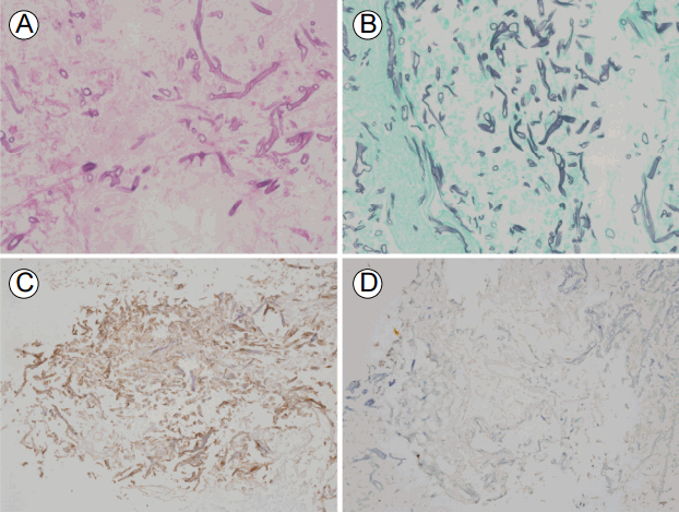
мЎ°м§Ғ мғқкІҖ: мҳӨлҘёмӘҪ T5 мІҷ추 мҳҶмқҳ лҶҚм–‘ мң мӮ¬ лі‘ліҖм—җм„ң CT мң лҸ„н•ҳ мЎ°м§Ғ мғқкІҖмқ„ н•ҳмҳҖлӢӨ. мЎ°м§Ғ мғқкІҖмқҳ Hematoxylin and Eosin м—јмғүм—җм„ң л§Ҳл””к°Җ м—Ҷмңјл©° л‘җк»ҳк°Җ лӢӨм–‘н•ң лӘЁм–‘мқ„ ліҙмқҙл©° л‘”к°ҒмңјлЎң к°Ҳлқјм ё лӮҳмҳӨлҠ” к· мӮ¬к°Җ мһҲм—Ҳкі , мЈјліҖмңјлЎң нҳёмӨ‘кө¬ м№ЁмңӨ л°Ҹ мЎ°м§Ғмқҳ кҙҙмӮ¬к°Җ мһҲм—ҲлӢӨ(Fig. 3A). GomoriвҖҷs methenamine silver м—јмғүм—җм„ң нқ‘мғүмңјлЎң м—јмғүлҗң к· мӮ¬к°Җ лҡңл ·н•ҳкІҢ кҙҖм°°лҗҳм—ҲлӢӨ(Fig. 3B). мЎ°м§Ғ м§„к· л°°м–‘м—җм„ңлҠ” м§„к· мқҙ мһҗлқјм§Җ м•Ҡм•ҳм§Җл§Ң, кі°нҢЎмқҙмқҳ мў…лҘҳлҘј к°җлі„н•ҳкё° мң„н•ҙ aspergillosisмҷҖ mucormycosis л©ҙм—ӯнҷ”н•ҷ(immunohistochemistry, IHC) м—јмғүмқ„ мқҙм „ м—°кө¬м—җм„ң кё°мҲ н•ң кІғ[3]кіј к°ҷмқҙ мӢңн–үн•ҳмҳҖкі , mucormycosis IHC stainм—җм„ң м–‘м„ұмқ„ лӮҳнғҖлӮҙм—ҲлӢӨ(Fig. 3C and 3D).
м№ҳлЈҢ л°Ҹ мһ„мғҒ кІҪкіј: мЎ°м§Ғ мғқкІҖ кІ°кіјлҘј л°”нғ•мңјлЎң лӢӨмӢң нқүл¶Җ CTлҘј лҰ¬л·°н•ҳмҳҖмқ„ л•Ң 3мЈј м „м—җ reverse halo signмқҙ кҙҖм°°лҗҳм—Ҳкі , мқҙмҷҖ мқём ‘н•ң л¶Җмң„м—җ м—°л¶ҖмЎ°м§Ғкіј лјҲ к°җм—јмқҙ мғқкёҙ кІғмқ„ кі л Өн• л•Ң 3мЈј м „м—җ м„ёк· м„ұ нҸҗл ҙмңјлЎң к°„мЈјлҗҳм—ҲлҚҳ лі‘ліҖмқҙ нҸҗм—җ мғқкёҙ н„ёкі°нҢЎмқҙмҰқмқҳ к°ҖлҠҘм„ұмқҙ лҶ’мқҖ кІғмңјлЎң нҢҗлӢЁн•ҳмҳҖлӢӨ. к·ёлҹ¬лӮҳ нҸҗм—җ 추к°Җм Ғмқё м№ЁмҠөм Ғмқё кІҖмӮ¬лҠ” 진н–үн•ҳм§Җ м•Ҡм•ҳлӢӨ. Liposomal amphotericin B 5 mg/kg/day нҲ¬м•Ҫн•ҳл©ҙм„ң ліҙмЎҙм Ғмқё м№ҳлЈҢлҘј н•ҳмҳҖмңјлӮҳ 1к°ңмӣ”м§ё лҡңл ·н•ң нҳём „ кІҪкіјлҠ” м—ҶлҠ” мғҒнғңлЎң м—°кі м§Җ лі‘мӣҗм—җм„ң liposomal amphotericin BлҘј мң м§Җн•ҳкё° мӣҗн•ҳм—¬ м „мӣҗлҗҳм—Ҳкі , м№ҳлЈҢлҘј мӢңмһ‘н•ң м§Җ 3к°ңмӣ” м§ё нҳём „, м•…нҷ” м—Ҷмқҙ 비мҠ·н•ң кІҪкіјлҘј ліҙмқҙкі мһҲлӢӨ(мҠӨн…ҢлЎңмқҙл“ңлҠ” нҲ¬м•Ҫн•ҳм§Җ м•Ҡм•ҳлӢӨ).
н„ёкі°нҢЎмқҙмҰқмқҖ нҳҲкҙҖ м№ЁлІ”мқ„ нҶөн•ҙ кёүмҶҚн•ң кҙҙмӮ¬лҘј мқјмңјнӮӨлҠ” нҠ№м§•мқ„ лӮҳнғҖлӮҙл©°, мЈјлЎң нҸ¬мһҗ(spore)мқҳ нқЎмһ…мқ„ нҶөн•ҙ мҪ”лҢҖлҮҢ л¶Җмң„ л°Ҹ нҸҗм—җ к°җм—јмқ„ мқјмңјнӮӨкұ°лӮҳ н”јл¶Җм—җ м§Ғм ‘м ҒмңјлЎң нҸ¬мһҗк°Җ м ‘мў…лҗҳл©ҙм„ң н”јл¶Җ н„ёкі°нҢЎмқҙмҰқмқ„ мқјмңјнӮӨкІҢ лҗңлӢӨ[1].
н„ёкі°нҢЎмқҙмҰқмқҳ кІ°м •м Ғмқё 진лӢЁмқҖ мЎ°м§Ғнҳ•нғңн•ҷм Ғ 진лӢЁкіј м§„к· л°°м–‘мқ„ мЎ°н•©н•ҳм—¬ мқҙлЈЁм–ҙм§Җм§Җл§Ң, нҠ№нһҲ н„ёкі°нҢЎмқҙмҰқм—җм„ңлҠ” л°°м–‘мқҙ мһҳ лҗҳм§Җ м•ҠлҠ”лӢӨ. л”°лқјм„ң мқҙлҹ¬н•ң кІҪмҡ°м—җлҠ” мЎ°м§Ғнҳ•нғңн•ҷм Ғ 진лӢЁм—җ мқҳмЎҙн•ҳкІҢ лҗҳм§Җл§Ң, лӘЁм–‘ мһҗмІҙк°Җ м„ңлЎң 비мҠ·н• л•ҢлҸ„ мһҲм–ҙ кө¬л¶„мқҙ л¶ҲлӘ…нҷ•н• мҲҳ мһҲлӢӨ[4]. к·ёлҹ¬лҜҖлЎң л©ҙм—ӯмЎ°м§Ғнҷ”н•ҷм—јмғү, мӨ‘н•©нҡЁмҶҢм—°мҮ„л°ҳмқ‘(polymerase chain reaction, PCR) л“ұмқҳ лӢӨлҘё ліҙмЎ°м Ғмқё 진лӢЁ л°©лІ•мқҙ м—°кө¬лҗҳкі мһҲмңјл©°, мөңк·ј л©ҙм—ӯмЎ°м§Ғнҷ”н•ҷм—јмғүмқ„ мқҙмҡ©н•ҳм—¬ 진лӢЁмқҳ м •нҷ•м„ұмқ„ лҶ’мқј мҲҳ мһҲлӢӨкі ліҙкі н•ҳмҳҖлӢӨ[3]. мқҙлІҲ мҰқлЎҖм—җм„ңлҸ„ GomoriвҖҷs methenamine sliver м—јмғүмқ„ нҶөн•ң мЎ°м§Ғнҳ•нғңн•ҷм Ғ 진лӢЁлҝҗл§Ң м•„лӢҲлқј mucormycosis IHC stain м–‘м„ұ л°Ҹ aspergillosis IHC stain мқҢм„ұ мҶҢкІ¬мқҙ нҷ•мқёлҗҳм–ҙ, 비лЎқ л°°м–‘м—җм„ң мқҢм„ұмңјлЎң нҷ•мқёлҗҳм—Ҳм§Җл§Ң н„ёкі°нҢЎмқҙмҰқмңјлЎң 진лӢЁн• мҲҳ мһҲм—ҲлӢӨ.
м№ЁмҠөм„ұ кі°нҢЎмқҙ к°җм—јм—җм„ң м•„мҠӨнҺҳлҘҙкёёлЈЁмҠӨмҰқ(aspergillosis)кіј н„ёкі°нҢЎмқҙмҰқмқ„ м •нҷ•нһҲ кө¬лі„н•ҳлҠ” кІғмқҖ м№ҳлЈҢ м•Ҫм ңк°Җ лӢ¬лқјм§Җкё° л•Ңл¬ёмқҙлӢӨ. м•„мҠӨнҺҳлҘҙкёёлЈЁмҠӨмҰқм—җм„ңлҠ” мқјм°Ё м№ҳлЈҢлЎң voriconazoleмқ„ нҲ¬м•Ҫн•ҳлҠ” кІғмқҙ amphotericin B deoxycholateлҘј нҲ¬м•Ҫн•ҳлҠ” кІғм—җ 비н•ҙ м№ҳлЈҢ м„ұкіөлҘ мқҙ лҚ” лҶ’лӢӨкі ліҙкі лҗҳм—ҲлӢӨ[5]. мқҙм—җ 비н•ҙ н„ёкі°нҢЎмқҙмҰқм—җм„ңлҠ” amphotericin B deoxycholateмқҳ мөңмҶҢм–өм ңлҶҚлҸ„(minimal inhibitory concentration)к°Җ 0.33 Вөg/mLмһ„м—җ 비н•ҙм„ң voriconazoleмқҳ мөңмҶҢм–өм ңлҶҚлҸ„лҠ” 41.14 Вөg/mLлЎң лҶ’м•„ voriconazoleмқҙ мқҳлҜё мһҲлҠ” нҡЁкіјлҘј лӮҳнғҖлӮҙм§Җ лӘ»н•ңлӢӨ[6]. л”°лқјм„ң м Җмһҗл“ӨмқҖ нҷ•мқёлҗң н„ёкі°нҢЎмқҙмҰқм—җ лҢҖн•ҳм—¬ liposomal amphotericin BлҘј нҲ¬м•Ҫн•ҳмҳҖлӢӨ.
н„ёкі°нҢЎмқҙмҰқм—җ мқҳн•ң мІҷ추 кіЁмҲҳм—јмқҖ л§Өмҡ° л“ңл¬ё м§Ҳлі‘мңјлЎң 1970л…„л¶Җн„° 2016л…„ 1мӣ”к№Ңм§Җ м „ м„ёкі„м ҒмңјлЎң 3мҳҲк°Җ ліҙкі лҗҳм—ҲлӢӨ(Table 1). Buruma л“ұ[7]мқҖ м•”мңјлЎң мқён•ҳм—¬ нӣ„л‘җм Ҳм ңмҲ л°Ҹ л¶Җ분 н•ҳмқёл‘җм Ҳм ңмҲ (laryngectomy with partial hypophyaryngectomy) л°Ҹ л°©мӮ¬м„ м№ҳлЈҢлҘј л°ӣм•ҳлҚҳ м Ғ мһҲлҠ” нҷҳмһҗм—җм„ң лӘ© нҶөмҰқмқҙ мһҲмңјл©° мІңмІңнһҲ 진н–үн•ҳлҠ” к·јл Ҙ м Җн•ҳ(м–‘мӘҪ нҢ”л¶Җн„° мӢңмһ‘н•ҳм—¬ мӮ¬м§Җл§Ҳ비лЎң 진н–ү) нҷҳмһҗлҘј ліҙкі н•ҳмҳҖлӢӨ. мқҙ нҷҳмһҗлҠ” 추нӣ„ кҙ‘лІ”мң„ нҸҗмғүм „мҰқмңјлЎң мқён•ҳм—¬ мӮ¬л§қн•ҳмҳҖлҠ”лҚ°, л¶ҖкІҖмқ„ нҶөн•ҳм—¬ мЎ°м§Ғн•ҷм ҒмңјлЎң л„“кі non-septateлҗң к· мӮ¬к°Җ ліҙм—¬ н„ёкі°нҢЎмқҙмҰқмңјлЎң 진лӢЁн•ҳмҳҖкі (л°°м–‘ кІҖмӮ¬лҠ” мӢңн–үн•ҳм§Җ м•Ҡм•ҳмқҢ), мқҙлЎң мқён•ң мІҷ추 кіЁмҲҳм—ј л°Ҹ кІҪл§үмҷё лҶҚм–‘мқҙ мӢ кІҪн•ҷм Ғ кІ°мҶҗмқҳ мӣҗмқёмһ„мқ„ нҷ•мқён•ҳмҳҖлӢӨ. Chen л“ұ[8]мқҖ мҡ”추 мІңмһҗ л°Ҹ radiofrequency nucleoplastyлҘј мӢңн–үл°ӣмқҖ мқҙнӣ„ н„ёкі°нҢЎмқҙмҰқм—җ мқҳн•ң мІҷ추 кіЁмҲҳм—ј л°Ҹ мІҷ추 추간нҢҗм—јмқ„ ліҙкі н•ҳмҳҖлӢӨ. н„ёкі°нҢЎмқҙмҰқмқҖ лҶҚм–‘м—җм„ң мӢңн–үн•ң л°°м–‘ кІҖмӮ¬мғҒ Rhizopus rhizopodoformisк°Җ лҸҷм •лҗҳм—Ҳкі , мЎ°м§Ғн•ҷм ҒмңјлЎң н„ёкі°нҢЎмқҙмҰқм—җ н•©лӢ№н•ң к· мӮ¬к°Җ нҷ•мқёлҗҳм–ҙ 진лӢЁлҗҳм—ҲлӢӨ. мқҙ нҷҳмһҗлҠ” liposomal amphotericin B нҲ¬м•Ҫкіј н•Ёк»ҳ л‘җ м°ЁлЎҖмқҳ мҲҳмҲ м Ғ ліҖм—°м Ҳм ңмҲ мқ„ мӢңн–үн•ҳм—¬ мҷ„м „нһҲ м№ҳлЈҢлҗҳм—ҲлӢӨ. л§Ҳм§Җл§үмңјлЎң 2014л…„м—җ Navanukroh л“ұ[9]мқҖ мӢ мһҘмқҙмӢқмқ„ л°ӣмқҖ м§Җ 1к°ңмӣ”мқҙ м§ҖлӮң нҷҳмһҗм—җм„ң н„ёкі°нҢЎмқҙмҰқм—җ мқҳн•ң нҸҗ к°җм—ј л°Ҹ м—үм№ҳлјҲ(sacral spine)мқҳ кіЁмҲҳм—ј, н—ҲлҰ¬м—үм№ҳлјҲ(lumbosacral spine)мқҳ кІҪл§үмҷё лҶҚм–‘мқ„ ліҙкі н•ҳмҳҖлӢӨ. н„ёкі°нҢЎмқҙмҰқмқҖ лҶҚм–‘ л°Ҹ кё°кҙҖм§ҖнҸҗнҸ¬м„ёмІҷм•Ў л°°м–‘кІҖмӮ¬м—җм„ң Cunninghamella spp.мқҙ лҸҷм •лҗҳм—Ҳкі , мӨ‘н•©нҡЁмҶҢм—°мҮ„л°ҳмқ‘кІҖмӮ¬лҘј нҶөн•ҙ Cunninghamella bertholletiaeлЎң нҷ•мқёлҗҳм–ҙ 진лӢЁлҗҳм—Ҳмңјл©°, мҲҳмҲ м Ғ ліҖм—°м Ҳм ңмҲ л°Ҹ liposomal amphotericin B нҲ¬м•ҪмңјлЎң нҳём „лҗҳм—ҲлӢӨ.
м•һм„ң кё°мҲ н•ң 3мҳҲмҷҖ ліё мҰқлЎҖлҘј мў…н•©н•ҳм—¬ ліҙл©ҙ, м•”, мӢ мһҘ мқҙмӢқкіј к°ҷмқҖ л©ҙм—ӯ м Җн•ҳ нҷҳмһҗмқҙкұ°лӮҳ мІҷ추м—җ м№ЁмҠөм Ғмқё мӢңмҲ мқ„ л°ӣмқҖ нҷҳмһҗм—җм„ң мІҷ추 кіЁмҲҳм—јмқҙ л°ңмғқн•ҳмҳҖлӢӨ. мІҷ추 кіЁмҲҳм—јмқҳ л°ңмғқ л¶Җмң„лҠ” лӢӨм–‘н•ҳмҳҖкі , 진лӢЁ л°©лІ•мқҖ мЈјлЎң мЎ°м§Ғнҳ•нғңн•ҷм Ғ 진лӢЁ лҳҗлҠ” л°°м–‘ кІҖмӮ¬лҘј н•ҳмҳҖкі , PCR кІҖмӮ¬лҘј мӢңн–үн•ң кІҪмҡ°лҸ„ мһҲм—ҲлӢӨ. 3мҳҲ мӨ‘ 2мҳҲм—җм„ң мҲҳмҲ м Ғ м№ҳлЈҢлҘј мӢңн–үн•ң нӣ„ н•ӯм§„к· м ң лі‘мҡ©мқ„ нҶөн•ҳм—¬ м„ұкіөм ҒмңјлЎң м№ҳлЈҢлҗҳм—ҲлӢӨ.
н„ёкі°нҢЎмқҙмҰқм—җ лҢҖн•ң к°ҖмһҘ мўӢмқҖ м№ҳлЈҢ л°©лІ•м—җ лҢҖн•ҙм„ңлҠ” м •лҰҪлҗң л°”к°Җ м—ҶмңјлӮҳ, Roden л“ұ[1]мқҙ мӢңн–үн•ң м—°кө¬м—җм„ң amphotericin B deoxycholate нҲ¬м•Ҫкіј н•Ёк»ҳ мҲҳмҲ м Ғ м Ҳм ңлҘј мӢңн–үн•ң кІҪмҡ° мғқмЎҙмңЁмқҙ к°ҖмһҘ лҶ’м•ҳмңјл©°(70%), amphotericin B deoxycholate лӢЁлҸ…(61%), мҲҳмҲ лӢЁлҸ…(57%)мңјлЎң м№ҳлЈҢн•ң кІҪмҡ° мғқмЎҙмңЁмқҙ лӮ®м•ҳлӢӨ. лҳҗн•ң мһҘкё°к°„мқҳ нҲ¬м•Ҫмқҙ н•„мҡ”н•ңлҚ° liposomal amphotericin Bмқҳ нҲ¬м•Ҫмқҙ кё°мЎҙмқҳ amphotericin B deoxycholateліҙлӢӨ мӢ лҸ…м„ұмқҳ л°ңмғқ л№ҲлҸ„к°Җ лӮ®м•„ м„ нҳёлҗңлӢӨ[10]. ліё мҰқлЎҖм—җм„ңлҸ„ liposomal amphotericin BлҘј нҲ¬м•Ҫн•ҳмҳҖмңјлӮҳ, мқҙлҜё к·јл Ҙмқҙ grade 0мңјлЎң л°ңмғқн•ң м§Җ 2мЈј мқҙмғҒ м§ҖлӮң мӢңм җмңјлЎң к°җм••мҲ мқҖ мҲҳмҲ лЎң м–»мқ„ мҲҳ мһҲлҠ” мқҙл“қкіј мң„н—ҳмқ„ кі л Өн•ҙм„ң мӢңн–үн•ҳм§Җ м•Ҡм•ҳлӢӨ.
ліё мҰқлЎҖлҠ” лӢ№лҮЁ л°Ҹ кёүм„ұ л°ұнҳҲлі‘ лі‘л Ҙмқҙ мһҲлҠ” нҷҳмһҗм—җм„ң н„ёкі°нҢЎмқҙм—җ мқҳн•ҙ мІҷ추 кіЁмҲҳм—јмқҙ л°ңмғқн•ҳмҳҖлӢӨ. мқҙлҹ¬н•ң кІҪн—ҳмқ„ л°”нғ•мңјлЎң мІҷ추 кіЁмҲҳм—ј нҷҳмһҗм—җм„ң м•…м„ұ мў…м–‘, лӢ№лҮЁ л“ұмқҳ кі°нҢЎмқҙ к°җм—ј мң„н—ҳм„ұмқҙ мһҲлҠ” кІҪмҡ° м„ёк· л°Ҹ кІ°н•ө мқҙмҷём—җ лӢӨлҘё мӣҗмқём—җ лҢҖн•ң к°ҖлҠҘм„ұмқ„ кі л Өн•ҳм—¬ мЎ°м§Ғ л°°м–‘ л°Ҹ мЎ°м§Ғ кІҖмӮ¬к°Җ л°ҳл“ңмӢң н•„мҡ”н•ҳлӢӨкі мғқк°Ғн•ңлӢӨ.
REFERENCES
1. Roden MM, Zaoutis TE, Buchanan WL, et al. Epidemiology and outcome of zygomycosis: a review of 929 reported cases. Clin Infect Dis 2005;41:634вҖ“653.


2. Mete B, Kurt C, Yilmaz MH, et al. Vertebral osteomyelitis: eight years' experience of 100 cases. Rheumatol Int 2012;32:3591вҖ“3597.


3. Jung J, Park YS, Sung H, et al. Using immunohistochemistry to assess the accuracy of histomorphologic diagnosis of aspergillosis and mucormycosis. Clin Infect Dis 2015;61:1664вҖ“1670.

4. Jensen HE, Salonen J, Ekfors TO. The use of immunohistochemistry to improve sensitivity and specificity in the diagnosis of systemic mycoses in patients with haematological malignancies. J Pathol 1997;181:100вҖ“105.


5. Herbrecht R, Denning DW, Patterson TF, et al. Voriconazole versus amphotericin B for primary therapy of invasive aspergillosis. N Engl J Med 2002;347:408вҖ“415.


6. Sun QN, Fothergill AW, McCarthy DI, Rinaldi MG, Graybill JR. In vitro activities of posaconazole, itraconazole, voriconazole, amphotericin B, and fluconazole against 37 clinical isolates of zygomycetes. Antimicrob Agents Chemother 2002;46:1581вҖ“1582.


7. Buruma OJ, Craane H, Kunst MW. Vertebral osteomyelitis and epidural abcess due to mucormycosis, a case report. Clin Neurol Neurosurg 1979;81:39вҖ“44.


8. Chen F, LГј G, Kang Y, et al. Mucormycosis spondylodiscitis after lumbar disc puncture. Eur Spine J 2006;15:370вҖ“376.


Three weeks before admission, axial (A) and coronal (B) chest computed tomography (CT) scans show consolidation with air bronchogram and aвҖҳreversed halo signвҖҷ in the right upper lobe (RUL). At the time of admission, the axial (C) and coronal (D) chest CT scans showed no change of cavitary lesion in the RUL.

FigureВ 1.
Sagittal Gd-enhancement T2-weighted (A) and sagittal Gd-enhancement fat-suppressed T1-weighted (B) magnetic resonance images of the thoracic vertebrae show enhancement of T4 and T5 vertebral bodies. Infiltrative prevertebral and epidural inflammatory masses at these levels cause cord compression and bilateral foraminal encroachment.

FigureВ 2.
(A) The vertebral body showed acute and chronic inflammation with fungal hyphae (H&E, Г—400). (B) GomoriвҖҷs methenamine silver (GMS) stain showed thick and sparsely septated fungal hyphae that branched at right angles, which is consistent with mucormycosis (GMS, Г—400). (C) These hyphae showed positive for anti-Rhizopus immunohistochemical staining (Г—200), (D) but negative for anti-Aspergillus immunohistochemical staining (Г—200).

FigureВ 3.
TableВ 1.
Cases of vertebral osteomyelitis caused by mucormycosis
| Authors, yr | Age/Sex | Underlying disease or condition | Spine region affected | Diagnostic methods | Surgery | Antifungal agents | Treatment duration | Prognosis |
|---|---|---|---|---|---|---|---|---|
| Buruma, et al. (1979) [7] | 60/M | Laryngectomy with partial hypophyaryngectomy and radiotherapy due to hypopharyngeal carcinoma | C34 | Histomorphology (autopsy) | No | No | . | Death |
| Chen, et al. (2006) [8] | 57/F | L4-5 disc puncture and radiofrequency nucleoplasty | L4-5 | Abscess culture, histomorphology | Yes | ABCD, followed by itraconazole | 8 months | Survival |
| Navanukroh, et al. (2014) [9] | 42/F | Kidney transplantation | S1 | BAL culture, abscess culture, PCR | Yes | LAMB, followed by posaconazole | 3 months | Survival |
| Present case (2016) | 67/F | AML, DM | T4-5 | Histomorphology, IHC | No | LAMB | On treatment | On treatment |


 PDF Links
 PubReader
 ePub Link
 Full text via DOI
 Download Citation
 Print
Print


