INTRODUCTION
Pheochromocytomas and paragangliomas are rare neuroendocrine tumors with an incidence of 2-8 per million persons per year [1]. Susceptibility to pheochromocytomas and paragangliomas is an established component of four genetic syndromes: multiple endocrine neoplasia types 2A and 2B, neurofibromatosis 1 (NF-1), von Hippel Lindau, and Carney-Stratakis dyad. NF-1 is an autosomal dominant genetic disorder of the nervous system primarily affecting the development of neural cell tissues. Vascular abnormalities are frequently associated with NF-1 and are reportedly seen in the renal, gastrointestinal, coronary, and cerebral vasculature [2,3]. Cerebrovascular manifestations include stenosis or occlusion of major vessels, arterio-venous fistulae, arterio-venous malformations, and aneurysms [4]. Spontaneous intracerebral hemorrhage is occasionally reported in patients with either NF-1 or pheochromocytoma [5]. A careful review of the literature, however, has not revealed a case of ruptured cerebral aneurysms caused by concurrent pheochromocytoma and NF-1, requiring urgent intervention.
We report a case of subarachnoid hemorrhage (SAH) caused by multiple ruptured cerebral aneurysms in a patient with concurrent NF-1 and pheochromocytoma.
CASE REPORT
A 57-year-old woman was admitted to our hospital for a 1-day history of headache followed by drowsiness. She did not report any history of hypertension. Her blood pressure (BP) on admission was 200/148 mmHg.
A computed tomography (CT) scan of the head revealed SAH more than 1-mm thick (Fisher grade 3) (Fig. 1A). Conventional cerebral angiography revealed ruptured bilateral cerebral aneurysms in the internal carotid artery (ICA), one large-sized fusiform aneurysm in the right distal ICA, and another saccular one in the left distal ICA (Fig. 1B-1 and 1B-2). Immediate coil embolization was performed for both aneurysms without complication (Fig. 1C-1 and 1C-2).
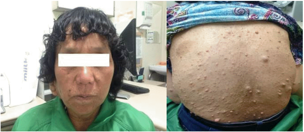
Physical examination revealed numerous pea-sized bumps distributed over the entire body (Fig. 2). Lisch nodules were observed under slit lamp examination. She had neither caf├σ-au-lait spots nor bony abnormalities on skull x-ray. She was diagnosed with NF-1 with two criteria: neurofibromas and Lisch nodules. She denied any familial history of NF-1.
Genetic analysis of the NF-1 gene was performed at the Department of Laboratory Medicine and Genetics, Samsung Medical Center, in Seoul Korea. Briefly, total RNA was extracted from peripheral blood leukocytes, and the entire NF-1 mRNA (reference sequence: NM_001042492.2) was amplified by reverse-transcription polymerase chain reaction (RT-PCR). After RT-PCR, Sanger sequencing of NF-1 mRNA followed by genomic DNA confirmation revealed a premature stop codon at the 328th residue encoding cysteine (p.Cys328*). This variant has previously been reported as NF-1 [6].
During hospitalization, fluctuation in BP was noted, and 24-hr ambulatory BP usually varied from 110 to 230 mmHg systolic and from 60 to 110 mmHg diastolic (Fig. 3A), requiring antihypertensive medication. Hypertension in the form of paroxysmal attacks led us to suspect pheochromocytoma. In a 24-hr urine collection, the patientέΑβs vanillylmandelic acid level was 22.8 mg/day (normal value; < 8 mg/day), and her metanephrine level was 6.0 mg/day (normal value; < 0.8 mg/day). Abdominal CT revealed an ovoid-shaped, heterogeneously well-enhancing mass in the medial limb of the right adrenal gland (Fig. 3B).
Elective surgical resection of the adrenal mass was planned. Ten milligrams of non-competitive, non-selective alpha-adrenergic blocker (phenoxybenzamine) and subsequent additional beta-adrenergic blocker were administered for 2 weeks prior to surgery. The mass was excised via a laparoscopic approach and proved to be a pheochromocytoma (Fig. 4). The patient was uneventful perioperatively. After resection, she was normotensive and discharged without antihypertensive medication.
DISCUSSION
NF-1 vasculopathy is a potentially serious but less well-known, under-recognized component of multisystemic genetic disorders. Based on a review of literature, the ICA is the most commonly affected (44%) followed by the middle cerebral artery (19.3%), anterior cerebral artery (16.4%), and posterior cerebral artery (8.2%). These data are consistent with our case, which affected both internal carotid arteries [7]. The precise mechanism of cerebral vasculopathy in NF-1 is not clearly understood but is likely related to the function of neurofibromin, the protein product of the NF-1 gene [8].
Pheochromocytoma is a rare manifestation of NF-1, estimated in 0.1-5.7% of people with NF-1. Mutation of the NF-1 gene is known to cause familial pheochromocytoma [9]. Although pheochromocytoma accounts for less than 0.5% of patients with paroxysmal or sustained hypertension [10], it is imperative to diagnose it early to avoid its devastating complications. Among these complications, cerebral hemorrhage is unusual but potentially fatal. In our case, we assumed that fluctuating blood pressure from pheochromocytoma predisposed the cerebral aneurysmal vessels to rupture. This case demonstrates that the delayed diagnosis and treatment of pheochromocytoma in patients with NF-1 can cause serious complications from fluctuating blood pressure. Thus, in patients with NF-1 and hypertension, screening for the early detection of cerebrovascular abnormalities and pheochromocytoma is crucial to prevent a life-threatening event.
To our knowledge, the present report is the first concerning SAH due to ruptured multiple cerebral aneurysms associated with pheochromocytoma in a patient with NF-1.


 PDF Links
 PubReader
 ePub Link
 Full text via DOI
 Download Citation
 Print
Print


